Achalasia
Clinical Snapshot
Achalasia is a primary esophageal motility disorder defined by two abnormalities occurring together: failure of the lower esophageal sphincter (LES) to relax during swallowing, and absent normal (peristaltic) contraction in the body of the esophagus. Both result from progressive degeneration of the inhibitory, nitric-oxide- and VIP-releasing neurons of the myenteric (Auerbach) plexus in the distal esophagus and LES — the cause of that degeneration is idiopathic in the large majority of North American cases, though it is thought to involve an autoimmune process, possibly triggered by a prior viral infection, in a genetically susceptible person. With the inhibitory input lost, cholinergic (excitatory) tone goes unopposed: the LES stays persistently contracted instead of relaxing to let a swallowed bolus pass, and the esophageal body loses its coordinated peristaltic wave.[1]
Achalasia is uncommon but not rare, and it is important precisely because it is a treatable cause of progressive dysphagia and weight loss that is frequently mistaken — sometimes for years — for reflux disease, given how routinely both present with retrosternal discomfort and regurgitation. There is currently no drug that reverses or halts the underlying neuronal loss: every available therapy, pharmacologic or procedural, works by mechanically or pharmacologically reducing resistance at the LES, not by restoring peristalsis. This chapter is written to be read alongside — not instead of — the Gastroesophageal Reflux Disease (GERD) and Dyspepsia and Peptic Ulcer Disease chapters, both of which already flag achalasia as the differential to consider once an adequate PPI trial has failed to explain a patient's symptoms.
Recognize the Condition
The defining symptom pattern
Progressive dysphagia to both solids and liquids is the hallmark of achalasia and the single most useful feature separating it from a mechanical cause of dysphagia. A structural narrowing — a peptic stricture, a Schatzki ring, or an esophageal cancer — typically causes dysphagia for solids first, progressing to liquids only as the lumen narrows further. Because achalasia is a functional outflow problem rather than a fixed narrowing, liquids are often impaired from early in the disease course, sometimes even before solids become difficult. Onset is usually insidious, developing over months to years, and patients frequently develop compensatory behaviours long before seeking care — eating very slowly, drinking large amounts of fluid with each bite to help push the bolus through, standing and stretching or arching the neck and back after swallowing (sometimes performing a Valsalva-like maneuver), and avoiding solid or dry foods in social settings. A history of these adaptive behaviours is itself a useful clue.[1]

Other common features
- Regurgitation of undigested food — typically bland or only mildly sour rather than frankly acidic, since it is esophageal content rather than refluxed gastric acid; can occur well after a meal, including during sleep, and is a genuine aspiration risk.
- Unintentional weight loss — occurs in a meaningful proportion of patients by the time of diagnosis and should always prompt consideration of pseudoachalasia (see Red Flags & Refer) rather than being assumed to be a routine feature.
- Retrosternal chest pain, sometimes cramping or squeezing in character — more prominent in Chicago Classification type III (spastic) disease, and occasionally severe enough to mimic cardiac chest pain.
- Nocturnal cough, choking, or recurrent aspiration pneumonia — from regurgitated esophageal contents entering the airway while supine.
- Heartburn-like symptoms occur in a substantial minority and are a major reason achalasia is initially misdiagnosed and treated as GERD — the mechanism here is retained, fermenting food producing lactic acid in the esophagus, not true gastric acid reflux, and these symptoms characteristically do not improve with a PPI trial.
Risk Factors
- No strong sex predilection; can present at any age, most commonly diagnosed between 30 and 60 years, though pediatric and older-adult presentations both occur.
- An autoimmune, possibly virally-triggered process in a genetically susceptible person is the leading theory for idiopathic achalasia — the large majority of North American cases.
- Chagas disease (Trypanosoma cruzi infection, endemic to parts of Latin America) causes a secondary achalasia that is clinically and manometrically indistinguishable from idiopathic disease, through direct destruction of the same myenteric neurons — ask specifically about birth or extended residence in an endemic region.
- Allgrove (triple A) syndrome — a rare autosomal recessive disorder combining Achalasia, Addisonian adrenal insufficiency, and Alacrima (absent tearing) — is an important cause in a child presenting with achalasia (see Special Populations).
- Chronic, high-dose opioid use can produce a manometric pattern that mimics achalasia or esophagogastric junction outflow obstruction (opioid-induced esophageal dysfunction) by directly increasing LES tone and disrupting peristalsis — always review the medication list before attributing a new manometric diagnosis to primary achalasia.
- A first-degree relative with achalasia is a very weak and inconsistent risk factor — true familial clustering is rare and should prompt consideration of a distinct genetic syndrome rather than being treated as meaningfully increasing an individual's risk on its own.
Red Flags & Refer
Pseudoachalasia — a malignancy (most often an adenocarcinoma of the gastric cardia or distal esophagus, occasionally a paraneoplastic process from a tumour elsewhere) infiltrating the myenteric plexus or mechanically obstructing the gastroesophageal junction — can reproduce the identical symptom pattern, and in some cases the identical manometric pattern, as primary achalasia. This is the single most important diagnosis not to miss in this chapter: proceeding directly to a myotomy or pneumatic dilation in a patient with an unrecognized malignancy delays cancer treatment and can worsen outcomes. Have a low threshold for pursuing the features below, and never let a manometry result alone be the final word.[1]
- Rapid symptom onset — dysphagia progressing to significant severity in under 6 months is atypical for idiopathic achalasia, which is typically insidious over a much longer course.
- Age 50-60 years or older at first presentation.
- Substantial, rapid weight loss out of proportion to the reported duration and severity of dysphagia.
- A short overall disease duration before diagnosis is sought, particularly when combined with either of the above.
- Any mass, irregular narrowing, or infiltrative-appearing mucosa seen at endoscopy, especially at the gastroesophageal junction or gastric cardia — biopsy any such finding before proceeding further.
Upper endoscopy — including careful retroflexed inspection of the gastroesophageal junction and cardia, with biopsy of anything suspicious — must be performed before high-resolution manometry in every new suspected achalasia diagnosis specifically to exclude this possibility; manometry cannot reliably distinguish primary achalasia from pseudoachalasia on its own.[1]
Other reasons to escalate
- Suspected aspiration pneumonia from regurgitation — treat the acute infection and reinforce positional/dietary precautions (see Non-Pharmacologic Treatment) while arranging definitive workup.
- Severe, unrelenting chest pain or any suspicion of an alternative cardiac cause — investigate cardiac causes first rather than assuming an esophageal spasm pattern.
- Suspected esophageal perforation after pneumatic dilation, Heller myotomy, or POEM (new severe chest/back pain, fever, subcutaneous emphysema, or hemodynamic instability following the procedure) — this is a surgical emergency requiring immediate assessment, not routine post-procedure discomfort.
- A patient with long-standing (roughly 10-15 years or more), poorly controlled achalasia reporting a new or changed symptom pattern — long-standing achalasia carries an increased long-term risk of esophageal squamous cell carcinoma from chronic stasis and mucosal irritation; a low threshold for repeat endoscopy is appropriate even though no fixed surveillance interval is established in current guidance.
Assessment & Diagnosis
Diagnostic sequence
Follow this order deliberately — each step exists to exclude the step before it from being wrong, not merely to add confirmation.[1]
- Upper endoscopy first, with careful retroflexed inspection of the gastroesophageal junction and biopsy of any abnormal-appearing mucosa, to exclude a mechanical or malignant cause (pseudoachalasia) before anything else. Endoscopic clues supporting achalasia itself include retained food or saliva in a dilated esophagus despite a fasting patient, and a puckered, tight-appearing gastroesophageal junction that nonetheless allows the scope to pass with gentle pressure (a finding that helps distinguish a functional from a truly fixed mechanical obstruction).
- Barium esophagram, looking for esophageal body dilation, a smoothly tapered "bird's beak" narrowing at the gastroesophageal junction, and poor or absent emptying of contrast into the stomach. A timed barium esophagram (measuring the height of the retained barium column at 1, 2, and 5 minutes after swallowing) adds an objective, reproducible measure of esophageal emptying that is also useful later for assessing treatment response.
- High-resolution manometry (HRM) — the diagnostic gold standard. HRM confirms the diagnosis by demonstrating an elevated integrated relaxation pressure (IRP, reflecting incomplete LES relaxation) together with absent normal peristalsis in the esophageal body, and assigns the Chicago Classification subtype that most directly guides treatment selection (see below). Esophageal pressure topography (the modern HRM display) is preferred over older conventional line-tracing manometry for this diagnosis.[1]
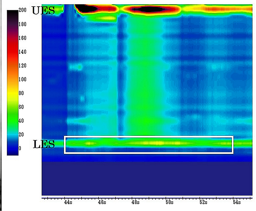
Chicago Classification v4.0 — subtypes and what they predict
| Subtype | Manometric pattern | Practical implication |
|---|---|---|
| Type I (classic) | Elevated IRP with 100% failed peristalsis and no significant esophageal pressurization | Effectively treated by any definitive modality — pneumatic dilation, Heller myotomy, or POEM. |
| Type II | Elevated IRP with 100% failed peristalsis and panesophageal pressurization on at least 20% of swallows | The subtype with the best overall response to treatment across all three definitive modalities — the most favourable prognosis of the three. |
| Type III (spastic) | Elevated IRP with premature (spastic) contractions on at least 20% of swallows and no normal peristalsis | Poorest response to pneumatic dilation specifically — favour POEM (with an extended myotomy covering the spastic segment) or Heller myotomy with an extended myotomy. |
A related but distinct manometric diagnosis: esophagogastric junction outflow obstruction (EGJOO)
EGJOO shows an elevated IRP similar to achalasia, but with some preserved, non-failed peristalsis — it does not meet full criteria for achalasia. Causes include a subtle mechanical process, early/evolving achalasia, and — relevant to pharmacists specifically — chronic opioid use, which can produce this exact pattern by directly raising LES tone. Confirmed EGJOO in a patient on regular opioids should prompt a trial of opioid reduction or discontinuation, with repeat manometry, before committing to an achalasia-directed invasive procedure.
Distinguishing achalasia from its key mimics
| Feature | Achalasia | GERD | Pseudoachalasia (malignancy) | Diffuse esophageal spasm |
|---|---|---|---|---|
| Dysphagia pattern | Progressive, solids and liquids from early on | Usually absent unless a stricture has developed, typically solids first | Progressive, often more rapid course than idiopathic achalasia | Intermittent, not reliably progressive |
| Regurgitation | Undigested food, bland/mildly sour, can occur well after eating | Acidic or bilious, related to position and meal timing | Can resemble achalasia | Uncommon |
| Response to an adequate PPI trial | No meaningful improvement | Improves | No meaningful improvement | No meaningful improvement |
| Barium esophagram | Bird's beak narrowing, dilated esophagus, delayed emptying | Usually normal | Narrowing can look similar but is often more irregular or shouldered | Corkscrew pattern possible |
| Endoscopy | Retained food/saliva, puckered gastroesophageal junction; mucosa itself normal | Erosive esophagitis, or normal in NERD | Mass, irregular stricture, or infiltrative mucosa; biopsy positive | Normal |
| HRM | Elevated IRP with absent normal peristalsis (subtyped I/II/III) | Normal, or a hypotensive LES with reflux on pH-impedance testing | Can mimic achalasia's manometric pattern exactly — endoscopic exclusion of malignancy is still required | Normal IRP; premature contractions without the LES outflow obstruction of achalasia |
Treatment Targets
- Relieve dysphagia and regurgitation and durably reduce functional outflow resistance at the LES — no available therapy restores normal peristalsis or reverses the underlying neuronal loss, so every treatment goal here is symptomatic and mechanical, not curative.
- Prevent and reverse malnutrition and unintentional weight loss.
- Reduce aspiration risk from regurgitation.
- Select the definitive therapy most likely to succeed for the patient's specific Chicago Classification subtype and overall fitness for a procedure.
- Minimize treatment-related complications, particularly esophageal perforation during dilation or myotomy.
- Anticipate and manage the reflux that Heller myotomy and POEM predictably create by weakening the LES barrier.
- Maintain long-term symptom control through monitoring, since recurrence after any single treatment is common and further intervention is often eventually needed.
Choose Initial Treatment
There is no disease-modifying drug therapy for achalasia
Pharmacologic therapy — sublingual nitrates or calcium channel blockers — bridge only, never definitive treatment. Efficacy is modest, inconsistent, and typically wanes within weeks as tolerance develops, and both drug classes carry a meaningful hypotension/headache burden at the doses required to lower LES pressure. Reserve pharmacologic bridging for a patient awaiting a scheduled definitive procedure, or one who is a genuinely poor candidate for any procedural option and has explicitly declined or cannot access one — it should never be used as an ongoing substitute for definitive therapy in a patient who is, or could become, a reasonable procedural candidate.[1]
Type I or Type II pneumatic dilation, laparoscopic Heller myotomy (with a partial fundoplication to reduce post-myotomy reflux), and POEM are comparably effective — selection is guided by local expertise, the patient's fitness for a procedure, and patient preference rather than by strong subtype-specific superiority of one modality over another.[1][3]
Type II specifically shows the best overall response to treatment of the three subtypes, regardless of which of the three modalities is chosen.
Type III (spastic) pneumatic dilation has a materially lower success rate in this subtype — favour POEM with an extended myotomy that covers the spastic segment, or Heller myotomy with a similarly extended myotomy, over pneumatic dilation as the initial approach.[4]
Poor surgical/procedural candidate, or awaiting a scheduled definitive procedure a sublingual nitrate or calcium channel blocker taken before meals, and/or endoscopic botulinum toxin injection into the LES, are reasonable temporizing measures (see Drug Tables for dosing). Botulinum toxin's effect is temporary (weeks to a few months) and repeated injections cause submucosal fibrosis that measurably increases the technical difficulty and complication risk of a later myotomy — favour it specifically in patients unlikely to ever proceed to surgery (very frail, limited life expectancy) rather than as a routine bridge in someone who will eventually have a myotomy or POEM.
Start a once-daily PPI routinely after Heller myotomy or POEM given the substantial, well-documented risk of post-procedure gastroesophageal reflux from the weakened LES barrier — reflux esophagitis was seen in 57% of patients at 3 months after POEM versus 20% after Heller myotomy with fundoplication in the largest randomized comparison to date.[4] Symptom severity correlates poorly with objective esophagitis on endoscopy after these procedures, so counsel patients that the absence of heartburn does not reliably mean the absence of reflux, and do not rely on symptoms alone to decide whether PPI therapy is still needed.[5]
Non-Pharmacologic Treatment
Definitive procedures — the primary treatment for achalasia
These are drug-free interventions and, for most patients who are reasonable candidates, represent the actual goal of treatment rather than a "non-drug adjunct" to pharmacologic therapy — the reverse of how most other chapters in this library are organized.
The 3 definitive modalities
Pneumatic dilation (PD)
Endoscopic, fluoroscopically-guided graded balloon dilation of the LES, typically starting with a 30 mm balloon and progressing to 35 mm and then 40 mm on subsequent sessions if needed. Effective for type I and II achalasia; materially less effective for type III. The main serious risk is esophageal perforation (roughly 2-5% across published series, most often at the time of a larger initial dilation), which is why PD is generally performed where surgical backup is immediately available.[1][3]
Laparoscopic Heller myotomy (LHM)
Surgical division of the circular muscle fibres of the LES and distal esophagus, almost always paired with a partial fundoplication (Dor or Toupet) in the same operation specifically to reduce the reflux that an unprotected myotomy would otherwise cause. Durable long-term symptom relief in a large majority of appropriately selected patients.
Peroral endoscopic myotomy (POEM)
An endoscopic, natural-orifice technique: a submucosal tunnel is created in the esophageal wall and the circular muscle layer is divided from within, without any external incision. No anti-reflux procedure is added in standard POEM, which is the main reason its post-procedure reflux rate is higher than LHM's. Comparably effective to LHM for type I/II disease, and generally preferred for type III given the ability to extend the myotomy further proximally to cover the spastic segment.[4]
Reserved for refractory or end-stage disease
Esophagectomy is a last-resort option for a small minority of patients with progressive, treatment-refractory megaesophagus (a markedly dilated, tortuous "sigmoid" esophagus) who have failed dilation, myotomy, and POEM — it carries substantially higher morbidity than any of the above and is reserved for genuinely refractory disease.
Dietary and positional counselling (adjunctive, alongside definitive therapy)
- Eat slowly, chew thoroughly, and take small, frequent meals rather than large ones.
- Drink fluids with meals to help the bolus pass, and remain upright during and for a period after eating.
- Avoid lying down soon after a meal, and elevate the head of the bed to reduce nocturnal regurgitation and aspiration risk.
- Refer for a formal nutrition assessment if unintentional weight loss is significant, pending definitive treatment.
Special Populations
Breastfeeding: PPIs and H2RAs are compatible with breastfeeding at usual doses. Published safety data specific to nifedipine, isosorbide dinitrate, or botulinum toxin use for achalasia during breastfeeding are limited — review each agent's own product-specific lactation guidance individually rather than assuming class-wide safety.
Pediatrics
Achalasia is rare in children but does occur, and a child presenting with achalasia should prompt screening for Allgrove (triple A) syndrome — the combination of Achalasia, Addisonian adrenal insufficiency, and Alacrima (absent tearing) caused by AAAS gene mutations. Missing the adrenal insufficiency component can be life-threatening if an adrenal crisis develops unrecognized, so this screen matters even when the achalasia itself seems isolated. Diagnosis and management should be directed by pediatric gastroenterology; pharmacologic bridge dosing in this chapter is written for adults and is not established for routine pediatric use.
Older adults
The age group with the highest incidence of new achalasia diagnoses. Increased sensitivity to the hypotensive effects of nitrates and calcium channel blockers argues for particular caution with pharmacologic bridging in this group. POEM and Heller myotomy are both well tolerated even in carefully selected older patients and age alone should not be used to withhold definitive therapy; regurgitation-related aspiration risk is also higher in this group, reinforcing the positional/dietary counselling above.
Secondary achalasia — Chagas disease
In a patient with a relevant travel or residency history in an endemic region of Latin America, consider Trypanosoma cruzi serology. Chagasic achalasia is managed with the same definitive-therapy framework above once the diagnosis is confirmed and a mechanical/malignant cause has still been excluded by endoscopy.
Opioid-induced esophageal dysfunction
Chronic, regular opioid use can itself produce an achalasia-like or EGJOO manometric pattern by directly increasing LES tone. Review and, where feasible, taper the opioid regimen and repeat manometry before proceeding to an achalasia-directed invasive procedure in a patient on significant chronic opioid therapy.
Monitoring & Follow-Up
Reassess objectively rather than relying on symptoms alone at every stage of follow-up — this theme recurs throughout achalasia management because subjective symptom reporting has repeatedly been shown to correlate poorly with objective esophageal emptying and with post-procedure reflux.[5]
- Use the Eckardt symptom score (a simple 0-12 composite of dysphagia frequency, regurgitation frequency, chest pain frequency, and weight loss, each scored 0-3) at follow-up visits as an objective, reproducible measure of treatment success — a score of 3 or less generally indicates good symptom control, and this is the outcome measure used in essentially all major achalasia trials, making it a useful common language with the treating gastroenterologist or surgeon.
- A timed barium esophagram, repeated at intervals after treatment, gives an objective measure of esophageal emptying independent of how the patient is currently feeling, and is useful both to confirm initial treatment success and to detect early, pre-symptomatic recurrence.
- Track weight and nutritional status at every visit until dysphagia and regurgitation are well controlled.
- After Heller myotomy or POEM, reassess for reflux with endoscopy (or pH monitoring where available) rather than by symptoms alone, particularly if PPI discontinuation is being considered — silent esophagitis is common enough after these procedures that a symptom-free patient cannot be assumed to be reflux-free.[4][5]
- All achalasia treatments are palliative rather than curative — plan for long-term follow-up and counsel patients explicitly that symptom recurrence years after an initially successful treatment is a recognized part of the disease course, not a sign that something was done wrong the first time.
- In long-standing, poorly controlled disease, maintain a low threshold for repeat endoscopy given the increased long-term risk of esophageal squamous cell carcinoma, even though no fixed surveillance interval is established in current guidance.
Common Errors to Avoid
- Attributing progressive dysphagia to solids and liquids to GERD and repeatedly escalating PPI therapy without considering achalasia — dysphagia to liquids in particular should prompt reconsideration of a motility disorder rather than a reflux diagnosis.
- Ordering high-resolution manometry before endoscopy has excluded a mechanical or malignant cause — manometry alone cannot reliably distinguish primary achalasia from pseudoachalasia, and pursuing a myotomy in an unrecognized malignancy delays cancer treatment.[1]
- Treating nitrates or calcium channel blockers as ongoing, definitive therapy rather than a temporary bridge — efficacy is modest and typically wanes within weeks as tolerance develops, and continuing them delays effective definitive treatment.
- Prescribing a pharmacologic bridge without appreciating the real hypotension/headache burden of the doses required to meaningfully lower LES pressure, particularly in older adults.
- Using repeated botulinum toxin injections in a patient who is actually a good candidate for myotomy — the resulting submucosal fibrosis measurably increases the technical difficulty and complication risk of the later procedure.
- Choosing pneumatic dilation as the primary approach in confirmed Chicago Classification type III (spastic) achalasia despite its materially lower success rate in this subtype.
- Omitting routine PPI counselling after Heller myotomy or POEM, or discontinuing PPI therapy post-procedure based on the absence of symptoms alone — objective esophagitis correlates poorly with how the patient feels after these procedures.[4][5]
- Missing Allgrove (triple A) syndrome — and specifically the adrenal insufficiency component — in a child presenting with achalasia.
- Assuming a single successful treatment means the condition is "cured" rather than continuing long-term, objective follow-up — recurrence years later is a recognized part of the disease course, not a treatment failure to be alarmed by.
- Missing an opioid-induced esophagogastric junction outflow obstruction pattern and proceeding straight to an achalasia-directed invasive procedure without first reviewing the opioid regimen.
Clinical Decision Algorithm
Drug Tables
How to use this table: Doses are adult doses and require patient-specific confirmation (renal/hepatic function, weight, allergy history). Pediatric dosing is weight-based per the current product monograph unless stated. Drug pricing is intentionally omitted. Select any drug name to open its complete Pharmasist Drug Database page.
Pharmacologic Bridge Therapy — Temporary Only, Never Definitive Treatment
Reduces LES pressure directly (calcium channel blockers) or compensates for the missing inhibitory neurotransmitter (nitrates). Efficacy is partial even initially and typically wanes further within weeks as tolerance develops. Reserve for a patient awaiting a scheduled definitive procedure, or one who is a genuinely poor procedural candidate and has declined or cannot access pneumatic dilation, Heller myotomy, or POEM.
| Drug | Dosage | Adverse effects | Comments |
|---|---|---|---|
| Isosorbide dinitrate | Adults: 5 mg sublingually, 10-15 minutes before meals | Headache, hypotension, dizziness, reflex tachycardia | Faster onset and generally more potent LES-pressure reduction than nifedipine in comparative studies, but tolerance develops quickly with regular use.[1] |
| Nifedipine | Adults: 10-20 mg sublingually, 30-45 minutes before meals | Hypotension and headache in up to about 30% of patients; peripheral edema; dizziness | The most widely used bridge agent historically. Also used as a tocolytic in obstetrics — discuss timing with the obstetric team if used late in pregnancy.[1] |
| Diltiazem | Adults: 60-90 mg PO three times daily, before meals | Hypotension, headache, bradycardia, constipation | A reasonable alternative calcium channel blocker when nifedipine is not tolerated; achalasia-specific comparative efficacy data are more limited than for nifedipine. |
Endoscopic Botulinum Toxin — Temporary LES Chemodenervation
Injected directly into the LES at upper endoscopy. Provides temporary symptom relief lasting weeks to a few months; repeated injections cause submucosal fibrosis that measurably increases the technical difficulty and complication risk of a later myotomy — favour this option specifically in patients unlikely to ever proceed to surgery.
| Drug | Dosage | Adverse effects | Comments |
|---|---|---|---|
| OnabotulinumtoxinA | Adults: 100 units total, endoscopic LES injection, divided into 4 quadrants | Transient post-injection chest discomfort; rare mediastinitis | 100 units total, divided into 4 quadrants of the LES, injected endoscopically. Effect typically lasts 2-3 months before repeat injection is needed; efficacy tends to diminish with repeated courses.[1] |
Post-Procedure Reflux Management (After Heller Myotomy or POEM)
Both procedures work by deliberately weakening the LES barrier, which predictably increases gastroesophageal reflux — reflux esophagitis was seen in 57% of patients at 3 months after POEM versus 20% after Heller myotomy with fundoplication in the largest randomized comparison to date. Start a PPI routinely rather than waiting for reflux symptoms, since symptom severity correlates poorly with objective esophagitis after these procedures. See the Gastroesophageal Reflux Disease (GERD) chapter for the full acid-suppression ladder if standard-dose therapy is not sufficient.
| Drug | Dosage | Adverse effects | Comments |
|---|---|---|---|
| Omeprazole (or equivalent PPI) | Standard post-procedure dose: 20-40 mg PO once daily | Headache, nausea, diarrhea, abdominal pain | Take 30 minutes before a meal. Start routinely after Heller myotomy or POEM rather than only if reflux symptoms develop.[4] |
| Pantoprazole | Standard post-procedure dose: 40 mg PO once daily | Headache, nausea, diarrhea, abdominal pain | An interchangeable first-line PPI alternative to omeprazole for this indication. |
References
- ACG Clinical Guidelines: Diagnosis and Management of Achalasia — American College of Gastroenterology (Vaezi MF, Pandolfino JE, Yadlapati RH, Greer KB, Kavitt RT). Am J Gastroenterol 2020;115(9):1393-1411
- Esophageal motility disorders on high-resolution manometry: Chicago classification version 4.0 — Neurogastroenterology & Motility (Yadlapati R, Kahrilas PJ, Fox MR, et al.). Neurogastroenterol Motil 2021;33(1):e14058
- Pneumatic dilation versus laparoscopic Heller's myotomy for idiopathic achalasia — New England Journal of Medicine (Boeckxstaens GE, Annese V, des Varannes SB, et al.; European Achalasia Trial Investigators). N Engl J Med 2011;364(19):1807-1816
- Endoscopic or Surgical Myotomy in Patients with Idiopathic Achalasia — New England Journal of Medicine (Werner YB, Hakanson B, Martinek J, et al.). N Engl J Med 2019;381(23):2219-2229
- The Canadian POEM experience: The first 50 patients — Journal of the Canadian Association of Gastroenterology (Rai M, Woo M, Bechara R). J Can Assoc Gastroenterol 2021;4(3):110-114
Professional reference only. Apply clinical judgment and verify patient-specific decisions against current authoritative guidance and product information.