Actinic Keratosis
Clinical Snapshot
Actinic keratosis (AK) is a discrete area of epidermal keratinocyte dysplasia caused by cumulative ultraviolet (UV) radiation exposure, most often Trichophyton-unrelated — it is a purely UV-driven, non-infectious lesion. AK is the most common precancerous skin lesion seen in practice and is widely regarded as the earliest clinically recognizable step on a continuum of keratinocyte atypia that can progress through the epidermis to invasive squamous cell carcinoma (SCC); up to 80% of SCCs are thought to arise from a pre-existing AK.[1] Individually, the annual malignant-transformation risk of any single lesion is low — on the order of 0.6% of lesions per year progress to in situ or invasive SCC, while roughly half spontaneously regress on their own — but this reassuring per-lesion statistic is exactly why AK management is population/field-based rather than lesion-by-lesion: a patient with dozens of lesions across a field of chronic sun damage carries a materially higher cumulative risk, and it is impossible to predict in advance which specific lesion will be the one that progresses.[1] This is the single most important counselling point: AK is a marker of field cancerization and a precancerous diagnosis, not a cosmetic blemish — the goal of treatment is cancer prevention, and every patient with AK should be offered treatment rather than reassurance alone.[2] Classic AK is often easier to feel than to see: a rough, dry, sandpaper-like texture on palpation, on skin that otherwise looks only mildly discoloured, is a hallmark presentation and a reminder to examine chronically sun-exposed skin by touch as well as by sight.
Recognize the Condition
Clinical variants of actinic keratosis
Classic (typical) AK
An ill-defined, erythematous macule, papule, or thin plaque with a rough, dry, adherent scale, typically 3-6 mm, on chronically sun-exposed skin — the face, ears, balding scalp, dorsal hands and forearms, and the "V" of the neck. Often felt before it is seen: a fine, gritty, sandpaper-like texture on palpation is frequently the first clue, even when the lesion is barely visible by colour alone.[1]
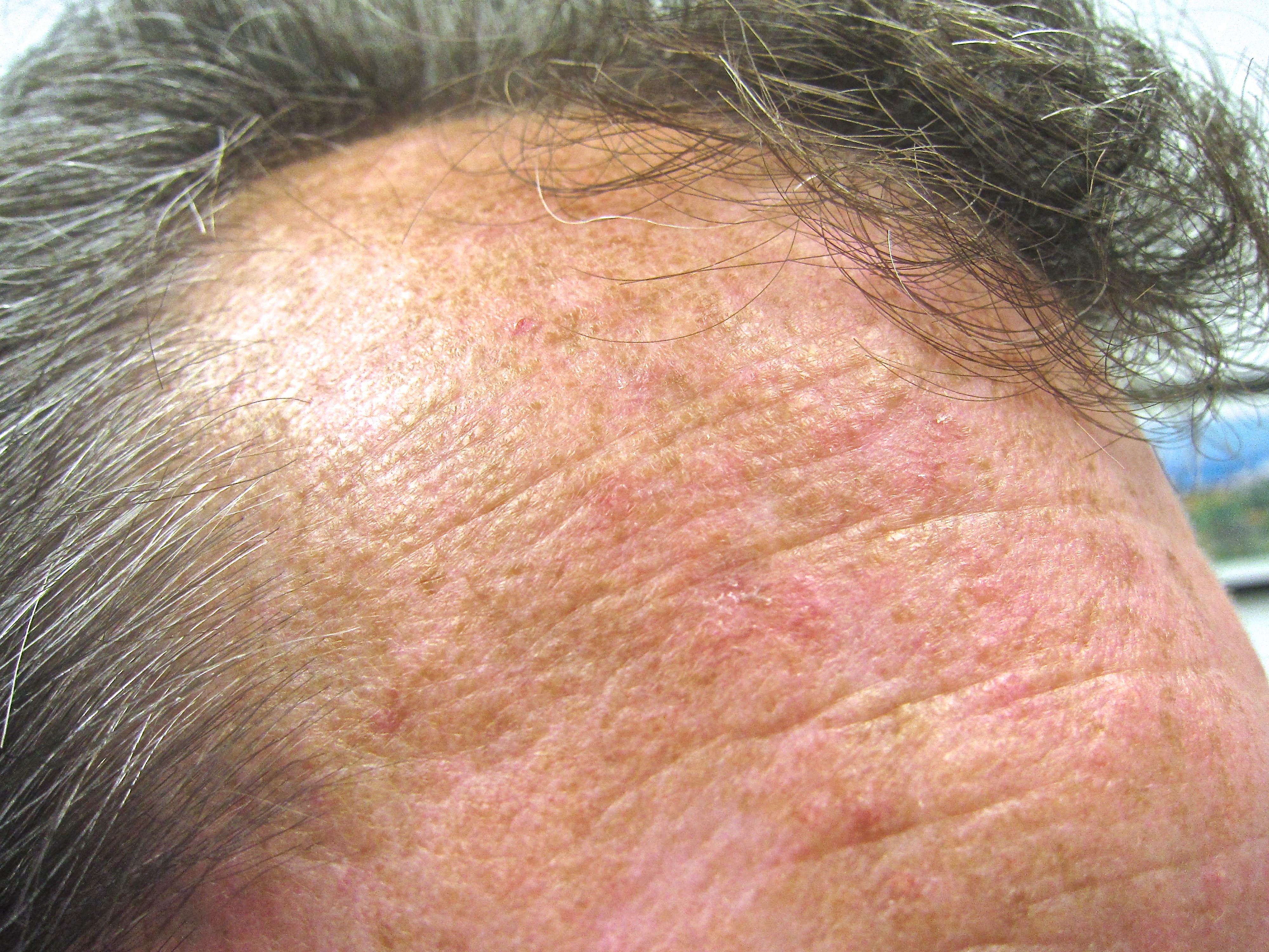
Hypertrophic (hyperkeratotic) AK
A thicker, more adherent, warty or verrucous scale than classic AK. This variant carries closer clinical suspicion because significant thickness and induration can mask early invasion underneath — a very thick or indurated lesion should prompt biopsy rather than an empiric topical trial.[3]
Pigmented AK
Brown pigmentation within an otherwise typical scaly AK, which can mimic a solar lentigo or, more concerningly, lentigo maligna. Dermoscopy or biopsy is warranted whenever the diagnosis is not clinically certain.
Actinic cheilitis
The AK variant affecting the lower lip vermilion: diffuse dryness, scaling, and blurring or loss of the sharp vermilion border ("lipstick sign"). Because lip SCC carries a disproportionately higher risk of metastasis than SCC at most cutaneous sites, actinic cheilitis should be treated proactively rather than watched.[3]
Cutaneous horn
A conical, hyperkeratotic projection that can sit atop an AK, an SCC, a wart, or a seborrheic keratosis — the horn's appearance alone does not tell you which. A meaningful proportion of cutaneous horns overlie a malignant or premalignant lesion at the base, so the base must always be examined and, in most cases, biopsied rather than simply pared or frozen.[3]
Risk Factors
- Cumulative lifetime ultraviolet exposure — the dominant driver; risk tracks with years of sun exposure and geographic UV index.
- Fair skin (Fitzpatrick phototype I-II), light eyes, and red or blond hair.
- Older age — prevalence rises steadily with each decade of cumulative sun exposure.
- Male sex, and outdoor occupation or recreation (agriculture, construction, outdoor sport).
- History of significant sunburns, and indoor tanning bed use.
- Immunosuppression — the single largest risk-amplifying factor. Solid organ transplant recipients on chronic immunosuppression develop AK earlier, in far greater numbers, and progress to SCC at a substantially higher rate and faster pace than immunocompetent patients; this population needs a fundamentally more aggressive approach (see Special Populations).[3]
- A personal history of AK, SCC, or basal cell carcinoma (BCC).
- Genetic photosensitivity disorders (e.g., xeroderma pigmentosum), which cause AK and skin cancer to present at an unusually young age.
- Living at lower latitude or higher altitude, where ambient UV exposure is higher.
Red Flags & Refer
Most AK is appropriate for standard field- or lesion-directed treatment, but biopsy or refer instead of treating empirically for any of the following — these are the features that distinguish AK from a lesion that has already progressed to SCC:
- Induration — a lesion that feels firm or indurated rather than simply rough and scaly.
- Ulceration or non-healing erosion that is not explained by recent treatment.
- Rapid growth or a lesion that has notably enlarged over weeks to a few months.
- Bleeding, whether spontaneous or with minor trauma.
- Pain or tenderness — most AK is asymptomatic or mildly irritating; a tender lesion is atypical.
- A lesion that is unusually thick, a cutaneous horn (biopsy the base), or diameter over roughly 1 cm.
- Any lesion that has failed to respond to an adequate course of appropriate treatment — persistence despite correct therapy needs histologic confirmation, not another empiric round.
- Diagnostic uncertainty, including possible lentigo maligna, BCC, or Bowen's disease (SCC in situ).
- Organ transplant recipients and other immunosuppressed patients — apply a lower threshold for biopsy given their substantially higher rate and pace of progression to invasive SCC.[3]
Assessment & Diagnosis
History
- Cumulative sun exposure history: outdoor occupation or recreation, history of sunburns, tanning bed use, and geographic/residential UV exposure.
- Duration and evolution of the lesion(s): new versus longstanding, any recent change in size, texture, colour, or symptoms.
- Immunosuppression status — transplant history, chronic immunosuppressive medication, HIV status — since this changes both urgency and treatment intensity.
- Personal or family history of skin cancer (AK, SCC, BCC, melanoma).
Examination
Examine chronically sun-exposed skin (face, ears, scalp, dorsal hands/forearms, neck, lower lip) by both inspection and palpation — many AKs are more apparent on touch than on sight. Grade clinical severity using the Olsen system where useful for documentation and treatment selection:
| Olsen grade | Description |
|---|---|
| Grade I (mild) | Slightly palpable AK, better felt than seen. |
| Grade II (moderate) | Moderately thick AK, both easily visible and palpable. |
| Grade III (severe) | Very thick, hyperkeratotic, and/or obviously visible AK. |
The Olsen grade matters clinically: some agents (tirbanibulin/Onakta in Canada) are Health Canada-approved only for non-hyperkeratotic, non-hypertrophic AK — Olsen grade I — and are not appropriate first choices for thicker, grade II-III lesions.[4]
Dermoscopy
Where available, dermoscopy of AK classically shows a "strawberry pattern": background erythema with a network of prominent, dilated follicular openings surrounded by a white-to-yellow halo, often with a fine, white surface scale. Dermoscopy is particularly useful for pigmented lesions and for distinguishing AK from early lentigo maligna.
Differential diagnosis
| Condition | Features that point away from uncomplicated AK |
|---|---|
| Squamous cell carcinoma in situ (Bowen's disease) | A sharply demarcated, persistent, often larger erythematous scaly plaque; can look identical to a thick AK clinically — biopsy when uncertain, since management differs. |
| Invasive SCC | Induration, ulceration, rapid growth, bleeding, or pain — any of the red flags above. |
| Basal cell carcinoma | Pearly, telangiectatic papule or nodule, sometimes with central ulceration ("rodent ulcer") — a different morphology from the flat, scaly AK plaque. |
| Seborrheic keratosis | A "stuck-on," waxy, well-demarcated papule/plaque, often more pigmented and greasy-appearing than the dry, rough scale of AK. |
| Lentigo maligna | An asymmetric, variably pigmented macule with irregular borders on sun-damaged skin — consider strongly for any pigmented, changing lesion, and biopsy if uncertain. |
| Discoid lupus erythematosus | Well-demarcated, erythematous, scaly plaques with follicular plugging and scarring/dyspigmentation — a photosensitive but non-neoplastic autoimmune process. |
| Verruca vulgaris (wart) | Discrete, hyperkeratotic papule with a rough, papillomatous surface and, classically, thrombosed capillaries ("black dots") on paring — a viral rather than UV-driven process. |
| Porokeratosis | An annular plaque with a distinctive thread-like, raised keratotic border — the border pattern, not the centre, is the distinguishing feature. |
Biopsy (shave or punch) any lesion where the diagnosis is uncertain, that shows a red-flag feature, or that fails to respond to an adequate treatment course — do not rely on clinical impression alone when SCC cannot be confidently excluded.[3]
Treatment Targets
- Eradicate visible AK lesions and treat the surrounding field of subclinical, UV-damaged skin ("field cancerization") that will otherwise continue producing new lesions.
- Reduce the risk of progression to invasive SCC — the central, cancer-prevention purpose of treating AK, not a secondary benefit.
- Relieve associated irritation, tenderness, or cosmetic concern where present.
- Identify and biopsy any lesion where SCC cannot be confidently excluded, rather than treating it as routine AK.
- Establish durable sun-protection habits and a lifelong skin-surveillance plan, since AK reflects an ongoing field effect rather than a one-time problem to be solved and forgotten.
Choose Initial Treatment
Step 1 — decide field-directed vs. lesion-directed based on lesion count and pattern
Lesion-directed, 1st line for isolated lesions a small number of discrete, easily identified AKs (roughly under 6-10, well-defined) are appropriately treated lesion by lesion — cryotherapy with liquid nitrogen is the most common and most accessible lesion-directed procedure.[1]
Field-directed, 1st line for multiple/diffuse lesions numerous or ill-defined AKs across a visibly photodamaged area (face, scalp, forearms) reflect field cancerization — subclinical lesions exist between the visible ones — and are better addressed with a field-directed topical agent or photodynamic therapy (PDT) than by freezing each visible lesion individually, since spot treatment alone leaves the subclinical field untreated and new lesions continue to appear.[3]
Combination therapy combining lesion-directed cryotherapy with a field-directed topical agent (e.g., cryotherapy followed by imiquimod or 5-fluorouracil) improves clearance over either modality alone and is a reasonable default for patients with both discrete thick lesions and a broader field of surrounding damage.[3]
Step 2 — select the specific field-directed agent
5-fluorouracil 5% cream applied twice daily for 2-4 weeks (face/scalp; up to 6 weeks may be needed at other sites) until the treated area reaches the erosion stage of the expected inflammatory reaction — a strong-evidence, guideline-endorsed field agent.[1][3]
Imiquimod 5% cream applied 2 times per week at bedtime for 16 weeks to a single treatment area up to approximately 25 cm² on the face or scalp (not both concurrently) — achieves the best durable (12-month) clearance of the commonly used field agents in comparative data.[1]
Imiquimod 3.75% cream applied once daily at bedtime using the "2-2-2" regimen — 2 weeks of treatment, a 2-week treatment-free interval, then 2 more weeks of treatment — to the entire face or scalp; a shorter, better-tolerated alternative to the 5% formulation for patients who need broader coverage.
Diclofenac 3% gel applied twice daily for 60-90 days — a milder-tolerability field option, useful when the more inflammatory reactions of 5-FU or imiquimod are not acceptable to the patient, though evidence support is somewhat less robust than for 5-FU or imiquimod.[3]
Tirbanibulin 1% ointment (Onakta in Canada) applied once daily for only 5 consecutive days to an area up to 100 cm² on the face or balding scalp — the shortest field-therapy course available, but Health Canada has approved it specifically for non-hyperkeratotic, non-hypertrophic AK (Olsen grade I); it is not the right choice for thick, hyperkeratotic lesions.[4]
Photodynamic therapy (PDT) is a further field-directed option, particularly suited to large, cosmetically sensitive areas — see the dedicated overview below.
Ingenol mebutate (Picato) — withdrawn, do not use was formerly a short-course (2-3 day) field-directed topical option. Health Canada requested its withdrawal from the Canadian market, effective October 2020, after a safety review linked it to an increased risk of non-melanoma skin cancer at treated sites; it is no longer available in Canada and should not be sourced or substituted from elsewhere.[5]
Non-Pharmacologic Treatment
- Sun protection is foundational to every AK treatment plan, not an afterthought — it reduces both the development of new lesions and, likely, progression risk in existing ones, and should be discussed at every visit regardless of which active therapy is chosen.[2]
- Daily broad-spectrum sunscreen, SPF 30 or higher, reapplied every 2 hours with outdoor exposure and after swimming or sweating.
- Photoprotective clothing (tightly woven, UPF-rated where available), wide-brimmed hats, and UV-blocking sunglasses.
- Avoid peak-UV hours (typically 10 am-4 pm) for prolonged outdoor exposure when practical, and seek shade.
- Avoid indoor tanning beds entirely.
- Perform regular self-skin examination and report new, changing, or non-healing lesions promptly rather than waiting for the next scheduled visit.
- Oral nicotinamide (vitamin B3) 500 mg twice daily is a reasonable adjunct for chemoprevention in patients with a history of multiple AKs or prior skin cancers — it is not a treatment for existing lesions but has been shown to reduce the rate of new AK and non-melanoma skin cancer development in high-risk patients; it is well tolerated and does not require the photoprotective precautions used with the systemic retinoids sometimes used in transplant patients.
Special Populations
Organ transplant recipients and other immunosuppressed patients
This is the highest-risk population for AK-to-SCC progression and needs a fundamentally more intensive approach, not the standard pathway: lower threshold for biopsy, more aggressive field-directed treatment, closer surveillance intervals (often every 3-6 months rather than annually), and consideration of adjunctive chemoprevention (e.g., oral nicotinamide, or systemic retinoids under dermatology/transplant-team direction) in patients with a heavy lesion burden or prior skin cancers.[3]
Pregnancy and breastfeeding
AK is not an urgent diagnosis, so non-urgent topical field therapy (5-fluorouracil, imiquimod, diclofenac, tirbanibulin) is reasonable to defer until after pregnancy and breastfeeding given limited safety data for these agents in this population. Where treatment cannot reasonably wait, cryotherapy is the preferred modality, since it is a physical/procedural treatment without systemic drug exposure.
Older adults
AK incidence rises steadily with age, and treatment decisions should account for polypharmacy, skin fragility, and the practical burden of an inflammatory topical course (application technique, caregiver support) rather than defaulting to the same regimen used in younger patients.
Pediatrics
AK from cumulative sun damage essentially does not occur in children. A precocious AK-like lesion in a child should raise suspicion for an underlying photosensitivity or DNA-repair genodermatosis (e.g., xeroderma pigmentosum) and warrants dermatology referral rather than routine management.
Monitoring & Follow-Up
For field-directed topical therapy, set the expectation up front that a visible inflammatory reaction (erythema, erosion, crusting) is the expected mechanism of action, not a complication — reassess at the end of the planned course (2-4 weeks for 5-FU, 16 weeks for imiquimod 5%, 5 days for tirbanibulin) once the reaction has settled, typically several weeks after the last application, to judge clearance.[3]
Common Errors to Avoid
- Treating AK as a purely cosmetic complaint and not explaining the SCC-prevention rationale — patients who understand why treatment matters are more likely to complete an uncomfortable topical course.
- Spot-freezing only the visible lesions in a patient with diffuse field damage, leaving the subclinical field untreated and allowing new lesions to keep appearing.
- Stopping a field-directed topical agent as soon as the expected inflammatory reaction appears, mistaking the mechanism of action for an adverse effect.
- Using tirbanibulin (Onakta) on thick, hyperkeratotic (Olsen grade II-III) lesions — it is approved and studied specifically for grade I disease.
- Treating an indurated, ulcerated, rapidly growing, bleeding, or treatment-refractory lesion as routine AK instead of biopsying it.
- Failing to examine and biopsy the base of a cutaneous horn, assuming the horn itself is diagnostic.
- Applying the standard (non-intensified) surveillance interval and treatment intensity to an organ transplant recipient or other immunosuppressed patient.
- Omitting sun-protection counselling because an active treatment was prescribed — the two are complementary, not alternatives.
- Assuming ingenol mebutate (Picato) is still an available option — it was withdrawn from the Canadian market in 2020.
Clinical Decision Algorithm
Drug Tables
How to use this table: Doses are adult doses and require patient-specific confirmation. Select any linked drug name to open its complete Pharmasist Drug Database page.
Antimetabolites (topical)
Strong-evidence, guideline-endorsed field-directed therapy.
| Drug / intervention | Dosage | Adverse effects | Drug interactions | Comments |
|---|---|---|---|---|
| Fluorouracil 5% cream | Face/scalp: Apply BID for 2-4 weeks Other body sites: Apply BID for up to 6 weeks | Marked local erythema, burning, erosion, crusting, photosensitivity of treated skin | None clinically significant given minimal systemic absorption at labelled dosing | Apply until the treated area reaches the erosion stage of the expected inflammatory reaction — counsel patients this reaction is the mechanism of action, not a complication. |
Immune response modifiers (topical)
Field-directed agents that work by stimulating a local immune response against dysplastic keratinocytes.
| Drug / intervention | Dosage | Adverse effects | Drug interactions | Comments |
|---|---|---|---|---|
| Imiquimod 5% cream | Dosing: Apply 2x/week at bedtime for 16 weeks; leave on ~8 hours then wash off | Local erythema, erosion, pruritus; flu-like symptoms (fatigue, myalgia, headache) with extensive application | None clinically significant given minimal systemic absorption at labelled dosing | Achieves the best 12-month durable clearance among the commonly used topical field agents in comparative data. Treatment area limited to ~25 cm² on the face or scalp, not both concurrently. |
| Imiquimod 3.75% cream | Dosing: Apply once daily at bedtime for 2 weeks, then a 2-week treatment-free interval, then once daily for 2 more weeks | Local erythema, erosion; generally milder than the 5% formulation | None clinically significant given minimal systemic absorption at labelled dosing | Shorter, better-tolerated "2-2-2" regimen intended for treating the entire face or scalp as one field. |
NSAIDs (topical)
A milder-tolerability field-directed alternative to fluorouracil or imiquimod.
| Drug / intervention | Dosage | Adverse effects | Drug interactions | Comments |
|---|---|---|---|---|
| Diclofenac 3% gel | Dosing: Apply BID for 60-90 days | Local irritation, dryness, contact dermatitis | None clinically significant given minimal systemic absorption at labelled dosing | Somewhat lower evidence strength than fluorouracil or imiquimod; reasonable when a gentler local reaction is needed to support adherence. |
Microtubule inhibitors (topical)
The newest and shortest-course field-directed agent; Canadian brand Onakta (tirbanibulin), Health Canada Notice of Compliance issued May 12, 2023.
| Drug / intervention | Dosage | Adverse effects | Drug interactions | Comments |
|---|---|---|---|---|
| Tirbanibulin 1% ointment (Onakta) | Dosing: Apply once daily for 5 consecutive days to an area up to 100 cm²; wash hands immediately after application; avoid washing/touching the treated area for ~8 hours | Local erythema, flaking/scaling, crusting, swelling; less commonly vesiculation/pustulation or erosion/ulceration | None clinically significant given minimal systemic absorption at labelled dosing | Health Canada-approved specifically for non-hyperkeratotic, non-hypertrophic AK (Olsen grade I) of the face or balding scalp — not indicated for thicker, hyperkeratotic lesions. Avoid the periocular area. |
Procedural / physical therapies
Lesion-directed and field-directed procedures performed in office, not systemically dosed drugs.
| Drug / intervention | Dosage | Adverse effects | Drug interactions | Comments |
|---|---|---|---|---|
| Cryotherapy (liquid nitrogen) | Technique: Spray or cotton-tip applicator; single freeze-thaw cycle (~5-10 sec) for thin lesions, longer or double freeze-thaw for thicker lesions, with a 1-2 mm margin of visibly normal skin | Local pain, blistering, temporary or permanent hypo/hyperpigmentation (higher risk in darker skin types), rare scarring | Not applicable | The most common and most accessible lesion-directed procedure. High short-term clearance but recurrence at 1 year is common — reassess rather than assuming a single treatment is curative. |
| Photodynamic therapy (ALA or MAL + light) | Technique: Photosensitizer applied after lesion preparation, incubated (~30 min for daylight PDT, longer for conventional PDT), then illuminated; typically 1-2 sessions | Significant pain during illumination (conventional PDT more than daylight PDT), erythema, crusting | Not applicable | Field-directed option well suited to large or cosmetically sensitive areas, with excellent cosmetic outcomes. Daylight PDT reduces pain versus conventional lamp-based PDT while maintaining comparable efficacy. |
Withdrawn agent — do not use
Listed to state explicitly that this agent is no longer available in Canada, not as a routine option.
| Drug / intervention | Dosage | Adverse effects | Drug interactions | Comments |
|---|---|---|---|---|
| Ingenol mebutate (Picato) | Use in actinic keratosis: Not available — withdrawn from the Canadian market | Associated with increased reporting of squamous cell carcinoma at treated sites | Not applicable — withdrawn | Health Canada requested withdrawal from the Canadian market effective October 2020 after a safety review linked it to increased non-melanoma skin cancer risk. Do not use or source from outside Canada. |
Chemoprevention adjunct (oral, not a treatment for existing lesions)
Reduces the rate of new lesion formation in high-risk patients; does not treat existing AK.
| Drug / intervention | Dosage | Adverse effects | Drug interactions | Comments |
|---|---|---|---|---|
| Nicotinamide (vitamin B3) | Dosing: 500 mg PO BID | Generally well tolerated; mild GI upset uncommon | None clinically significant | Reasonable adjunct for patients with a history of multiple AKs or prior skin cancers, particularly where systemic retinoid chemoprevention is not appropriate. Does not require the photoprotective precautions used with systemic retinoids. |
References
- Non-melanoma Skin Cancer in Canada Chapter 3: Management of Actinic Keratoses — Journal of Cutaneous Medicine and Surgery (Poulin Y, Lynde CW, Barber K, Vender R, Claveau J, Bourcier M, Ashkenas J). J Cutan Med Surg 2015;19(3):228-243
- Actinic Keratoses — Canadian Dermatology Association. Public patient education, Canadian Dermatology Association
- Guidelines of care for the management of actinic keratosis — Journal of the American Academy of Dermatology (Eisen DB, Asgari MM, Bennett DD, Connolly SM, Dellavalle RP, Freeman EE, Goldenberg G, Leffell DJ, Peschin S, Sligh JE, Wu PA, Frazer-Green L, Malik S, Schlesinger TE). J Am Acad Dermatol 2021;85(4):e209-e233
- Summary Basis of Decision for Onakta (tirbanibulin) — Health Canada, Drug and Health Products Portal. Summary Basis of Decision, Notice of Compliance issued May 12, 2023
- PICATO (ingenol mebutate gel, 0.015% and 0.05%) — Product Withdrawal in Canada due to Potential Increased Risk of Skin Cancer — Health Canada. Health professional risk communication, October 27, 2020
Professional reference only. Apply clinical judgment and verify patient-specific decisions against current authoritative guidance and product information.