Athlete's Foot (Tinea Pedis)
Clinical Snapshot
Tinea pedis ("athlete's foot") is a dermatophyte infection of the skin of the foot, most often caused by Trichophyton rubrum and Trichophyton interdigitale, and is the most common dermatophyte infection encountered in practice.[1] It presents in several distinct patterns — interdigital, moccasin (hyperkeratotic), vesiculobullous, and the rare ulcerative type — each with a different appearance, a different natural history, and in some cases a different treatment approach, so recognizing which subtype is in front of you matters more than reflexively reaching for "the athlete's foot cream."[1] Most uncomplicated tinea pedis is genuinely a self-care condition: a topical antifungal matched to the subtype and continued for the full course — even after itching resolves — cures the great majority of cases without prescription therapy being required.[2] The pharmacist's core value here is threefold: confirming this really is tinea pedis and not one of its many mimics, matching the right topical agent and duration to the presentation, and recognizing the minority of patients — diabetes, peripheral vascular disease, immunosuppression, or spreading erythema — who need to be moved out of self-care and into medical assessment.
Recognize the Condition
The 4 clinical subtypes of tinea pedis
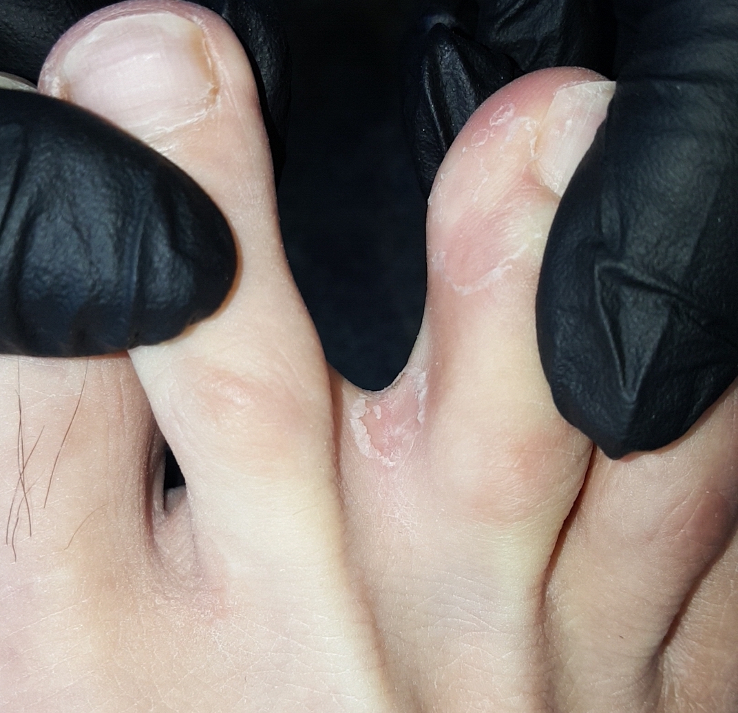
Interdigital (most common)
Erythema, scaling, peeling, and maceration in the web spaces, classically starting between the 4th and 5th toes. The skin can look white and soggy from retained moisture. Pruritus is the dominant symptom; fissuring at the base of the web space causes pain and burning and is a common portal for secondary bacterial infection.[1]
Moccasin (chronic hyperkeratotic)
Diffuse, chronic scaling with variable underlying erythema across the sole, heel, and the medial and lateral borders of the foot, in a distribution resembling a moccasin shoe, often with a fine "collarette" of scale at the edge. Usually caused by T. rubrum, typically minimally symptomatic, and notably resistant to short courses of topical therapy — this is the subtype most likely to need an extended topical course or oral therapy.[1]

Vesiculobullous (inflammatory)
Intensely pruritic, sometimes painful, 1-3 mm vesicles or bullae on an erythematous base, typically on the instep or medial foot. Vesicles rupture to leave a raw, weeping surface. A remote, symmetric vesicular eruption on the hands (a dermatophytid or "id" reaction) accompanies a meaningful minority of cases and resolves on its own once the foot infection is treated — it is not a separate infection and does not need its own antifungal therapy.[1]
Ulcerative (uncommon)
Rapidly spreading interdigital erosions and ulcers, more aggressive than the vesiculobullous pattern and frequently complicated by secondary bacterial infection (cellulitis, lymphangitis). Seen disproportionately in patients with diabetes, peripheral vascular disease, or immunodeficiency — this presentation should prompt the red-flag assessment below rather than routine self-care.[1]
A fifth pattern, occult tinea pedis, shows no visible lesions at all despite active infection and is most often identified incidentally or in athletes — a reminder that a normal-looking foot does not exclude dermatophyte carriage in a patient with recurrent reinfection, or unexplained spread to the nails or groin.[1]
Risk Factors
- Occlusive, non-breathable footwear and prolonged shoe-wearing without rotation between pairs.
- Communal bathing facilities, public pool decks, and shared gym or locker-room showers.
- Hyperhidrosis (excess sweating), particularly plantar hyperhidrosis.
- Male sex — roughly 3 times the risk of females — and age 16-45, though any age can be affected.[1]
- Diabetes mellitus, peripheral vascular disease, and immunodeficiency.
- Concurrent atopic dermatitis or psoriasis.
- Obesity.
- Occupational or living exposure — athletes, military personnel, manual laborers, and long-term care or shelter residents.[1]
- A personal history of tinea pedis or onychomycosis, or a household contact with either.
Red Flags & Refer
Most tinea pedis is appropriate for self-care, but escalate to medical assessment — and treat as possible cellulitis rather than simple tinea — for any of the following:
- Spreading erythema, warmth, swelling, or pain extending beyond the original lesion, red streaking up the leg (lymphangitis), or fever/chills — these suggest secondary bacterial cellulitis rather than uncomplicated fungal infection and need urgent assessment, not an antifungal alone.[3]
- The ulcerative subtype (rapidly spreading interdigital erosions/ulcers) — this pattern is frequently complicated by bacterial superinfection and disproportionately affects patients with diabetes, peripheral vascular disease, or immunodeficiency.[1]
- Diabetes or peripheral vascular disease with any active tinea pedis — fungal and secondary bacterial foot infections carry up to a 3-fold higher risk of complications such as cellulitis, and up to a 5-fold higher risk of gangrene, in patients with diabetes; treat these patients more actively and with a lower threshold for referral and prescription oral therapy rather than a standard self-care trial.[3]
- Immunocompromise (including poorly controlled HIV, transplant recipients, or active chemotherapy) — presentation can be extensive or atypical, and self-care is not appropriate as the sole management.
- Extensive disease covering a large surface area, or disease that has already spread to the nails (onychomycosis), the groin (tinea cruris), or the hands (tinea manuum) — a topical trial limited to the foot will not address the reservoir driving reinfection.
- No improvement, or worsening, after a full and adherent course of an appropriately chosen topical antifungal — this needs diagnostic reassessment, not simply switching to another topical agent empirically.
- Diagnostic uncertainty — a presentation that does not fit typical tinea pedis (symmetric, bilateral, sharply demarcated plaques favor psoriasis or contact dermatitis over tinea) warrants confirmation before treating.
Assessment & Diagnosis
History
- Characterize the distribution (interdigital, sole/heel/moccasin, vesicular, or ulcerative) and ask specifically about pruritus, burning, pain, and how long the presentation has been evolving.
- Ask about footwear habits, communal shower/pool exposure, hyperhidrosis, and whether household contacts have similar symptoms.
- Screen for diabetes, peripheral vascular disease, and immunosuppression — these change both the urgency of assessment and the treatment plan.
- Ask about prior self-treatment: which product, how it was applied, and for how long — premature discontinuation once itching resolves is the most common reason an apparent "treatment failure" is really an incomplete course.[1]
Examination
Confirm the distribution matches one of the 4 subtypes above, and specifically check for fissuring, maceration, or frank drainage suggesting secondary bacterial infection, and for spreading erythema, warmth, or lymphangitic streaking suggesting cellulitis.
Differential diagnosis
| Condition | Features that point away from tinea pedis |
|---|---|
| Irritant or allergic contact dermatitis (footwear dyes, adhesives, rubber accelerators) | Distribution often matches the shoe/sock contact pattern rather than the classic web-space or moccasin pattern; frequently symmetric and bilateral. |
| Atopic or dyshidrotic (pompholyx) eczema | Symmetric, bilateral involvement; personal or family history of atopy; deep-seated palmar/plantar vesicles without the web-space predilection of interdigital tinea. |
| Palmoplantar psoriasis | Well-demarcated, symmetric, often thicker plaques with a silvery scale; look for psoriatic nail changes or plaques elsewhere on the body. |
| Pitted keratolysis | Punched-out pits on the weight-bearing sole with a distinctive malodor — a bacterial, not fungal, process. |
| Erythrasma | Interdigital, but caused by Corynebacterium minutissimum; coral-red fluorescence under Wood's lamp helps distinguish it (tinea pedis does not fluoresce). |
| Candidal intertrigo | Beefy-red erythema with satellite pustules; more maceration/occlusion; needs an anti-yeast rather than a dermatophyte-only agent. |
| Bacterial infection (impetigo, early cellulitis) | Honey-crusted lesions, or spreading warm erythema with pain/fever, rather than the chronic scaling typical of tinea. |
| Scabies | Intense nocturnal pruritus, burrows, and involvement of finger web spaces, wrists, and genitals in addition to the feet. |
Most classic interdigital or moccasin-pattern disease can be diagnosed clinically without testing. Confirm the diagnosis with a potassium hydroxide (KOH) preparation of a skin scraping from the active, scaling border (or a fungal culture, or an in-office dermatophyte test strip where available) whenever the presentation is atypical, before starting oral therapy, or when an initial topical course has failed — this avoids weeks of ineffective self-treatment for what may actually be psoriasis, contact dermatitis, or a bacterial process.[1]
Treatment Targets
- Achieve mycological cure of the dermatophyte infection, not just symptom relief.
- Resolve pruritus, fissuring, and discomfort.
- Prevent secondary bacterial infection and, in at-risk patients, cellulitis.
- Prevent spread to the nails, groin, hands, or household contacts.
- Reduce recurrence through footwear and foot-hygiene measures, since tinea pedis is one of the more frequently recurrent dermatophyte infections.
Choose Initial Treatment
Step 1 — most uncomplicated tinea pedis is topical, first-line self-care
Interdigital tinea pedis, 1st line topical terbinafine 1% cream applied twice daily for 1 week — a 1-week course of terbinafine achieves comparable or superior cure rates to 4 weeks of an azole, with markedly better adherence given the shorter course.[2][4]
Azole alternative clotrimazole 1% or miconazole 2% cream applied twice daily for 4 weeks — effective, inexpensive, and widely available without a prescription, but needs a longer, harder-to-complete course and achieves a somewhat lower cure rate than terbinafine in head-to-head trials.[2]
Tolnaftate 1% applied twice daily for 2-4 weeks is another nonprescription option, but it is fungistatic rather than fungicidal and has no activity against Candida — avoid it when the presentation could represent candidal intertrigo rather than a pure dermatophyte infection.
Step 2 — the moccasin (hyperkeratotic) pattern needs a longer course
Use the same topical agents but plan for a longer course — typically 4 weeks at minimum, sometimes longer — since the thickened stratum corneum limits drug penetration and this subtype is the most treatment-resistant.[1] A salicylic acid keratolytic applied concurrently can improve penetration and speed clinical response by softening hyperkeratotic scale.
Step 3 — reserve oral (systemic) therapy for the right patients
Move to oral therapy for extensive or widespread disease, chronic moccasin-type disease that has not responded to an adequate topical course, immunocompromised patients, or when tinea pedis coexists with onychomycosis — the infected nail acts as an ongoing fungal reservoir that will keep reseeding the skin.[1]
Oral terbinafine, 1st line oral agent 250 mg once daily for 2 weeks — effective, has fewer clinically significant drug interactions than the oral azoles, and outperforms both itraconazole and griseofulvin in comparative evidence.[1]
Oral itraconazole 200 mg twice daily for 1 week (pulse dosing) is an effective alternative — screen carefully for CYP3A4 interactions before prescribing, since itraconazole is a potent CYP3A4 inhibitor.[1]
Oral fluconazole 150 mg once weekly for 2-6 weeks is a further alternative when terbinafine and itraconazole are not suitable.[1]
Avoid oral griseofulvin and oral ketoconazole as routine choices — griseofulvin has poor bioavailability, needs a longer course, and carries more adverse effects (photosensitivity, GI upset, hepatic effects) than terbinafine, and oral ketoconazole has been largely abandoned given its hepatotoxicity and adrenal/neurologic risk relative to the alternatives above.[1]
Step 4 — treat secondary bacterial infection if present
A macerated, malodorous, or frankly infected interdigital space (the "dermatophytosis complex") needs treatment directed at the bacterial component as well — an antifungal with combined antibacterial activity (e.g., ciclopirox) or a short course of an oral antibiotic for true cellulitis, in addition to antifungal therapy for the underlying tinea.[1]
Non-Pharmacologic Treatment
- Dry the feet thoroughly, especially between the toes, after every bath, shower, or swim — moisture is the single biggest driver of both initial infection and recurrence.
- Wear moisture-wicking or cotton socks and change them at least daily, more often if the feet sweat heavily.
- Choose breathable, non-occlusive footwear; alternate between at least 2 pairs of shoes so each can fully dry out between wears.
- Wear sandals or shower shoes in communal showers, pool decks, and gym locker rooms rather than going barefoot.
- Apply a desiccating or antifungal powder (e.g., miconazole or tolnaftate powder) to the feet and inside footwear to control moisture and reduce recurrence, particularly with hyperhidrosis.[1]
- Disinfect or discard heavily infected footwear — hot laundering of socks, and UV-C or ozone shoe sanitizing where available, reduce the fungal reservoir that drives reinfection.[1]
- Do not share towels, socks, shoes, or nail-care tools with household members.
- Treat any concurrent onychomycosis or tinea at another body site (groin, hands) at the same time — an untreated nail or skin reservoir is one of the most common causes of "recurrent" tinea pedis.
- Manage plantar hyperhidrosis directly (for example, an antiperspirant containing aluminum chloride) if it is a recurring contributor.
Special Populations
Pregnancy
Topical antifungal therapy is preferred in pregnancy given minimal systemic absorption — topical terbinafine, clotrimazole, and miconazole are all reasonable choices.
Avoid oral therapy where possible: oral fluconazole and itraconazole carry teratogenicity signals at the doses used for dermatophyte infections, and griseofulvin is specifically contraindicated in pregnancy. If oral therapy is genuinely unavoidable, this decision should be made by the prescriber weighing the specific clinical picture, not defaulted to.
Breastfeeding
Topical antifungals are compatible with breastfeeding given minimal systemic absorption. Data on the oral agents in lactation are more limited; apply the same benefit/risk judgment as in pregnancy if oral therapy is being considered.
Pediatrics
Topical therapy is preferred in children — no dose adjustment or laboratory monitoring is needed and adverse effects are minimal. Where oral therapy is genuinely needed, oral terbinafine can be dosed by weight under prescriber direction (broadly: 62.5 mg/day for 10-20 kg, 125 mg/day for 20-40 kg, and the adult 250 mg/day dose above 40 kg, each for 2 weeks); oral itraconazole and fluconazole can also be used off-label with weight-based dosing.[1]
Diabetes and peripheral vascular disease
Treat actively rather than deferring to a routine self-care trial — these patients carry a materially higher risk of secondary bacterial infection, cellulitis, and, in diabetes specifically, limb-threatening complications arising from an infected foot.[3] Favor a lower threshold for oral therapy, closer follow-up, and prompt escalation if any sign of secondary infection develops; podiatry involvement is reasonable for recurrent or hyperkeratotic disease in this group.
Immunosuppression
Presentation can be more extensive or atypical, and self-care alone is not appropriate as the sole management — involve a prescriber for diagnosis confirmation and consider oral therapy or combination oral-plus-topical treatment.
Monitoring & Follow-Up
Reassess at the end of the planned course — 1 week for topical terbinafine in interdigital disease, 4 weeks (or longer) for azoles, tolnaftate, or moccasin-type disease, and 2 weeks for oral terbinafine — rather than waiting for the patient to bring it up. Confirm symptom resolution, and where the diagnosis was uncertain or treatment was extended, consider repeat KOH microscopy to confirm mycological cure rather than relying on appearance alone, since clinical improvement can lag behind, or mask, incomplete eradication.[1]
Common Errors to Avoid
- Treating a symmetric, bilateral, sharply demarcated plaque as tinea pedis without first considering psoriasis or contact dermatitis.
- Using tolnaftate when a candidal component is plausible — it has no anti-yeast activity.
- Under-treating moccasin (hyperkeratotic) tinea pedis with a 1-week course intended for interdigital disease — this subtype needs a longer course, and often more.
- Stopping treatment as soon as itching or visible scaling improves, before the full course is complete — the leading cause of apparent treatment failure and early recurrence.
- Treating the skin but ignoring a concurrent fungal nail or groin infection that continues to reseed the foot.
- Missing early cellulitis in a patient with diabetes or peripheral vascular disease by treating spreading erythema, warmth, or pain as "just fungal."
- Prescribing oral itraconazole without screening for CYP3A4 drug interactions first.
- Defaulting to oral griseofulvin or ketoconazole out of habit rather than using terbinafine, itraconazole, or fluconazole, which have better efficacy and/or safety profiles for this indication.[1]
- Not counselling on footwear and sock hygiene, leaving the environmental driver of recurrence unaddressed even after a technically successful antifungal course.
Clinical Decision Algorithm
Drug Tables
How to use this table: Doses are adult doses unless a separate pediatric line is given, and require patient-specific confirmation, including renal/hepatic adjustment. Select any drug name to open its complete Pharmasist Drug Database page.
Allylamines (topical and oral)
First-line class for most tinea pedis given the shortest effective topical course and, for oral use, the fewest clinically significant interactions.
| Drug | Dosage | Adverse effects | Drug interactions | Comments |
|---|---|---|---|---|
| Terbinafine, topical 1% cream | Interdigital: Apply BID for 1 week Moccasin (hyperkeratotic): Apply BID for up to 4 weeks or longer | Local irritation, burning, stinging, dryness | None clinically significant given minimal systemic absorption | 1st-line topical agent for interdigital disease given its short course. Continue for the full duration even after itching resolves. |
| Terbinafine, oral | Adults: 250 mg PO once daily for 2 weeks Children 10 to under 20 kg: 62.5 mg PO once daily for 2 weeks Children 20-40 kg: 125 mg PO once daily for 2 weeks Children over 40 kg: 250 mg PO once daily for 2 weeks | GI upset, headache, transient taste/smell disturbance, rash; rare hepatotoxicity, blood dyscrasias | Terbinafine inhibits CYP2D6, raising levels of tricyclic antidepressants, SSRIs (fluoxetine, paroxetine), tramadol, atomoxetine, and beta-blockers metabolized via CYP2D6; cimetidine decreases and rifampin increases terbinafine clearance | 1st-line oral agent for extensive, refractory, or moccasin-type disease, or when tinea pedis coexists with onychomycosis. Obtain baseline liver function tests if other hepatotoxicity risk factors are present. |
Azoles (topical and oral)
Effective, widely available OTC topical options; a longer course than terbinafine is needed for cure.
| Drug | Dosage | Adverse effects | Drug interactions | Comments |
|---|---|---|---|---|
| Clotrimazole, topical 1% cream | Dosing: Apply BID for 4 weeks | Local irritation, burning, contact dermatitis (uncommon) | None clinically significant given minimal systemic absorption | OTC alternative to terbinafine; adherence over the full 4-week course is the main limiting factor for cure. |
| Miconazole, topical 2% cream/powder/spray | Dosing: Apply BID for 4 weeks | Local irritation, burning | None clinically significant given minimal systemic absorption | Comparable efficacy and dosing rationale to clotrimazole. Powder/spray forms are also used inside footwear for prevention. |
| Itraconazole, oral (pulse) | Adults: 200 mg PO BID for 1 week | GI upset, headache; rare hepatotoxicity, heart failure | Potent CYP3A4 inhibitor — avoid or use with caution with statins metabolized via CYP3A4 (atorvastatin/lovastatin/simvastatin), calcium channel blockers, digoxin, warfarin, and other CYP3A4 substrates; H2-antagonists and proton pump inhibitors reduce absorption | Alternative oral agent to terbinafine. Screen the full medication list for CYP3A4 interactions before dispensing. |
| Fluconazole, oral | Adults: 150 mg PO once weekly for 2-6 weeks | Nausea, headache; rare hepatotoxic reactions | Inhibits CYP2C9/CYP2C19/CYP3A4 — raises levels of warfarin, phenytoin, sulfonylureas, and several other substrates; monitor for toxicity | Further oral alternative when terbinafine and itraconazole are unsuitable; lower comparative efficacy than either. |
Thiocarbamates (topical)
Nonprescription option; no activity against Candida.
| Drug | Dosage | Adverse effects | Drug interactions | Comments |
|---|---|---|---|---|
| Tolnaftate, topical 1% cream/solution/powder | Treatment: Apply BID for 2-4 weeks Prevention: Apply once daily to feet and inside footwear | Local irritation (uncommon) | None clinically significant given minimal systemic absorption | Fungistatic rather than fungicidal. Not effective against Candida — avoid if a yeast component is suspected. Powder form is also used preventively inside footwear. |
Hydroxypyridones (topical)
Broader-spectrum option when secondary bacterial colonization accompanies the fungal infection.
| Drug | Dosage | Adverse effects | Drug interactions | Comments |
|---|---|---|---|---|
| Ciclopirox, topical 0.77% cream | Dosing: Apply BID for up to 4 weeks | Local irritation | None clinically significant given minimal systemic absorption | Combined antifungal, anticandidal, and antibacterial activity — a reasonable choice for the macerated, malodorous "dermatophytosis complex" presentation. |
Keratolytic adjunct
Not an antifungal — used alongside topical or oral therapy to improve penetration through hyperkeratotic scale.
| Drug | Dosage | Adverse effects | Drug interactions | Comments |
|---|---|---|---|---|
| Salicylic acid | Dosing: Apply per product directions concurrently with the antifungal | Local irritation, stinging | None clinically significant with topical foot use | Adjunct for moccasin (hyperkeratotic) tinea pedis; softens thickened scale so the antifungal penetrates more effectively. |
Oral agents generally not recommended for this indication
Listed to state explicitly that these should not be first choices for tinea pedis, not as routine options.
| Drug | Dosage | Adverse effects | Drug interactions | Comments |
|---|---|---|---|---|
| Griseofulvin, oral | Use in tinea pedis: Not a preferred agent — use terbinafine, itraconazole, or fluconazole first | Photosensitivity, GI upset, headache, hepatic dysfunction; contraindicated in pregnancy | Induces hepatic CYP enzymes — reduces efficacy of warfarin and hormonal contraceptives | Poor bioavailability and a longer required course than terbinafine, with a less favourable adverse-effect profile. Reserve for situations where the preferred oral agents cannot be used. |
| Ketoconazole, oral | Use in tinea pedis: Not recommended — do not use | Hepatotoxicity, adrenal suppression at higher exposure | Potent CYP3A4 inhibitor with an extensive interaction list | Largely abandoned for dermatophyte infections given hepatotoxicity and neurologic/endocrine risk relative to terbinafine, itraconazole, or fluconazole. |
References
- Tinea pedis: an updated review — Drugs in Context (Leung AKC, Barankin B, Lam JM, Leong KF, Hon KL). Drugs Context 2023;12:1-16
- Topical management of tinea pedis — Canadian Family Physician, Tools for Practice (adapted from the Alberta College of Family Physicians' PEER evidence program) (Thomas B, Falk J, Allan GM). Can Fam Physician 2021;67(1):30
- Fungal foot infection, cellulitis and diabetes: a review — Diabetic Medicine (Bristow IR, Spruce MC). Diabet Med 2009;26(5):548-551
- Consensus for the treatment of tinea pedis: a systematic review of randomised controlled trials — Journal of Fungi (Ward H, Parkes N, Smith C, Kluzek S, Pearson R). J Fungi (Basel) 2022;8(4):351
Professional reference only. Apply clinical judgment and verify patient-specific decisions against current authoritative guidance and product information.