Benign Paroxysmal Positional Vertigo
Benign Paroxysmal Positional Vertigo
References
- Clinical Practice Guideline: Benign Paroxysmal Positional Vertigo (Update) — Otolaryngology-Head and Neck Surgery, American Academy of Otolaryngology-Head and Neck Surgery Foundation (Bhattacharyya N, Gubbels SP, Schwartz SR, et al.). Otolaryngol Head Neck Surg 2017;156(3_suppl):S1-S47
- Epidemiology of benign paroxysmal positional vertigo: a population based study — Journal of Neurology, Neurosurgery & Psychiatry (von Brevern M, Radtke A, Lezius F, Feldmann M, Ziese T, Lempert T, Neuhauser H). J Neurol Neurosurg Psychiatry 2007;78(7):710-715
- Benign paroxysmal positional vertigo: Diagnostic criteria - Consensus document of the Committee for the Classification of Vestibular Disorders of the Bárány Society — Journal of Vestibular Research (von Brevern M, Bertholon P, Brandt T, Fife T, Imai T, Nuti D, Newman-Toker D). J Vestib Res 2015;25(3-4):105-117
- Otology/Neurotology recommendations - Choosing Wisely campaign — Journal of Otolaryngology - Head & Neck Surgery, Otology/Neurotology subspecialty group of the Canadian Society of Otolaryngology - Head & Neck Surgery (Ma AK, Nedzelski J, Chen J, et al.). J Otolaryngol Head Neck Surg 2019;48:60
- HINTS to Diagnose Stroke in the Acute Vestibular Syndrome: Three-Step Bedside Oculomotor Examination More Sensitive Than Early MRI Diffusion-Weighted Imaging — Stroke (Kattah JC, Talkad AV, Wang DZ, Hsieh YH, Newman-Toker DE). Stroke 2009;40(11):3504-3510
- Guidelines for reasonable and appropriate care in the emergency department 3 (GRACE-3): Acute dizziness and vertigo in the emergency department — Academic Emergency Medicine (Edlow JA, Carpenter C, Akhter M, et al.). Acad Emerg Med 2023;30(5):442-486
- Effectiveness of betahistine as an add-on therapy to Epley maneuver for benign paroxysmal positional vertigo: A systematic review and meta-analysis — World Journal of Otorhinolaryngology - Head and Neck Surgery (Alsolamy R, Alaraifi AK, Aloqaili Y). World J Otorhinolaryngol Head Neck Surg 2024;11(1):116-124
- SERC (betahistine dihydrochloride tablets) Product Monograph — Health Canada. Health Canada Drug Product Database, Product Monograph
- Comparison of outcomes of the Epley and Semont maneuvers in posterior canal BPPV: A randomized controlled trial — Laryngoscope Investigative Otolaryngology (Sinsamutpadung C, Kulthaveesup A). Laryngoscope Investig Otolaryngol 2021;6(4):866-871
- Randomized Controlled Trial Assessing Vitamin D's Role in Reducing BPPV Recurrence in Older Adults — Otolaryngology-Head and Neck Surgery (De Chua KW, Huang X, Koh XH, et al.). Otolaryngol Head Neck Surg 2025;172(1):127-136
Clinical Snapshot
Benign paroxysmal positional vertigo (BPPV) is the most common peripheral vestibular disorder and the most common identifiable cause of vertigo seen in clinical practice. It results from canalithiasis — otoconia (calcium carbonate crystals normally embedded in the utricular macula) that have become dislodged and migrated into one of the semicircular canals, most often settling in the posterior canal. Once free-floating debris is present in a canal, a change in head position relative to gravity causes the otoconia to shift under gravity, dragging endolymph with them and deflecting the cupula — the sensory structure at the base of the canal — producing a brief, intense, and genuinely disorienting spinning sensation together with a characteristic pattern of nystagmus.

The 3 canals are affected with very different frequency, and the canal involved determines both the diagnostic test and the treatment maneuver used:[1]
| Canal involved | Approximate share of cases | Nystagmus pattern on positional testing |
|---|---|---|
| Posterior semicircular canal | 85-95% | Upbeating and torsional (the top pole of the eye beats toward the affected, downward ear) on the Dix-Hallpike test |
| Lateral (horizontal) semicircular canal | 5-15% | Purely horizontal, geotropic (toward the ground) or apogeotropic (away from the ground), on the supine roll test |
| Anterior (superior) semicircular canal | Under 2% | Downbeating and torsional on the Dix-Hallpike test — uncommon enough, and similar enough in appearance to a central lesion, that it deserves extra diagnostic caution |
BPPV is genuinely common and genuinely disruptive: a population-based German study found a 1-year prevalence of 1.6% and a lifetime prevalence of 2.4%, with cumulative incidence rising to almost 10% by age 80 — women are affected roughly 1.5 to 2.2 times as often as men, and onset peaks in the 5th to 7th decades of life.[1][2] The functional impact is real and frequently underestimated: an estimated 86% of patients report that BPPV interrupts their daily activities and 68% report reduced work capacity, with U.S. diagnostic and management costs estimated at roughly $2 billion annually.[1] Despite this burden, BPPV is also one of the few vertigo syndromes that is both fully diagnosable and fully treatable at the bedside, without imaging, without vestibular function testing, and without medication — a canalith repositioning maneuver resolves the great majority of cases in 1 to a few treatment sessions. This is the central message for pharmacist counselling: BPPV is a mechanical, plumbing-style problem best fixed by moving the debris back out of the canal, not a condition to suppress chemically.
Recognize the Condition
The classic pattern
Patients describe brief, intense episodes of true rotational vertigo — the room spinning, or the sensation of spinning themselves — triggered reliably by a specific change in head position: rolling over in bed toward the affected side, looking up at a high shelf ("top-shelf vertigo"), bending forward to tie a shoe, or tipping the head back at the hairdresser or dentist. Each episode is brief, classically lasting under 1 minute (often only 10-30 seconds), though the associated nausea, unsteadiness, and anxiety about the next episode can linger well beyond the vertigo itself. Between episodes and with the head still, the patient is typically symptom-free, though some report a vaguer, low-grade unsteadiness that persists between attacks. Nausea is common; vomiting is possible with a severe episode but is not the norm.
What is conspicuously absent — and why that matters
BPPV does not cause hearing loss, tinnitus, or aural fullness, and it does not cause a headache, focal neurologic deficit, or truly continuous (non-positional) vertigo. The presence of any of these should prompt a different diagnosis rather than atypical BPPV — see Red Flags & Refer and Assessment & Diagnosis for the differential.
Canalithiasis vs. cupulolithiasis
Two mechanisms, two clinical signatures
Canalithiasis (the great majority of cases)
Free-floating debris in the canal lumen. On positional testing: a short latency (a few seconds up to about 20-30 seconds) before nystagmus begins, nystagmus and vertigo build then resolve within about 1 minute, and the response fatigues (weakens) with repeated positioning. This is the pattern the Epley and Semont maneuvers are designed to clear.
Cupulolithiasis (less common)
Debris adherent directly to the cupula itself, making it persistently gravity-sensitive. On positional testing: little or no latency, nystagmus that persists as long as the provoking position is held (often well over 1 minute) rather than resolving on its own, and little to no fatigue with repeated testing. This pattern responds less predictably to a standard repositioning maneuver and often needs specialist input — see Treatment Failure/Next Step.
Presentation by canal
Canal-specific clues
Posterior canal (85-95%)
Provoked by the Dix-Hallpike test; produces upbeating, torsional nystagmus. By far the most common presentation and the one most pharmacists and primary care clinicians will encounter.[1]
Lateral (horizontal) canal (5-15%)
Often provoked more by rolling over in bed than by looking up, and confirmed with the supine roll test rather than the Dix-Hallpike test; produces purely horizontal nystagmus, geotropic or apogeotropic depending on whether the debris is free-floating (canalithiasis) or cupula-adherent (cupulolithiasis).[3]
Anterior (superior) canal (under 2%)
Rare, and produces downbeating, torsional nystagmus on Dix-Hallpike — a pattern that overlaps enough with central causes of downbeat nystagmus that the diagnosis deserves particular caution and, often, specialist confirmation.[1]
Risk Factors
- Idiopathic in the majority of cases — no identifiable trigger is found in most patients, particularly older adults.
- Age — incidence rises steadily with age, peaking in the 5th to 7th decades of life.
- Female sex — affected roughly 1.5 to 2.2 times as often as men.
- Head trauma, even relatively minor, is the most common identifiable secondary cause, particularly in younger patients.
- A prior episode of vestibular neuritis or labyrinthitis — debris can dislodge during or after the acute illness (sometimes called Lindsay-Hemenway syndrome).
- Migraine, including vestibular migraine — associated with a higher rate of BPPV and of BPPV recurrence.
- Ménière's disease.
- Prolonged bed rest, immobility, or recent surgery (including dental, ENT, and unrelated surgical procedures involving prolonged supine positioning).
- Osteoporosis and vitamin D deficiency — both are associated with a higher risk of BPPV and, particularly, of recurrence after successful treatment.
- Diabetes.
- Prior ear surgery, including cochlear implantation.
Red Flags & Refer
BPPV is, by definition, episodic and strictly positional — vertigo present only with a specific head-position change and absent with the head still. Any of the following should prompt urgent reassessment for a central (posterior circulation stroke or TIA) cause rather than a routine BPPV work-up:
- Sudden, severe ("thunderclap" or "worst of my life") headache accompanying the vertigo.
- Any new focal neurologic deficit — diplopia, dysarthria, dysphagia, facial droop, limb weakness or numbness, or ataxia clearly out of proportion to the vertigo itself.
- Continuous vertigo lasting hours to days, rather than brief position-triggered episodes — this pattern is acute vestibular syndrome, not BPPV, and needs its own urgent work-up (see below).
- Inability to stand or walk unassisted between episodes.
- Significant cardiovascular risk factors (hypertension, diabetes, atrial fibrillation, smoking, prior stroke/TIA) in a patient with new vertigo of any pattern.
- Nystagmus on positional testing that is purely vertical (pure downbeating, without a torsional component) or direction-changing, that lacks the expected latency, that fails to fatigue with repeated testing, or that persists well beyond a minute — these patterns overlap with a central positional-vertigo mimic rather than typical canalithiasis.[3][4]
- The Dix-Hallpike and supine roll tests are repeatedly negative or atypical despite a clinical picture that otherwise sounds positional — the Canadian Choosing Wisely recommendation on this point is explicit: consider a less common BPPV variant or central positional vertigo rather than proceeding to a repositioning maneuver on a negative or atypical test.[4]
The HINTS exam — for continuous (acute vestibular syndrome), not for episodic positional vertigo
The HINTS exam (Head-Impulse, Nystagmus, Test-of-Skew) is a 3-part bedside oculomotor exam developed specifically to distinguish a peripheral cause (typically vestibular neuritis) from a central cause (typically a cerebellar or brainstem stroke) in a patient with acute vestibular syndrome — continuous vertigo, nausea, and unsteadiness lasting more than 24 hours, with nystagmus present even without positional triggering. In the hands of a trained examiner, a HINTS pattern fully consistent with a peripheral cause is more sensitive for excluding stroke than an early MRI with diffusion-weighted imaging obtained within the first 24-48 hours.[5] This is a different clinical scenario from typical, episodic, position-triggered BPPV — a patient with classic BPPV does not need a HINTS exam, because the Dix-Hallpike test itself is the appropriate bedside tool. HINTS becomes relevant when the presentation is genuinely continuous rather than truly positional, or when the distinction is not clear.[6]
| Component | Finding suggesting a peripheral cause | Finding suggesting a central cause |
|---|---|---|
| Head Impulse test | Abnormal — a corrective (catch-up) saccade is seen after a rapid head turn | Normal head impulse in a patient with obvious ongoing vertigo/nystagmus is paradoxically reassuring for a peripheral lesion and worrying for a central one |
| Nystagmus | Unidirectional, horizontal, and it does not change direction with gaze | Direction-changing (beats one way on right gaze, the other way on left gaze), or purely vertical/torsional |
| Test of Skew | Negative — no vertical correction on the alternating cover test | Positive — a vertical skew deviation is present |
The pattern most concerning for a central cause is often summarized as Normal head impulse, direction-changing nystagmus, Skew present — sometimes taught as "INFARCT." Any component result that fits the central column, in a patient with acute vestibular syndrome, warrants urgent neurologic assessment and consideration of posterior circulation imaging rather than being managed as BPPV or vestibular neuritis.[5][6]
Assessment & Diagnosis
History
Characterize the trigger (specific position change), duration (seconds to under a minute, not hours), and whether the vertigo is truly absent between episodes with the head still. Ask specifically about hearing loss, tinnitus, and aural fullness — their presence argues against BPPV and toward Ménière's disease or another otologic cause. Screen for every red flag in Red Flags & Refer, and take a full medication list, since some drugs (aminoglycosides, certain anticonvulsants, sedatives) can themselves cause or worsen dizziness or imbalance.
Dix-Hallpike test — the diagnostic standard for posterior and anterior canal BPPV
- Sit the patient upright on the exam table with legs extended, positioned so that when they are laid back, their head will hang slightly off the end of the table (or use a folded towel/pillow under the shoulders to achieve the same head-hanging position).
- Turn the patient's head 45° toward the ear being tested.
- Supporting the head, quickly recline the patient to supine with the head extended (tipped back) about 20° below horizontal, maintaining the 45° turn.
- Keep the patient's eyes open and observe for nystagmus and ask about vertigo for at least 30-60 seconds — a positive test for posterior canal BPPV shows upbeating, torsional nystagmus (top pole beating toward the downward/tested ear) after a short latency, building then resolving within about a minute, with the response weakening (fatiguing) on immediate repeat testing. Downbeating, torsional nystagmus instead suggests the much less common anterior canal variant.
- Slowly return the patient to sitting, then repeat on the opposite side to confirm laterality and rule out bilateral involvement.
Supine roll test (Pagnini-McClure test) — for lateral canal BPPV
If the Dix-Hallpike test is negative or the history suggests lateral canal involvement (vertigo more with rolling over in bed than with looking up), perform the supine roll test: with the patient supine and the head flexed about 30°, quickly turn the head 90° to one side and observe for about 30-60 seconds, then return to centre and repeat to the other side. Purely horizontal nystagmus, without the torsional component seen in posterior/anterior canal BPPV, confirms lateral canal involvement; the side that provokes the stronger nystagmus (geotropic pattern) or the weaker/more prolonged nystagmus (apogeotropic pattern) helps identify the affected ear.[3]
Bárány Society diagnostic criteria (summary)
The Bárány Society's international consensus criteria formalize the above bedside findings into a diagnosis, and explicitly recognize probable as well as definite BPPV for patients whose history is classic but who are not actively symptomatic at the time of testing.[3]
- Recurrent attacks of positional vertigo or dizziness provoked by lying down, turning over in bed, tilting the head back, or bending forward.
- Duration of individual attacks under 1 minute.
- Positional nystagmus elicited by the Dix-Hallpike or supine roll test with a latency of a few seconds and a characteristic pattern for the canal involved (torsional-upbeating for posterior; torsional-downbeating for anterior; purely horizontal, geotropic or apogeotropic, for lateral).
- Not attributable to another disorder.
Differential diagnosis
| Condition | Features that point away from BPPV |
|---|---|
| Central positional vertigo (posterior fossa stroke, tumour, or demyelinating lesion) | Nystagmus that is purely vertical (without torsion), direction-changing, lacks the expected latency, does not fatigue, or persists well beyond a minute; other neurologic signs; see Red Flags & Refer. |
| Vestibular neuritis / labyrinthitis | Continuous (not positional) vertigo lasting days, worsened but not solely triggered by movement; abnormal head impulse test on the affected side; hearing loss favours labyrinthitis over neuritis. |
| Ménière's disease | Fluctuating hearing loss, tinnitus, and aural fullness accompanying spontaneous vertigo attacks lasting 20 minutes to several hours — not brief, purely position-triggered episodes. |
| Vestibular migraine | A personal or family history of migraine; episodes lasting minutes to days; often accompanied by headache, photophobia, or phonophobia, though not always. |
| Orthostatic hypotension / presyncope | Lightheadedness rather than true rotational vertigo, triggered specifically by standing up rather than by lying down or turning over; associated with a measured postural blood pressure drop. |
| Cardiac arrhythmia | Presyncope or syncope, palpitations, exertional triggers; warrants an ECG if suspected. |
| Cervicogenic dizziness | Vague unsteadiness associated with neck movement and neck pain, without true rotational vertigo or the characteristic nystagmus of BPPV. |
| Medication-induced dizziness | Temporal relationship to a new or dose-increased sedating, antihypertensive, or ototoxic drug. |
Imaging and vestibular testing — routinely not needed
Do not obtain radiographic imaging in a patient who meets diagnostic criteria for BPPV and has no additional signs or symptoms inconsistent with the diagnosis, and do not order formal vestibular function testing (electronystagmography, video head impulse testing, or audiometry) in a patient who meets diagnostic criteria and has no additional vestibular symptoms warranting investigation — a positive Dix-Hallpike or supine roll test in a patient with a classic history is diagnostic on its own.[1][4] Reserve imaging and specialist vestibular testing for a red flag, an atypical or non-fatiguing nystagmus pattern, or a repeatedly negative positional test in a patient whose history still sounds positional.
Treatment Targets
- Confirm the diagnosis efficiently at the bedside with the Dix-Hallpike and/or supine roll test, without reflexive imaging or vestibular testing.
- Identify and urgently refer the rare patient whose "positional vertigo" is actually a central mimic.
- Resolve the vertigo with a canal-appropriate repositioning maneuver as the definitive, first-line treatment — not medication.
- Avoid inappropriate, prolonged, or first-line use of vestibular suppressant medications, which do not treat the underlying canalithiasis and can impair the central nervous system's own compensation.
- Correct the specific misconception that patients need to sleep upright or avoid certain positions after a successful repositioning maneuver.
- Reduce fall risk, particularly in older adults, through prompt treatment and appropriate home-safety counselling.
- Identify and address modifiable recurrence risk factors, particularly vitamin D deficiency.
- Reassess within about a month to confirm sustained resolution, and escalate appropriately if the diagnosis or treatment response is not as expected.
Choose Initial Treatment
Canalith repositioning is first-line — not medication
1st line, posterior or anterior canal BPPV a canalith repositioning procedure — the Epley maneuver, or the Semont maneuver as an equally reasonable alternative, particularly when neck extension is limited or not well tolerated — performed at the same visit the diagnosis is confirmed (see Non-Pharmacologic Treatment for full technique). This is a strong, guideline-level recommendation: canalith repositioning resolves symptoms faster and more reliably than observation alone.[1]
1st line, lateral canal BPPV the barbecue roll (Lempert) maneuver for the geotropic subtype, or the Gufoni maneuver for either subtype — typically performed by an audiologist, otolaryngologist, or vestibular physiotherapist given the added complexity of subtype identification.
Reasonable alternative observation with scheduled follow-up, since spontaneous resolution occurs in an estimated 27-50% of untreated posterior canal BPPV within several weeks — a legitimate option if a patient declines a maneuver, but repositioning resolves symptoms meaningfully faster and is preferred whenever available.[1]
Reasonable adjunct or alternative self-administered vestibular rehabilitation exercises (e.g., Brandt-Daroff exercises) — less reliably effective than a formal repositioning maneuver for immediate resolution, but a reasonable option when a trained provider is not readily available, or as an adjunct for residual imbalance after successful repositioning.[1]
No post-procedure restrictions
Do not recommend postprocedural postural restrictions after a successful canalith repositioning procedure — older teaching advised sleeping upright for 1-2 nights and avoiding lying on the affected side, but current evidence shows no benefit from these restrictions, and they add real inconvenience for no measurable gain. This is a specific, named point of outdated teaching worth correcting proactively with patients and colleagues.[1]
Where medication genuinely fits
Do not routinely treat BPPV with vestibular suppressant medications — antihistamines (e.g., meclizine, dimenhydrinate) or benzodiazepines. These drugs do not address the underlying canalithiasis, provide no more than brief symptomatic relief, and can measurably impair the central nervous system's own vestibular compensation process, prolonging recovery — a real cost, not merely a theoretical one, particularly with more than a few days of use.[1] Special situation only a single dose, or a very short course of no more than 1-2 days, may reasonably be used for severe acute nausea/vomiting that would otherwise prevent the patient from tolerating a repositioning maneuver or basic activity — stop as soon as the acute episode has settled rather than continuing through the recovery period (see Pharmacologic Treatment Options and Drug Tables).
Betahistine — an honest evidence picture
Special situation, not routine betahistine is sometimes trialled as an add-on for residual dizziness that persists after an otherwise successful repositioning maneuver. Current systematic review evidence is limited, heterogeneous, and methodologically uncertain — betahistine added to the Epley maneuver has not demonstrated a clear, consistent benefit over the Epley maneuver alone, and repositioning alone resolves symptoms faster than betahistine alone.[7] In Canada, betahistine (Serc) is formally indicated for Ménière's disease rather than BPPV specifically — using it for residual post-repositioning dizziness is an off-label, individualized trial, not a guideline-endorsed routine step.[8]
Non-Pharmacologic Treatment
Epley maneuver (canalith repositioning procedure) — posterior/anterior canal BPPV
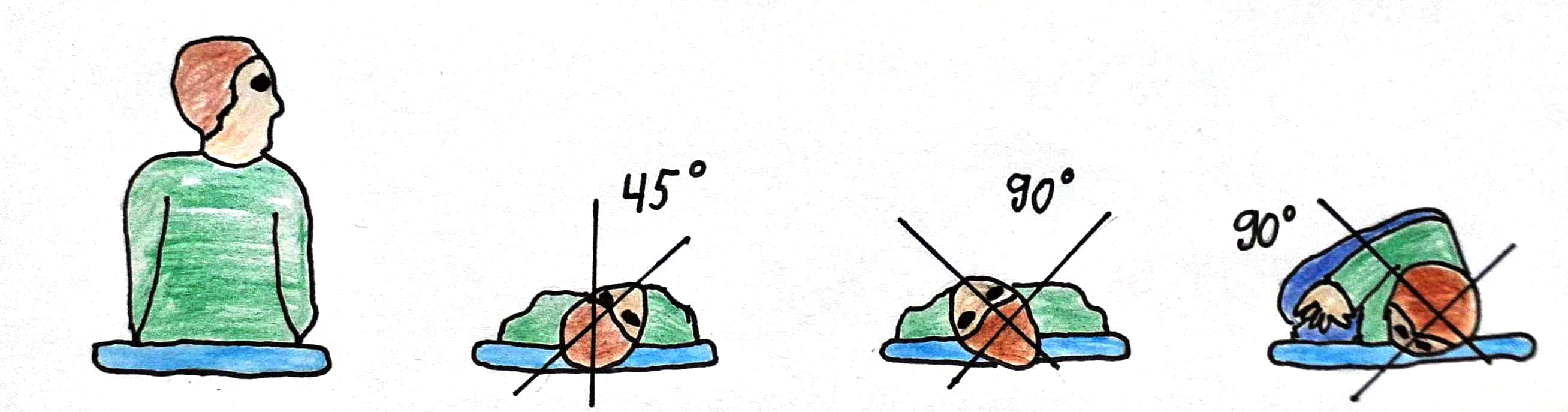
- Sit the patient upright with legs extended, head turned 45° toward the affected ear (the ear that produced a positive Dix-Hallpike test).
- Quickly recline the patient to the Dix-Hallpike position — supine with the head extended about 20° below horizontal, still turned 45° toward the affected ear. Hold until the provoked nystagmus and vertigo resolve, plus roughly another 20-30 seconds (typically about 1-2 minutes total).
- Turn the patient's head 90° to the opposite side (so it is now turned 45° toward the unaffected ear), keeping the neck extended. Hold for about 30-60 seconds, or until any nystagmus resolves.
- Roll the patient onto the unaffected side while turning the head a further 90° in the same direction, so the patient ends up looking down toward the floor. Hold for about 30-60 seconds.
- Slowly bring the patient back up to sitting, maintaining the head position through the transition, then bring the head to a neutral, chin-slightly-down position.
- After a brief rest, repeat the Dix-Hallpike test to confirm resolution. If nystagmus or vertigo is still provoked, repeat the full maneuver — up to 2-4 repetitions in the same visit is common and reasonable practice.
Semont (liberatory) maneuver — alternative for posterior/anterior canal BPPV
A reasonable alternative to the Epley maneuver, particularly useful when a patient cannot comfortably tolerate the neck extension the Epley maneuver requires. It relies on 2 rapid, sequential movements rather than the Epley's slower, staged repositioning, and has shown comparable effectiveness in randomized comparison.[9]
- Sit the patient upright at the edge of the table, head turned 45° away from the affected ear (toward the unaffected side).
- Rapidly lay the patient down onto the affected side, maintaining the 45° head turn, so the affected ear is down and the nose points up. Hold for about 1-3 minutes, or until nystagmus and vertigo resolve.
- In a single rapid, continuous movement — without pausing upright and without changing the head-to-trunk angle — move the patient through sitting and down onto the opposite side, ending with the nose angled down toward the floor. Hold for about 1-3 minutes.
- Slowly return the patient to sitting.
Lateral canal BPPV — barbecue roll and Gufoni maneuvers
For the geotropic subtype, the barbecue roll (Lempert) maneuver rotates the patient through a sequence of roughly 90° increments while supine — a full 270-360° roll away from the affected ear, pausing briefly at each position — using gravity to walk the debris out of the lateral canal and back into the utricle. The Gufoni maneuver is an alternative for either the geotropic or apogeotropic subtype, using a rapid lateral positioning sequence rather than a full roll. Both maneuvers require correctly identifying the affected ear and the geotropic/apogeotropic subtype first — this is typically performed by an audiologist, otolaryngologist, or vestibular physiotherapist rather than in routine primary care.
Brandt-Daroff exercises — self-administered adjunct or alternative
A habituation-based home exercise program, useful when a trained provider is not readily available or as an adjunct for residual dizziness after successful repositioning. Sit upright on the edge of the bed; quickly lie down onto one side with the head turned 45° toward the ceiling, remain until any dizziness subsides plus about 30 seconds; sit back up for about 30 seconds; then repeat lying down on the opposite side. A typical program is 5 repetitions per side, 3 times daily, continued until the patient is symptom-free for 2 consecutive days. These exercises are less reliably effective than a formal repositioning maneuver for immediate resolution and work through habituation rather than actually clearing the canal.[1]
Confirming resolution and safety notes
- Repeat the Dix-Hallpike or supine roll test after the maneuver, at the same visit, to confirm the nystagmus and vertigo no longer provoke — do not simply assume success.
- No post-procedure positional restrictions (sleeping upright, avoiding the affected side) are needed — resume normal activity immediately.[1]
- Relative or absolute contraindications to Dix-Hallpike/roll testing and repositioning maneuvers include unstable cervical spine disease, recent cervical spine surgery or trauma, severe cervical stenosis or limited neck mobility, vertebrobasilar insufficiency, unstable cardiac disease, severe or symptomatic carotid stenosis, and recent retinal detachment or eye surgery — in these patients, use a gentler, slower, modified technique (or a side-lying test in place of full Dix-Hallpike) or refer to a vestibular specialist rather than performing the standard rapid maneuver.
- Counsel every patient on fall-risk precautions for the period around diagnosis and treatment — get up slowly, avoid driving until confident vertigo is not being provoked by routine head movements, and use handrails/lighting at home, particularly for older adults (see Special Populations).
Pharmacologic Treatment Options
| Drug / intervention | Role | Dose / use | Important adverse effects | Teaching points |
|---|---|---|---|---|
| Dimenhydrinate Special situation | Brief use only, for severe acute nausea/vomiting preventing maneuver tolerance or basic activity | 50-100 mg PO/IM/IV/PR every 4-6 hours PRN, maximum 400 mg/day; limit to 1-2 days | Sedation, anticholinergic effects, paradoxical excitation in children | Not a treatment for BPPV itself — stop as soon as the acute episode settles; continued use can impair central compensation. |
| Betahistine Special situation | Off-label trial for residual dizziness after an otherwise successful repositioning maneuver | 16 mg PO 3 times daily (24-48 mg/day divided, per the Canadian product monograph) | GI upset, headache; caution in peptic ulcer disease and asthma given histaminergic activity | Evidence for this specific use is limited and heterogeneous; in Canada it is formally indicated for Ménière's disease, not BPPV. |
| Diazepam Special situation | Severe acute vertigo/anxiety with the initial episode only, brief use | 2-5 mg PO/IV as needed; avoid routine or repeated dosing | Sedation, ataxia, dependence with continued use; particular fall risk in older adults | A short-acting non-benzodiazepine or dimenhydrinate is generally preferable in older adults given fall and cognitive risk. |
| Lorazepam Special situation | Severe acute vertigo/anxiety with the initial episode only, brief use | 0.5-1 mg PO/SL/IV as needed; avoid routine or repeated dosing | Sedation, ataxia, dependence with continued use; particular fall risk in older adults | Reserve for a single severe episode, not maintenance therapy — impairs central compensation with continued use. |
| Ondansetron Alternative | Acute nausea/vomiting when a sedating antihistamine is best avoided (e.g., older adults, need to preserve alertness for driving assessment) | 4-8 mg PO/IV/ODT, may repeat per the product monograph | Headache, constipation, QTc prolongation at higher/repeated doses | Less sedating than dimenhydrinate and does not carry the same vestibular-suppressant/central-compensation concern. |
| Prochlorperazine Alternative | Acute nausea/vomiting, alternative to dimenhydrinate | 5-10 mg PO/IM every 6-8 hours PRN, or 25 mg PR every 12 hours PRN | Sedation, extrapyramidal symptoms, anticholinergic effects | Brief use only, for the same reason as dimenhydrinate — treats the nausea, not the BPPV itself. |
Special Populations
Older adults
BPPV is disproportionately consequential in older adults because of fall risk: untreated or under-treated BPPV materially increases fall risk, and the medications most often reached for reflexively — antihistamines and benzodiazepines — compound that risk further through sedation, anticholinergic burden, and cognitive impairment. Preferred approach prompt canalith repositioning remains the treatment of choice and is both effective and safe in older adults; a modified, gentler technique (for example, using a reclining chair, or avoiding full neck extension if cervical spine disease is present) can be used if standard positioning is not well tolerated. Avoid benzodiazepines and minimize antihistamine duration specifically in this group. Assess gait and home fall-risk (lighting, throw rugs, grab bars) as part of routine management, and screen for and correct vitamin D deficiency given its association with BPPV recurrence in this population (see Monitoring & Follow-Up).[10]
Pregnancy
Canalith repositioning maneuvers are entirely nonpharmacologic and are the preferred, safe treatment in pregnancy at any trimester — there is no reason to withhold or delay a repositioning maneuver because of pregnancy. If an antiemetic is genuinely needed for associated nausea, dimenhydrinate is a reasonable and commonly used option in pregnancy; avoid benzodiazepines. Confirm any medication choice against the Nausea and Vomiting of Pregnancy chapter and the drug's own Drug Database entry for full pregnancy-specific detail.
Breastfeeding
Dimenhydrinate and ondansetron are both considered compatible with breastfeeding at standard short-term doses; confirm specifics against each drug's own Drug Database entry. Minimize benzodiazepine use given infant sedation risk with repeated maternal dosing.
Pediatrics
BPPV is uncommon in children — when episodic positional vertigo does occur in a child, vestibular migraine is a more common explanation and should be considered first. When BPPV is genuinely diagnosed in a child, the same canal-appropriate repositioning maneuvers are used, with technique modified for the child's size and cooperation; involve a pediatric otolaryngology or audiology specialist rather than performing an unmodified adult-technique maneuver.
Cervical spine disease, unstable cardiac disease, or recent eye surgery/retinal detachment
These are relative or absolute contraindications to the standard rapid Dix-Hallpike/Epley technique (see Non-Pharmacologic Treatment) — use a slower, gentler modified approach or refer to a vestibular specialist rather than performing the standard maneuver unmodified.
Renal and hepatic impairment
Any medication use in BPPV is intended to be brief (days, not weeks), which limits the practical significance of organ-impairment dose adjustment — but confirm standard renal/hepatic dosing guidance for dimenhydrinate, betahistine, diazepam, lorazepam, ondansetron, or prochlorperazine against each drug's own Drug Database entry if a course is being used.
Monitoring & Follow-Up
Reassess within about 1 month of the initial repositioning treatment to document that symptoms have genuinely resolved rather than assuming success from a single in-visit negative Dix-Hallpike — a meaningful minority of patients need more than 1 treatment session, and some need a second visit for a repeat maneuver.[1]
- At follow-up, confirm the specific trigger movements no longer provoke vertigo, not just that the patient "feels better" overall — residual imbalance without true positional vertigo is common in the days after a successful maneuver and does not necessarily mean the BPPV itself has failed to resolve.
- Educate every patient explicitly that recurrence is common — commonly cited figures range from roughly 15% to 50% over 1-5 years of follow-up — and that recurrence is not a sign of treatment failure or a more serious problem; the same repositioning maneuver is typically effective again.
- For a patient with confirmed vitamin D deficiency and recurrent BPPV, particularly an older adult, vitamin D supplementation is a reasonable, evidence-supported step: a randomized trial in this population found a substantially lower recurrence rate with vitamin D supplementation compared with placebo.[10] This is a genuinely actionable, low-risk intervention worth raising proactively rather than waiting for the patient to ask.
- Screen for and address osteoporosis in patients with recurrent BPPV and other osteoporosis risk factors, given the documented association between the two conditions.
- If a short course of a vestibular suppressant or antiemetic was used for the acute episode, confirm it was actually stopped rather than continued indefinitely by default.
Treatment Failure / Next Step
- Before assuming the maneuver failed, confirm it was performed correctly and completely — the correct canal and affected ear were identified, each position was held long enough (until nystagmus/vertigo resolved, plus the recommended additional time), and the patient was moved through the full sequence without an interrupting pause.
- Repeat the same repositioning maneuver — 2-4 repetitions in a single visit, or a second treatment visit a few days later, resolves the great majority of otherwise-typical posterior canal BPPV; a single attempt failing to fully resolve symptoms is common and does not by itself indicate a different diagnosis.
- Reconfirm the canal and subtype with a fresh Dix-Hallpike and supine roll test — canaliths can migrate into a different canal after a maneuver (canal conversion); if a different canal is now involved, treat with the canal-appropriate maneuver instead of repeating the original one.
- If nystagmus on retesting is non-fatiguing, lacks the expected latency, or persists well beyond a minute, reconsider cupulolithiasis (which responds less predictably to a standard maneuver) or a central positional-vertigo mimic — refer to a vestibular specialist (audiology, otolaryngology, or vestibular physiotherapy) or for neurologic assessment rather than repeating the same maneuver indefinitely.[3]
- For residual dizziness or imbalance after the positional nystagmus itself has resolved, refer for formal vestibular rehabilitation therapy rather than restarting or extending medication.
- For recurrent or refractory BPPV, especially in an older adult, screen for and correct vitamin D deficiency and assess for osteoporosis (see Monitoring & Follow-Up).[10]
- Any new red flag emerging during follow-up (see Red Flags & Refer) should prompt the same urgent central-cause work-up as at initial presentation — a prior BPPV diagnosis does not make a patient immune to an unrelated new central event.
Common Errors to Avoid
- Reaching for meclizine, dimenhydrinate, or a benzodiazepine as first-line or ongoing therapy instead of arranging a canalith repositioning maneuver — medication does not treat the underlying canalithiasis and can measurably impair central compensation.
- Recommending postprocedural positional restrictions (sleeping upright, avoiding the affected side) after a successful maneuver — this is outdated teaching not supported by current evidence.
- Ordering routine MRI/CT or formal vestibular/audiometric testing in a patient with a classic history and a clearly positive, typical Dix-Hallpike or roll test.
- Treating any position-triggered vertigo as BPPV without confirming the nystagmus pattern actually fits — purely vertical, direction-changing, non-fatiguing, or unusually prolonged nystagmus is a red flag for a central mimic, not routine BPPV.
- Applying the HINTS exam framework to episodic, position-triggered vertigo — HINTS is designed for continuous acute vestibular syndrome, not for typical BPPV, where the Dix-Hallpike test itself is the correct bedside tool.
- Using the posterior-canal Epley maneuver for lateral canal BPPV instead of the canal-appropriate barbecue roll or Gufoni maneuver.
- Assuming a single repositioning attempt is sufficient and not offering a repeat maneuver or follow-up visit when symptoms persist.
- Missing hearing loss, tinnitus, or aural fullness as clues pointing toward Ménière's disease rather than BPPV.
- Not screening a patient with recurrent BPPV, especially an older adult, for vitamin D deficiency and osteoporosis.
- Performing a full-speed Dix-Hallpike or Epley maneuver in a patient with an unrecognized contraindication (unstable cervical spine disease, recent retinal detachment, unstable cardiac disease) instead of using a modified technique or referring.
Clinical Decision Algorithm
Drug Tables
How to use this table: doses are adult doses and require patient-specific confirmation, including renal/hepatic adjustment. Select any drug name to open its complete Pharmasist Drug Database page. None of these agents is a substitute for a canal-appropriate repositioning maneuver, which remains the definitive treatment for BPPV.
Short-Term Antiemetic / Vestibular-Suppressant Therapy — Adjunct Only, Not Routine
Reserve for brief use during a severe acute episode, specifically when nausea/vomiting or intense vertigo prevents the patient from tolerating basic activity or a repositioning maneuver. Stop as soon as the acute episode settles — continued use can impair the central nervous system's own vestibular compensation and does not treat the underlying canalithiasis.
| Drug | Dosage | Adverse effects | Drug interactions | Comments |
|---|---|---|---|---|
| Dimenhydrinate | Adults: 50-100 mg PO/IM/IV/PR every 4-6 hours PRN, maximum 400 mg/day | Sedation, anticholinergic effects (dry mouth, blurred vision, urinary retention), confusion in older adults, paradoxical excitation in children | Additive sedation with other CNS depressants; may mask ototoxicity of a concurrently administered ototoxic drug | The most commonly used Canadian OTC option for this purpose; limit to 1-2 days. |
| Meclizine | Adults: 25-50 mg PO once, or every 24 hours PRN for brief use | Sedation, anticholinergic effects, dizziness | Additive sedation with other CNS depressants | The classic American vestibular-suppressant example; not a commonly marketed Canadian product — dimenhydrinate is the practical Canadian equivalent. |
| Ondansetron | Adults: 4-8 mg PO/IV/ODT, may repeat per the product monograph | Headache, constipation, QTc prolongation at higher/repeated doses | Additive QTc prolongation with other QTc-prolonging drugs | Less sedating than dimenhydrinate; a reasonable choice when preserving alertness matters (e.g., before a driving-safety assessment). |
| Prochlorperazine | Adults: 5-10 mg PO/IM every 6-8 hours PRN, or 25 mg PR every 12 hours PRN | Sedation, extrapyramidal symptoms, anticholinergic effects | Additive sedation with other CNS depressants; additive extrapyramidal risk with other dopamine antagonists | An alternative to dimenhydrinate for acute nausea; same brief-use caution applies. |
Benzodiazepines — Reserved for a Single Severe Acute Episode, Not Maintenance Therapy
Do not use routinely or repeatedly — benzodiazepines impair central vestibular compensation with continued use and carry meaningful fall and dependence risk, particularly in older adults.
| Drug | Dosage | Adverse effects | Drug interactions | Comments |
|---|---|---|---|---|
| Diazepam | Adults: 2-5 mg PO/IV as needed | Sedation, ataxia, dependence with continued use, respiratory depression at higher doses | Additive CNS depression with alcohol, opioids, or other sedatives | Reserve for a single severe episode; avoid repeated dosing. |
| Lorazepam | Adults: 0.5-1 mg PO/SL/IV as needed | Sedation, ataxia, dependence with continued use, respiratory depression at higher doses | Additive CNS depression with alcohol, opioids, or other sedatives | Reserve for a single severe episode; avoid repeated dosing. |
Betahistine — Off-Label, Special-Situation Adjunct
In Canada, formally indicated for Ménière's disease rather than BPPV. Sometimes trialled, off-label, for residual dizziness after an otherwise successful repositioning maneuver — current evidence for this specific use is limited and heterogeneous, and it is not a substitute for repositioning.
| Drug | Dosage | Adverse effects | Drug interactions | Comments |
|---|---|---|---|---|
| Betahistine | Adults: 24-48 mg/day PO in divided doses | GI upset, headache | None significant at standard doses; use with caution in peptic ulcer disease and asthma given histaminergic activity; contraindicated in pheochromocytoma | 16 mg tablets: half to 1 tablet 3 times daily; 24 mg tablets: 1 tablet twice daily. |
Professional reference only. Apply clinical judgment and verify patient-specific decisions against current authoritative guidance and product information.