Blepharitis
Clinical Snapshot
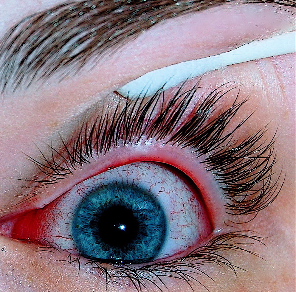
Blepharitis is chronic inflammation of the eyelid margin — crusting, scale, and debris at the base of the eyelashes, frequently mistaken for recurrent conjunctivitis when the real problem is the eyelids, not the conjunctiva.[1] It is classified as anterior (affecting the lash base and follicles — staphylococcal, seborrheic, or Demodex-related) or posterior (meibomian gland dysfunction, MGD — obstruction and altered secretion from the meibomian glands along the lid margin); many patients have a mixed picture. Blepharitis is a chronic, relapsing condition that is managed with ongoing lid hygiene, not cured with a single course of treatment — set this expectation with the patient from the first visit.
Recognize the Condition
The subtypes of blepharitis
Anterior blepharitis
Crusting, scale, and greasy debris at the base of the lashes; may have collarettes (cylindrical debris specifically encircling the lash base) suggesting Demodex mite involvement, missing or misdirected lashes, and recurrent styes (hordeola). Staphylococcal anterior blepharitis tends to have harder, fibrous scale with lash loss/misdirection; seborrheic anterior blepharitis tends to have greasier, softer scale and is often associated with seborrheic dermatitis elsewhere (scalp, eyebrows).
Posterior blepharitis (meibomian gland dysfunction)
Meibomian gland orifices along the lid margin become obstructed, with thickened, turbid, or absent secretion on gland expression; the tear film becomes unstable, causing evaporative dry eye symptoms — burning, foreign-body sensation, fluctuating vision. Often associated with ocular rosacea (facial flushing, telangiectasias, rhinophyma) and less itch-predominant than allergic conjunctivitis.
Demodex blepharitis
Collarettes at the lash base are the hallmark sign of Demodex mite infestation — a specific, now separately treatable subtype of anterior blepharitis (see Pharmacologic Treatment Options).
Risk Factors
- Ocular or facial rosacea — strongly associated with posterior blepharitis/MGD.
- Seborrheic dermatitis elsewhere (scalp, eyebrows, nasolabial folds).
- Older age — both Demodex prevalence and MGD increase with age.
- Contact lens wear.
- Prior styes (hordeola) or chalazia.
- Poor eyelid hygiene, or heavy eye makeup use without adequate removal.
Red Flags & Refer
Refer to ophthalmology for:
- Any corneal involvement (photophobia, reduced vision, or a corneal opacity on exam) — chronic blepharitis/MGD can cause secondary keratitis.
- A unilateral, asymmetric, or rapidly progressive lid-margin lesion, particularly with lash loss over a localized area or a firm, non-tender nodule (consider sebaceous gland carcinoma, a rare but important masquerade of chronic unilateral "blepharitis," especially in an older adult).
- Suspected preseptal or orbital cellulitis (lid swelling with warmth, tenderness, fever, or pain with eye movement) rather than simple blepharitis.
- Blepharitis not responding to a consistent, adequate trial of lid hygiene plus appropriate topical/oral therapy.[2]
Assessment & Diagnosis
Differentiating bacterial, viral, and allergic conjunctivitis, and blepharitis
These four conditions are the most commonly confused causes of a red, irritated eye in community practice. Distinguishing them correctly determines whether the patient needs an antibiotic, supportive care, an antihistamine, or eyelid hygiene — and getting it wrong means either an unnecessary antibiotic or a missed diagnosis.



| Feature | Bacterial conjunctivitis | Viral conjunctivitis | Allergic conjunctivitis | Blepharitis |
|---|---|---|---|---|
| Laterality | Often one eye first, may spread to the other | Usually starts in one eye, spreads to the other within days | Bilateral, essentially always | Bilateral, essentially always |
| Discharge | Purulent, thick, yellow-green; lids stuck together on waking | Watery to mucoid; no true pus | Watery, stringy/mucoid | Greasy debris and crusting at the lash base, not a true discharge |
| Itching | Minimal or absent | Mild, if present | The dominant symptom — if the patient does not report itching, reconsider allergic conjunctivitis | Mild irritation/burning more than itching |
| Associated findings | May follow otitis media in children; no preauricular node in most cases | Preauricular lymph node, recent URTI, conjunctival membranes, subconjunctival hemorrhage | Chemosis (conjunctival swelling), lid edema, history of atopy/seasonal pattern | Lash crusting, missing/misdirected lashes, recurrent styes, meibomian gland dysfunction |
| Typical course | Self-limited over 1-2 weeks even without treatment; antibiotics modestly shorten this | Self-limited over 1-3 weeks; highly contagious for the first 3-7 days | Persists as long as allergen exposure continues; recurs seasonally | Chronic, relapsing; managed rather than cured |
| 1st-line management | Often no treatment needed; a topical antibiotic modestly shortens recovery | Supportive care only — cold compresses, lubricants | Remove/avoid the allergen; topical antihistamine/mast-cell stabilizer | Lid hygiene: warm compresses plus lid margin cleansing |
History
- Chronicity and prior episodes — blepharitis is typically a long-standing, relapsing complaint, not an acute one.
- Burning/irritation versus true itching (itching more prominent in allergic conjunctivitis).
- Morning symptoms (crusting) versus symptoms that worsen through the day (more typical of evaporative dry eye from MGD).
- History of rosacea, seborrheic dermatitis, or recurrent styes.
- Contact lens wear and eye makeup habits.
Examination
- Inspect the lash base for scale/crusting and specifically for collarettes (Demodex sign).
- Assess lash position — missing, misdirected, or thinned lashes suggest chronic anterior blepharitis.
- Express the meibomian glands (gentle digital pressure on the lower lid margin) to assess secretion quality — clear and easily expressed is normal; thick, turbid, toothpaste-like, or absent secretion suggests MGD.
- Look for signs of ocular or facial rosacea.
- Fluorescein staining if corneal involvement is possible.
Treatment Targets
- Reduce lid margin inflammation and bacterial/Demodex load.
- Restore normal meibomian gland function and tear film stability.
- Prevent complications — recurrent styes, chalazia, secondary keratitis.
- Set realistic expectations: control, not cure.
Choose Initial Treatment
1st line, all blepharitis lid hygiene — warm compresses followed by lid margin cleansing — is the foundation of treatment for every subtype and must be maintained long-term, not just during a flare.
2nd line, anterior blepharitis with secondary bacterial infection a topical antibacterial ointment applied to the lid margin (not just used as eye drops) in addition to lid hygiene.
Confirmed Demodex blepharitis lotilaner ophthalmic solution, the first drug specifically approved for this indication, in addition to lid hygiene.
Moderate-to-severe posterior blepharitis/MGD low-dose oral doxycycline (anti-inflammatory dosing, not antibacterial dosing) for 1-3 months, in addition to lid hygiene and warm compresses, when topical measures alone are insufficient.[3]
Non-Pharmacologic Treatment
- Warm compresses — apply to closed lids for 5-10 minutes, 1-2 times daily; this softens debris and helps liquefy meibomian gland secretions. A reusable warming mask/device can help maintain adequate, sustained heat compared with a cooling washcloth.
- Lid margin cleansing — after warm compresses, gently clean the lash base with a commercial eyelid wipe, diluted baby shampoo on a cotton swab, or a dedicated lid-hygiene product, 1-2 times daily during flares and as maintenance thereafter.
- Lid massage — gentle massage along the lid margin after warm compresses helps express meibomian gland contents in posterior blepharitis/MGD.
- Omega-3 fatty acid supplementation — may support meibomian gland secretion quality as an adjunct; evidence is modest but the intervention is low-risk.
- Remove eye makeup thoroughly each day and replace eye makeup regularly to reduce contamination.
- Set the expectation up front that this is a chronic, relapsing condition managed with ongoing hygiene, not a one-time course of treatment.
Special Populations
Pregnancy/breastfeeding
Lid hygiene and topical antibacterial ointments are appropriate. Avoid oral doxycycline in pregnancy and breastfeeding — it is associated with fetal/infant tooth discoloration and bone growth effects; if oral therapy for severe posterior blepharitis is truly needed, discuss alternatives with the prescriber managing the pregnancy.
Children
Avoid oral tetracyclines (including doxycycline) under 8 years of age given the risk of permanent tooth discoloration — lid hygiene and topical measures are the mainstay in this age group; a macrolide (e.g., oral azithromycin) is sometimes used instead for significant pediatric blepharitis/MGD under specialist direction.
Contact lens wearers
Blepharitis increases the risk of contact-lens-related complications (including bacterial keratitis) — reinforce hygiene and consider temporarily reducing or stopping lens wear during a flare.
Ocular rosacea
Patients with posterior blepharitis and facial rosacea benefit from co-management of the skin disease (topical/oral rosacea therapy) alongside eyelid-directed treatment — treating one without the other often gives incomplete control.
Monitoring & Follow-Up
Blepharitis is a chronic, relapsing condition managed with ongoing lid hygiene rather than cured with a single course of treatment — reinforce this at every visit. Reassess after 4-6 weeks of consistent therapy; a lack of any improvement with genuinely consistent hygiene and appropriate topical/oral therapy should prompt reconsideration of the diagnosis (or a trial of the other treatment tier — e.g., adding oral doxycycline if topical measures alone have been tried, or testing specifically for Demodex if not already assessed).
Common Errors to Avoid
- Treating blepharitis with a single course of antibiotic ointment and no lid-hygiene plan — without ongoing hygiene, symptoms reliably recur.
- Missing Demodex as a treatable cause by not examining specifically for collarettes.
- Prescribing full antibacterial-dose doxycycline for MGD when the anti-inflammatory (low) dose is what is actually indicated and better tolerated for this purpose.
- Assuming chronic unilateral "blepharitis" in an older adult with progressive lash loss is simple blepharitis rather than considering sebaceous gland carcinoma, a rare masquerade.
- Using a topical corticosteroid/antibiotic combination long-term without specialist supervision and monitoring for glaucoma/cataract risk.
- Not addressing coexisting rosacea or seborrheic dermatitis, which drives ongoing flares if left untreated.
Clinical Decision Algorithm
Drug Tables
How to use this table: Doses are adult doses and require patient-specific confirmation (renal/hepatic function, weight, allergy history). Pediatric dosing is weight-based per the current product monograph unless stated. Drug pricing is intentionally omitted. Select any drug name to open its complete Pharmasist Drug Database page.
Topical antibacterials — lid margin application
Applied directly to the lid margin (not just instilled as drops) for anterior blepharitis with secondary bacterial infection.
| Drug | Dosage | Adverse effects | Comments |
|---|---|---|---|
| Erythromycin ophthalmic ointment | All ages: Apply to lid margin at bedtime for 2-4 weeks | Chronic use may cause corneal epithelial toxicity, allergy, resistance | Apply a small amount to the lid margin, not into the eye itself, using a clean fingertip or cotton swab. |
| Fusidic acid ophthalmic | All ages: Apply to lid margin BID for 2-4 weeks | Chronic use may cause corneal toxicity, allergy | BID dosing convenient for lid-margin application. |
Demodex-targeted therapy
For confirmed Demodex blepharitis (collarettes present at the lash base).
| Drug | Dosage | Adverse effects | Comments |
|---|---|---|---|
| Lotilaner ophthalmic solution | All ages: 1 drop each eye BID for 6 weeks | Instillation-site stinging/burning (~10%); rare chalazion/hordeolum, punctate keratitis | First drug specifically FDA-approved for Demodex blepharitis (2023). Complete the full 6-week course even if collarettes improve earlier. |
Oral therapy for posterior blepharitis/meibomian gland dysfunction
Anti-inflammatory (low) dosing, not antibacterial dosing — a different regimen from treating a systemic infection.
| Drug | Dosage | Adverse effects | Comments |
|---|---|---|---|
| Doxycycline (low-dose) | Adults: 50-100 mg PO once daily | GI upset, photosensitivity, rare esophageal irritation | 1-3 month course; longer in rosacea-associated disease. Avoid in pregnancy, breastfeeding, and children under 8 years (tooth discoloration). |
Topical antibacterial/corticosteroid combination
Short-term only, specialist-directed, for a significant inflammatory flare — corticosteroid component carries real long-term risk (glaucoma, cataracts) and may worsen undiagnosed herpetic/fungal disease.
| Drug | Dosage | Adverse effects | Comments |
|---|---|---|---|
| Tobramycin/dexamethasone | All ages: Suspension 1 drop TID-QID, or ointment TID-QID, short-term | Corneal toxicity, allergy, resistance; corticosteroid effects (stinging, may worsen herpetic/fungal keratitis, long-term glaucoma/cataracts) | Severe blepharitis, rosacea-associated eyelid disease. |
| Neomycin/polymyxin B/dexamethasone | All ages: Suspension 1 drop TID-QID, or ointment TID-QID, short-term | Corneal toxicity, allergy, resistance; corticosteroid effects | Same indications and cautions as tobramycin/dexamethasone. |
References
- Blepharitis Preferred Practice Pattern — Ophthalmology, American Academy of Ophthalmology (Lin A, Rhee MK, Akpek EK, et al.). Ophthalmology 2024;131(4):P54-P127
- Bedside diagnosis of the 'red eye': a systematic review — American Journal of Medicine (Narayana S, McGee S). Am J Med 2015;128(11):1220-1224
- The role of oral doxycycline in meibomian gland dysfunction — Cornea/Ocular Surface literature (summarized in Blepharitis PPP evidence base). See Blepharitis PPP, Ophthalmology 2024;131(4)
- Advances in the treatment of Demodex blepharitis: strategies for improving patient outcomes — American Journal of Managed Care. AJMC, 2024
Professional reference only. Apply clinical judgment and verify patient-specific decisions against current authoritative guidance and product information.