Dermatological Considerations in Skin of Colour
Clinical Snapshot
Skin of colour (SOC) refers to the deep or rich end of the skin-pigmentation spectrum (Fitzpatrick types IV-VI, though Fitzpatrick typing was developed for UV sensitivity and correlates imperfectly with constitutive colour). The clinical relevance is not cosmetic — melanin density changes how disease looks, how fast it's recognized, and how it heals. Erythema, the visual cue most treatment-severity decisions and scoring tools (SCORAD, EASI, PASI) are built around, is frequently attenuated or absent in darkly pigmented skin and instead appears violaceous, grey, or hyperpigmented — which means relying on redness alone systematically under-recognizes active inflammation and delays escalation. The single most consequential downstream effect of that delay, and of skin inflammation in this population generally, is post-inflammatory hyperpigmentation (PIH): a disproportionate, often prolonged, and distressing dyspigmentation that follows essentially any cutaneous inflammation or injury and disproportionately affects patients with darker skin.[1] This chapter covers the pharmacotherapy consequences of these differences — not a general dermatology review, but the specific prescribing, dosing, and counselling adjustments skin of colour requires.
Recognize the Condition
Why the same disease looks different
Melanocyte density does not differ meaningfully by skin colour — pigmentation differences come from the ratio of eumelanin (brown-black) to pheomelanin (yellow-red), total melanin output, and melanosome size/distribution/degradation rate. Because melanin absorbs light broadly, it visually competes with the red of erythema, muting or masking it. The practical result: inflammation in darkly pigmented skin commonly reads as violaceous, grey, or simply as new hyperpigmentation rather than as classic redness. This is true across essentially every inflammatory dermatosis — eczema/atopic dermatitis and psoriasis both follow this pattern (see those chapters for full disease-specific management; the presentation difference described here is what should prompt more careful, non-erythema-reliant severity assessment before applying their treatment algorithms in a patient with skin of colour), and it is equally true of the drug reactions and infections pharmacists are positioned to catch.
| Exam finding | What to look for in skin of colour |
|---|---|
| Erythema | May range from pink to red to a purplish or violaceous hue rather than classic red; a purplish rim around a lesion is a useful SOC-specific cue. Because it can be underestimated visually, actively ask about and examine for pain, burning, itching, warmth, swelling, scaling, crusting, and erosions as corroborating evidence of inflammation rather than relying on colour change alone. |
| Pigmentary change | Hypo- or hyperpigmented macules/patches after any inflammatory dermatosis, external injury, or topical irritation — this is post-inflammatory hyper/hypopigmentation, covered in depth below. |
| Pallor | May present as ashen, grey, or yellowish rather than the classic pale appearance, and can be subtle. Check for reduced darkness of the palmar creases; comparing the patient's palms to an unaffected family member's can help, allowing for limited sensitivity/specificity. |
| Cyanosis | Examine sites where the epidermis is thin or pigment is minimal: oral mucous membranes (buccal, sublingual), lips, palms, soles, nail beds. May present as a bluish tint to palms/conjunctiva, a greyish-white colour around the tongue/lips, or a maroon tinge to the nail beds; patients with yellow-toned skin may show a greyish-green appearance overall. |
Baseline assessment principles
Establish the patient's own baseline skin colour early (an objective tool is preferable to a subjective description) so later changes can be judged against it rather than against an assumed norm. Compare the affected area to adjacent healthy skin for colour, texture, shininess, and tightness (subtle swelling can present only as smoother or more widely spaced pores), and to touch for temperature, firmness, and texture. Ask directly about pain, itch, and altered sensation rather than assuming these track visible severity. Examine interdigital areas, the sides of fingers/toes, and palms/soles specifically — skin cancers, including melanoma, occur at these non-sun-exposed sites disproportionately in skin of colour (see Red Flags & Refer).
Nail findings
Longitudinal melanonychia (a brown/black band or streak running the length of the nail plate) is a common benign finding in darkly pigmented skin, generally involving multiple digits on more than one hand. This must be distinguished from subungual melanoma, which typically involves a single digit (most often a thumb in an adult) and carries specific warning features.
| Feature | Benign longitudinal melanonychia | Subungual melanoma — refer for biopsy |
|---|---|---|
| Number of digits | Usually multiple | Usually a single digit |
| Band width | Narrow, even | Broad (3 mm or more), often widening toward the base (proximal nail fold) |
| Border | Regular, sharply defined | Blurry, fuzzy, irregular |
| Colour | Uniform | Variegated (multiple shades within the same band) |
| Extension | Confined to the nail plate | Pigment extending onto the proximal or lateral nail fold (Hutchinson sign) |
Risk Factors
- Fitzpatrick skin type IV-VI (or simply darker constitutive pigmentation — Fitzpatrick typing was designed for UV/burn sensitivity and correlates imperfectly with true pigmentation).
- For post-inflammatory hyperpigmentation specifically: any active inflammatory dermatosis (acne, eczema, psoriasis, insect bites), mechanical or chemical skin irritation (including from topical acne/retinoid therapy itself), and delayed treatment of the underlying inflammation.
- For melasma: female sex, sun/UV and visible-light exposure, hormonal exposure (pregnancy, combined oral contraceptives, hormone replacement therapy), and family history.
- For keloid/hypertrophic scarring: family history, high-tension anatomic sites (chest, shoulders, upper back, earlobes), and any dermal injury including piercing, surgery, vaccination, and acne itself.
- For traction alopecia: tightly applied head coverings (turbans, hijabs, wig caps), and tight hairstyles maintained for a prolonged period — braids, ponytails, buns, weaves, and hair extensions.
- For central centrifugal cicatricial alopecia (CCCA): occurs almost exclusively in women of African descent; traumatic hair practices (heat styling, chemical relaxers, tight styling) are proposed contributing factors, though the exact cause is unknown and likely multifactorial.
- For pseudofolliculitis barbae: shaving of tightly curled/coiled facial or neck hair, which re-enters the skin as it grows and triggers a foreign-body-type inflammatory reaction.
- For acral lentiginous melanoma specifically: increasing age; this is the most common melanoma subtype in patients with darkly pigmented skin, occurring on palms, soles, and the nail apparatus — sites not primarily explained by cumulative UV exposure, so sun-exposure counselling alone does not reduce this risk.
Red Flags & Refer
Escalate or refer promptly for any of the following:
- A rapidly enlarging, newly painful/tender, or otherwise symptomatic keloid or hypertrophic scar — warrants dermatology or plastic surgery assessment rather than continued empiric topical/intralesional management alone.
- Any atypical or rapidly changing pigmented lesion — new asymmetry, irregular or blurry border, colour variegation, diameter growth, or evolution over time — needs biopsy to exclude malignancy. Acral lentiginous melanoma (palms, soles, nail apparatus) is easy to miss or delay in skin of colour because it occurs at sites not routinely examined and can be mistaken for benign findings (a stubborn "bruise" that doesn't resolve, a nail streak assumed to be benign melanonychia); melanoma is genuinely underdiagnosed and diagnosed at a later stage in this population, so a low threshold for biopsy referral at these sites is warranted.[2]
- A nail pigment band that is broad (3 mm or more), asymmetric to a single digit, has a blurry/irregular border, shows variegated colour, or extends onto the surrounding nail fold (Hutchinson sign) — refer immediately rather than presuming benign longitudinal melanonychia.
- Severe or rapidly progressive hair loss, especially with associated scalp burning, tenderness, or visible scarring/smoothing of the follicular openings — refer urgently to dermatology. Central centrifugal cicatricial alopecia and other scarring alopecias cause permanent follicle destruction; hair will not regrow once true scarring has occurred, so the entire treatment goal is halting progression before that point, which makes early referral itself the time-critical intervention, not a specific drug.
- Suspected exogenous ochronosis — paradoxical blue-grey or darker discolouration developing at a site being treated with hydroquinone, rather than the expected lightening — stop hydroquinone immediately and refer; continued use worsens the condition.
Assessment & Diagnosis
History
- Document Fitzpatrick type or another structured pigmentation descriptor at baseline, not just at the time of a pigmentary complaint — this is what later change gets compared against.
- For any inflammatory dermatosis: ask specifically about pain, itch, burning, and warmth in addition to inspecting for colour change, since erythema may be attenuated or absent.
- Ask about current and recent hair-care practices (relaxers, heat styling, weaves/braids/extensions, tension level, head coverings) when hair loss, scalp irritation, or scarring is a concern — this history frequently identifies a modifiable cause.
- Ask specifically about use of skin-lightening products, including unregulated or imported preparations, which may contain high-dose hydroquinone, corticosteroids, or heavy metals (mercury) — patients may be reluctant to disclose this due to stigma, and undisclosed concurrent use changes both the differential and the safe treatment plan.
- For suspected melasma: ask about hormonal exposures (pregnancy, hormonal contraception, hormone replacement therapy) and sun/visible-light exposure pattern.
Examination
Perform a full skin, hair, scalp, nail, and mucous-membrane examination using the non-erythema-reliant cues above. Examine palms, soles, and the nail apparatus specifically in any patient with darkly pigmented skin presenting with a new or changing pigmented lesion, given the disproportionate contribution of acral lentiginous melanoma at these sites in this population.[2] Where weaves, braids, or wig caps limit direct scalp inspection, or where personal, cultural, or religious beliefs make a physical scalp exam infeasible, a detailed verbal history of the hair-loss pattern and timeline is still obtainable and should be taken.
When biopsy is, and is not, indicated
Biopsy any atypical pigmented lesion per the Red Flags criteria above rather than presuming a benign process (dermatosis papulosa nigra, benign melanonychia, seborrheic keratosis) without examination — these benign entities are common in skin of colour, but their presumptive diagnosis should not substitute for biopsy when a lesion has any atypical feature. Biopsy is not routinely needed to diagnose PIH itself, which is a clinical diagnosis based on a documented preceding inflammatory event or injury at the same site plus the characteristic macular, non-palpable colour change.
Treatment Targets
- Recognize and treat the underlying inflammatory dermatosis promptly enough — and, when needed, more assertively than erythema-based severity assessment alone would suggest — to limit the duration and intensity of inflammation that drives post-inflammatory hyperpigmentation.
- Resolve or substantially fade established PIH and melasma using an evidence-based topical regimen, without causing new irritation-driven pigmentation or exogenous ochronosis in the process.
- Sustain photoprotection (UV and visible light) as the foundation of any pigmentary treatment plan, since ongoing sun/light exposure undermines every other therapy.
- Minimize keloid/hypertrophic scar risk when a procedural or injectable option is being chosen, and treat an established keloid with an approach matched to its risk of recurrence.
- Halt progression of scarring alopecia (CCCA) and traction alopecia before permanent follicle loss occurs — regrowth is not possible once true scarring has developed, so early intervention is the entire objective.
- Preserve adherence to irritating but necessary topical regimens (retinoids, benzoyl peroxide, hydroquinone) through deliberate titration and vehicle selection, since irritation itself can trigger the same PIH the regimen is meant to prevent.
Choose Initial Treatment
Any inflammatory dermatosis in a patient with skin of colour
Do not wait for classic erythema to judge severity or to decide to escalate therapy — assess pain, itch, warmth, swelling, scaling, and duration alongside whatever colour change is present, and treat promptly and, where clinical severity supports it, more assertively than a purely erythema-based assessment would suggest, because the time the dermatosis remains active is the single biggest driver of whether PIH develops and how severe it becomes.[1]
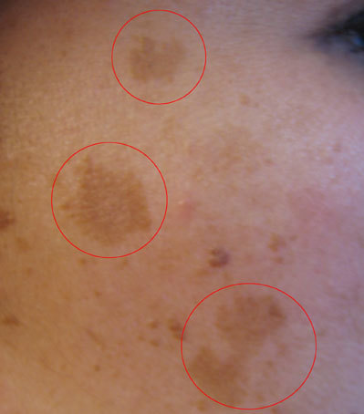
Established post-inflammatory hyperpigmentation
1st line combine strict, year-round broad-spectrum photoprotection with a topical agent — azelaic acid 15-20%, a topical retinoid (tretinoin or tazarotene), or time-limited hydroquinone 4% — chosen based on the patient's tolerance for irritation and the practicality of duration-limited hydroquinone use.
2nd line — when a single agent gives an inadequate response after an adequate trial combine two mechanistically different agents (e.g., a retinoid plus azelaic acid, or hydroquinone plus a retinoid) rather than simply extending a single agent past its recommended duration.
Special situation cysteamine cream is a reasonable non-hydroquinone option for a patient who cannot tolerate hydroquinone or who needs an option without ochronosis risk for longer-term use; topical tranexamic acid or, under specialist direction, oral tranexamic acid is an option for melasma-pattern pigmentation refractory to first-line topical therapy, after screening for thromboembolic risk factors.[3]
Melasma
1st line strict photoprotection (broad-spectrum SPF 30 or higher, reapplied, with a tinted iron-oxide-containing formulation) combined with a topical depigmenting agent — azelaic acid, a topical retinoid, or hydroquinone 4% used for a defined, time-limited course.[4]
2nd line — inadequate response after 8-12 weeks of adherent 1st-line therapy combination topical therapy (e.g., hydroquinone plus a retinoid, with or without a low-potency topical corticosteroid for a limited duration to reduce retinoid/hydroquinone irritation) or referral for a procedural option (chemical peel, laser).
Elevated keloid/hypertrophic scarring risk
When a procedure (biopsy, excision, piercing, cosmetic injection) is elective and a lower-risk alternative exists, factor keloid risk into that choice for a patient with a personal or family history of keloids, and counsel on early silicone gel/sheeting use starting once the wound has re-epithelialized. For an already-established keloid or hypertrophic scar, intralesional corticosteroid (triamcinolone) is the pharmacologic backbone of treatment, with 5-fluorouracil added for lesions responding inadequately to corticosteroid monotherapy.[5]
Retinoid or benzoyl peroxide irritation threatening adherence
Do not stop the agent outright at the first sign of irritation — step down frequency (alternate nights, then nightly as tolerated), apply over a moisturizer ("buffering") rather than to bare skin, and confirm a fragrance-free, non-comedogenic moisturizer is being used alongside it, since irritation-driven inflammation from the treatment itself can cause the same PIH the regimen is meant to prevent.
Non-Pharmacologic Treatment
Photoprotection — the essential adjunct to every pigmentary treatment
"Skin of colour doesn't need sunscreen" is a persistent and incorrect belief — melanin provides only partial, incomplete photoprotection, and UV/visible light is a major driver of both melasma and PIH severity in this population; broad-spectrum sunscreen SPF 30 or higher, reapplied through the day, is foundational to every pigmentary treatment plan here, not optional.[1]
- Recommend a tinted sunscreen containing iron oxide (in addition to zinc oxide/titanium dioxide) rather than an untinted mineral or chemical sunscreen for any patient being treated for melasma or PIH — untinted mineral sunscreens block UV but not visible light, and visible light (including from sun and from screens/indoor lighting) is a specific, underrecognized driver of hyperpigmentation in darker skin. Iron oxide blocks a large proportion of visible light and measurably improves outcomes on top of UV protection alone.[6]
- Address the practical adherence barrier directly: many mineral/physical sunscreens leave a visible white or ashy cast on darker skin, which is the actual reason many patients stop using them, not a lack of understanding that sunscreen matters. Recommending a tinted formulation solves both problems at once — it improves visible-light protection and blends into the skin tone, which is why tinted sunscreen counselling measurably improves real-world adherence in this population rather than being a cosmetic afterthought.[6]
- Reapplication every 2 hours with sun exposure, and after swimming/sweating, applies the same as for any patient — photoprotection benefit is dose- and consistency-dependent, and inconsistent reapplication is the most common reason topical pigmentary treatment fails to work despite correct drug selection.
Hair-care and hairstyling counselling (traction alopecia, CCCA, pseudofolliculitis barbae)
- Wear head coverings loosely where the head covering itself is the traction source, and loosen or remove braids/extensions immediately if pain or scalp irritation develops rather than continuing the style.
- Limit any single braided/woven style to a defined period (generally no more than 2 months) before removing and giving the scalp a rest interval.
- Prefer looser, shoulder-length styles over longer or tighter ones, and prefer sewn-in weaves over bonding glue, to reduce chronic tension.
- For pseudofolliculitis barbae: stop shaving for a defined period (commonly at least 8 weeks) to allow inflamed follicles to settle; when shaving resumes, use an electric razor rather than a blade where possible, shave in the direction of hair growth with gentle strokes, apply warm compresses before and a pre-shave oil/cream, and replace blades frequently.
Adjunctive scar-prevention measures
- Silicone gel or silicone sheeting applied to a healing wound once re-epithelialized, continued for an extended course, is the lowest-risk, best-evidence measure for reducing hypertrophic scar/keloid formation in a high-risk patient.
- Compression garments or dressings are used adjunctively at some anatomic sites at high risk for hypertrophic scarring.
Special Populations
Pregnancy
Melasma frequently develops or worsens in pregnancy ("mask of pregnancy," chloasma) and often fades spontaneously postpartum. During pregnancy, manage with photoprotection alone as the primary strategy. Hydroquinone, tretinoin, and tazarotene have meaningful systemic absorption/teratogenicity concerns and are not used in pregnancy — defer active topical pigment-lightening therapy until postpartum (and, if breastfeeding, until breastfeeding has finished, for the same agents) rather than treating through pregnancy. Azelaic acid has a more favourable pregnancy safety profile among the depigmenting agents and may be considered when treatment cannot reasonably wait, but confirm current product-specific guidance before use. Oral tranexamic acid is not used for melasma in pregnancy given pregnancy's own prothrombotic state.
Breastfeeding
As above, defer hydroquinone, tretinoin, and tazarotene until breastfeeding has finished unless the specific product's monograph states otherwise; azelaic acid is generally the more favourable option if treatment during lactation cannot reasonably wait.
Pediatrics
Pseudofolliculitis barbae and acne-associated PIH can both begin in adolescence. Hydroquinone and the retinoids used here are not established for routine use in young children; use in adolescents follows the same irritation-titration and duration-limit principles as in adults, started at the lower end of concentration/frequency. Traction alopecia can begin in early childhood from tight hairstyling and is fully preventable with the hairstyling counselling in Non-Pharmacologic Treatment — this is a genuine opportunity for early pharmacist counselling of caregivers before any permanent change occurs.
Patients using undisclosed skin-lightening products
Ask directly and non-judgmentally about use of imported or unregulated lightening products, since stigma can make patients reluctant to volunteer this. These products may contain high-concentration hydroquinone, potent topical corticosteroids, or mercury, each with its own toxicity profile (corticosteroid-induced acne, skin atrophy, or secondary infection; mercury causing systemic toxicity). Identifying this use changes both the differential for a presenting complaint and the safety of any new regimen prescribed on top of it.
Monitoring & Follow-Up
- Hydroquinone: reassess at 2 months; discontinue if no improvement, and do not continue past the product's labelled maximum duration even if response is ongoing — use a treatment-free interval before any further course. At every visit, examine the treated area specifically for early paradoxical darkening or a blue-grey tone, which signals exogenous ochronosis and requires immediate discontinuation and referral rather than a dose increase.[7]
- Melasma/PIH generally: reassess adherence to photoprotection at every visit — inadequate photoprotection is the most common reason a correctly chosen topical regimen appears to fail — and reassess pigment response at 6-8 weeks; maintain the regimen if a meaningful improvement is present, or step up to combination therapy/procedural referral if not.
- Retinoid/benzoyl peroxide-treated patients: ask specifically about irritation at follow-up, since irritation is the dominant reason for silent nonadherence in this population, and adjust frequency/vehicle before assuming the drug itself has failed.
- Keloid/hypertrophic scar under intralesional therapy: reassess lesion size, symptoms, and injection-site skin changes (atrophy, hypopigmentation) at each scheduled visit; a lesion enlarging or becoming newly symptomatic despite treatment needs specialist reassessment.
- Scarring alopecia (CCCA)/traction alopecia: monitor for continued progression with standardized photographs over time; any new area of smooth, scarred-appearing scalp without follicular openings signals irreversible loss in that area and should prompt an urgent rather than routine follow-up interval.
Common Errors to Avoid
- Judging the severity of an inflammatory dermatosis by redness alone in a patient with darkly pigmented skin, and under-treating or delaying escalation as a result — ask about and examine for pain, warmth, swelling, and duration instead.
- Telling a patient with skin of colour that sunscreen is unnecessary or less important for them — melanin provides only partial protection, and inadequate photoprotection is the leading cause of pigmentary-treatment failure in this population.[1]
- Recommending only untinted mineral sunscreen without addressing the visible white/ashy cast that is the actual reason many patients stop using it — offer a tinted, iron-oxide-containing formulation instead of simply repeating the same recommendation.
- Using hydroquinone continuously beyond its labelled duration, at high concentration, or without a treatment-free interval — this is the dominant preventable cause of exogenous ochronosis, which is itself very difficult to treat once established.
- Escalating a retinoid or benzoyl peroxide regimen too quickly, causing irritation that itself triggers new PIH — the very outcome the regimen was meant to prevent — rather than titrating frequency and using a buffering technique.
- Assuming a nail pigment band is benign melanonychia without checking for the specific atypical features (single digit, broad width, blurry border, variegated colour, nail-fold extension) that should trigger biopsy referral instead.
- Missing or delaying diagnosis of acral lentiginous melanoma by not examining palms, soles, and the nail apparatus as part of routine skin assessment in this population.
- Continuing to counsel tight braiding, weaves, or head coverings without modification once a patient reports scalp pain, irritation, or early hair thinning — this is the point at which the traction itself needs to change, not just be monitored.
- Treating an established keloid with excision alone as a first step, without adjunctive intralesional therapy or a discussion of recurrence risk.
Clinical Decision Algorithm
Drug Tables
How to use this table: Doses are adult doses and require patient-specific confirmation. Select any drug name to open its complete Pharmasist Drug Database page. Retinoid and benzoyl peroxide dosing for acne specifically is covered in full in the Acne Vulgaris chapter and is not repeated here beyond the irritation-titration notes relevant to PIH prevention.


Hydroquinone (established PIH and melasma)
The most effective single topical depigmenting agent, but the only one with a genuine dose- and duration-dependent risk of exogenous ochronosis (a paradoxical, difficult-to-treat blue-grey darkening) — this is why duration limits are not a formality.
| Drug | Dosage | Adverse effects | Comments |
|---|---|---|---|
| Hydroquinone 4% cream | Adults: Apply a thin layer to the affected area once or twice daily Duration: Time-limited course per product monograph, not indefinite maintenance | Local irritation, transient erythema, contact dermatitis. Exogenous ochronosis with prolonged/unsupervised use or concentrations over 5% — most commonly reported in the same darker Fitzpatrick types this agent is often used to treat, so counsel on the duration limit specifically rather than assuming higher risk means avoiding the drug entirely. | Reassess at 2 months; discontinue if no improvement. Do not use continuously beyond the labelled maximum duration (per product monograph, generally 2-3 months of continuous therapy) — use a treatment-free interval before any further course. Stop immediately and refer for any paradoxical darkening.<cite data-reference-id="4a3ff665-db57-4db9-9667-43338b67dced"></cite> |
Non-hydroquinone depigmenting/adjunct agents
Preferred where a longer-term or lower-irritation option is needed, or where hydroquinone is contraindicated or not tolerated.
| Drug | Dosage | Adverse effects | Comments |
|---|---|---|---|
| Azelaic acid 15-20% | Adults: Apply to affected area twice daily | Local burning, stinging, mild irritation, usually decreasing with continued use. | Efficacy approaches 4% hydroquinone with a more favourable long-term tolerability profile; suitable for combination regimens and for use where hydroquinone's duration limit is a barrier.<cite data-reference-id="33798a54-d835-4637-b9e3-225a5a75c7ed"></cite> |
| Cysteamine cream | Adults: Apply once daily for the short contact period specified by the product, then rinse off | Transient burning/stinging on application; distinctive sulfur-like odour can affect adherence. | No exogenous ochronosis risk, making it suitable for longer-term or maintenance therapy where hydroquinone cannot be continued. |
| Tranexamic acid, topical | Adults: Apply per product instructions as an adjunct to the primary regimen | Local irritation. | Adjunctive agent for melasma refractory to first-line topical therapy; not typically used as monotherapy. |
| Tretinoin | Adults: Pea-sized amount at bedtime, starting every other night and titrating to nightly as tolerated | Local irritation, dryness, peeling, photosensitivity. Irritation itself can trigger new PIH if titrated too aggressively. | Reduces both PIH/melasma pigmentation and, when acne-driven, the inflammatory lesion count generating new PIH. Titrate slowly (start alternate nights) and pair with a moisturizer to preserve adherence. Full acne-specific dosing is covered in the Acne Vulgaris chapter. |
| Tazarotene | Adults: Pea-sized amount at bedtime, starting on alternate nights | More irritating than tretinoin for most patients; photosensitivity. | Alternative retinoid when tretinoin is not tolerated or not effective; same slow-titration principle applies. |
Keloid and hypertrophic scar therapy
Intralesional corticosteroid remains the pharmacologic backbone for an established lesion; 5-fluorouracil is added, not substituted, for lesions responding inadequately to corticosteroid alone. Excision alone carries a high recurrence risk and is reserved for refractory lesions combined with adjunctive therapy.<cite data-reference-id="cd2e0491-e1a4-468f-a075-82a97f106d8e"></cite>
| Drug | Dosage | Adverse effects | Comments |
|---|---|---|---|
| Triamcinolone, intralesional | Adults: Intralesional injection per treatment protocol, repeated on a scheduled interval | Local dermal/subcutaneous atrophy, hypopigmentation, telangiectasia at the injection site with repeated dosing. | First-line pharmacologic treatment for an established keloid/hypertrophic scar, administered by a trained clinician on a scheduled interval. |
| Fluorouracil, intralesional | Adults: Intralesional injection, typically combined with triamcinolone, by a trained clinician | Local pain, ulceration, hyperpigmentation at the injection site. | Added to intralesional triamcinolone (not used alone) for lesions with an inadequate response to corticosteroid monotherapy. |
Traction alopecia and central centrifugal cicatricial alopecia (CCCA) adjuncts
Hair-practice modification (see Non-Pharmacologic Treatment) is the primary intervention for traction alopecia. For CCCA, evidence for any single agent is limited and response is often modest — the overriding goal is halting progression, since established scarring is permanent.<cite data-reference-id="3ece3a9e-37cc-4914-a8be-57e8099d7195"></cite>
| Drug | Dosage | Adverse effects | Comments |
|---|---|---|---|
| Doxycycline | Adults: Per current specialist-directed dosing for this indication — not a standardized fixed regimen | GI upset, photosensitivity. | Used for its anti-inflammatory rather than antimicrobial effect in progressive CCCA with inflammatory features; specialist-directed. |
| Minoxidil, topical | Adults: Apply per product instructions to the affected scalp area | Local irritation, unwanted facial hair with drift/inadequate application technique. | Adjunct to support hair density in areas without established scarring; will not regrow hair from permanently destroyed follicles. |
| Betamethasone (potent topical corticosteroid) | Adults: Apply to affected scalp per specialist-directed course; avoid prolonged continuous use | Local atrophy, telangiectasia, striae with prolonged use; systemic absorption with extensive/occlusive use. | Used for active inflammatory CCCA (erythema, tenderness, pustules) under specialist direction, generally for a limited course. |
Pseudofolliculitis barbae adjuncts
Shaving modification (see Non-Pharmacologic Treatment) is the primary intervention; topical therapy is adjunctive for inflammatory lesions and secondary infection prevention.<cite data-reference-id="9c42fe03-e120-4f9a-872e-7e2b426850e1"></cite>
| Drug | Dosage | Adverse effects | Comments |
|---|---|---|---|
| Benzoyl peroxide | Adults: Use as a wash per product instructions | Local dryness, irritation, bleaching of fabric/hair. | Antimicrobial cleanser use helps prevent secondary infection of injured follicles; full acne-specific dosing is covered in the Acne Vulgaris chapter. |
| Betamethasone (low-potency alternative preferred for facial use) | Adults: Short course per specialist-directed guidance; avoid prolonged continuous facial use | Local atrophy, telangiectasia with prolonged facial use. | Short courses only for inflammatory papules/pustules; prolonged use on the face is avoided given atrophy risk — use the lowest effective potency for the shortest effective course. |
References
- Global consensus on the management of melanin hyperpigmentation disorders — Journal of the European Academy of Dermatology and Venereology (Passeron T, Desai SR, Abdallah M, et al.). J Eur Acad Dermatol Venereol 2026;40(4):760-772
- Clinical Presentations of Melanoma in African Americans, Hispanics, and Asians — Dermatologic Surgery (Higgins S, Nazemi A, Feinstein S, Chow M, Wysong A). Dermatol Surg 2019;45(6):791-801
- Post-inflammatory hyperpigmentation: A systematic review of treatment outcomes — Journal of the European Academy of Dermatology and Venereology (Kashetsky N, Feschuk A, Pratt ME). J Eur Acad Dermatol Venereol 2024;38(3):470-479
- Latin American consensus on the treatment of melasma — International Journal of Dermatology (Ocampo-Candiani J, Alas-Carbajal R, Bonifaz-Araujo JF, et al.). Int J Dermatol 2025;64(3):499-512
- The most current algorithms for the treatment and prevention of hypertrophic scars and keloids: a 2020 update of the algorithms published 10 years ago — Plastic and Reconstructive Surgery (Ogawa R). Plast Reconstr Surg 2022;149(1):79e-94e
- Guide to tinted sunscreens in skin of color — International Journal of Dermatology (Zhou C, Lee C, Salas J, Luke J). Int J Dermatol 2024;63(3):272-276
- ERFA Hydroquinone Product Monograph — Health Canada (ERFA Canada 2012 Inc.). Health Canada Drug Product Database, approved product monograph
- Treatment for central centrifugal cicatricial alopecia-Delphi consensus recommendations — Journal of the American Academy of Dermatology (Jackson T, Sow Y, Dinkins J, et al.). J Am Acad Dermatol 2024;90(6):1182-1189
- Traction alopecia: the root of the problem — Clinical, Cosmetic and Investigational Dermatology (Billero V, Miteva M). Clin Cosmet Investig Dermatol 2018;11:149-159
- Pseudofolliculitis barbae; current treatment options — Clinical, Cosmetic and Investigational Dermatology (Ogunbiyi A). Clin Cosmet Investig Dermatol 2019;12:241-247
Professional reference only. Apply clinical judgment and verify patient-specific decisions against current authoritative guidance and product information.