Hematuria
Clinical Snapshot
Hematuria — blood in the urine — is classified as gross (visible) hematuria, blood visible to the naked eye ranging from a faint pink tinge to frank, clot-laden blood, or microscopic (non-visible) hematuria, detected only on urine dipstick or laboratory microscopy. Current guidance defines significant microscopic hematuria as ≥3 red blood cells per high-power field (RBC/HPF) on microscopic evaluation of a single, properly collected urine specimen — a positive dipstick alone, no matter how strongly positive, is never sufficient on its own to diagnose hematuria or to trigger a full workup; it must first be confirmed by laboratory microscopy.[1][2]
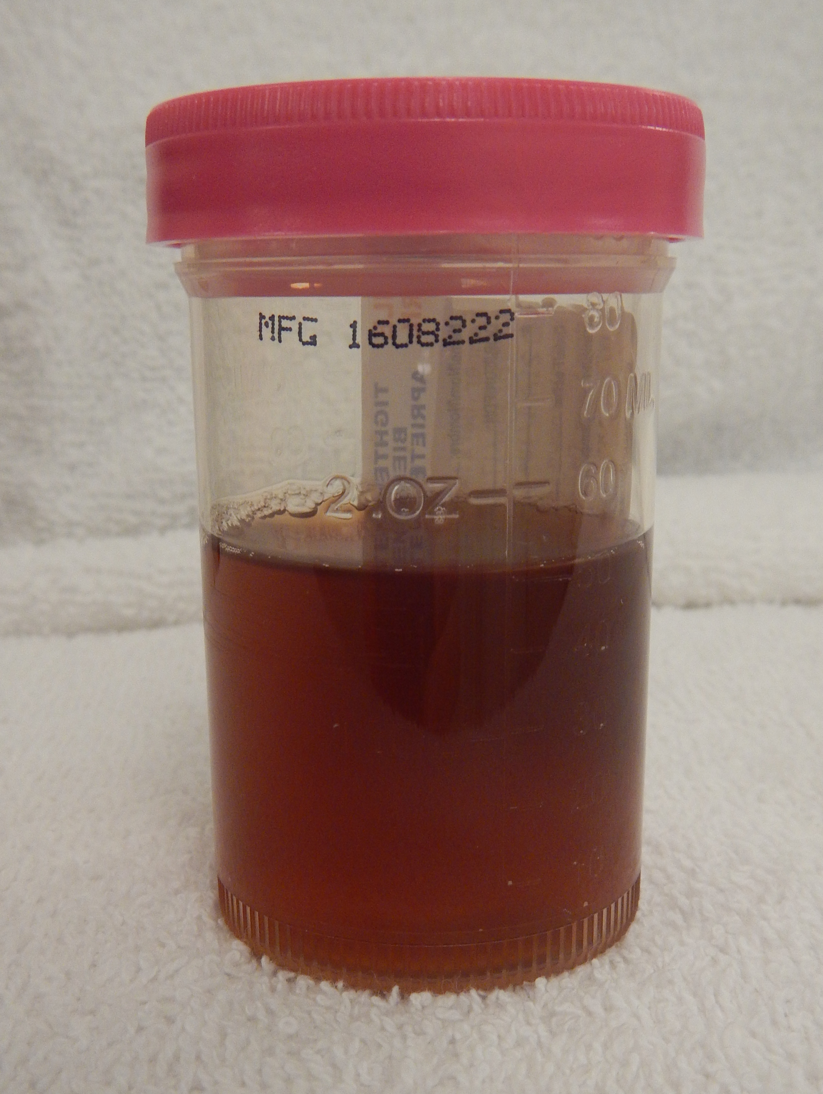
Hematuria is common — population screening studies report a prevalence of microscopic hematuria ranging from 2.4% to 31.1%, depending on the population studied and the threshold used.[2] The great majority of cases have a benign or self-limited explanation, but hematuria is a sign, not a diagnosis: it may be associated with an underlying urologic malignancy in up to 10% of cases,[2] and the entire purpose of a structured workup is to reliably separate that minority from the many transient or benign causes without over-testing everyone. The single most important framing point for pharmacists, returned to throughout this chapter: hematuria in a patient taking an anticoagulant or antiplatelet agent still needs the same evaluation as hematuria in anyone else — patients on a DOAC or warfarin very reasonably, but often incorrectly, assume their medication fully explains the bleeding.[1][2]
Recognize the Condition
Benign, transient causes — common, and worth actively ruling out
A meaningful share of positive dipsticks, and even some confirmed microscopic hematuria, resolve completely once a transient contributing factor is addressed. Guidance is explicit that these should be actively assessed for and, where present, retested after resolution — before further workup begins, not instead of it if hematuria persists.[2]
- Urinary tract infection — the most common benign explanation; confirm with urine culture, treat, and re-test the urinalysis with microscopy after treatment to document that the hematuria itself has actually resolved — it cannot simply be assumed to have resolved along with the infection.[1]
- Menstrual or vaginal contamination — repeat a midstream, clean-catch specimen once menses has ended if contamination is possible.[2]
- Vigorous exercise ("athletic" or exercise-induced hematuria) — typically resolves within 48-72 hours of rest; retest after abstaining from strenuous activity.[2]
- Recent urethral trauma, sexual activity, or urologic instrumentation/catheterization — retest once the precipitating event is well in the past.[2]
- A recent viral illness.[2]
Causes that genuinely need investigation
| Category | Examples | Clue on history/exam |
|---|---|---|
| Urothelial or renal malignancy | Bladder cancer (by far the most common finding on a positive urologic workup), upper tract urothelial carcinoma, renal cell carcinoma, prostate cancer | Painless gross hematuria (the classic, non-negotiable red flag), age over 40, a smoking history, irritative voiding symptoms without infection, occupational exposure (see Risk Factors) |
| Nephrolithiasis | Renal or ureteral calculus | Colicky flank pain radiating to the groin, often with nausea; hematuria is frequently microscopic even during an acute stone episode |
| Glomerular (medical renal) disease | IgA nephropathy, thin basement membrane nephropathy, post-infectious or other glomerulonephritis, lupus nephritis | Dysmorphic RBCs or red cell casts on microscopy, proteinuria, an abnormal or declining creatinine/eGFR, new hypertension — see Assessment & Diagnosis and Special Populations |
| Benign prostatic hyperplasia, urethral stricture, or a bladder/urethral diverticulum | — | Lower urinary tract symptoms, a known BPH diagnosis, prior urethral instrumentation or infection |
| Chronic inflammation of the lower urinary tract | A chronic indwelling catheter, ureteric stent, bladder stone, or chronically infected foreign body | Known device or chronic infection history |
Painless gross hematuria deserves special emphasis: it is the single most classic presenting sign of bladder cancer, and the absence of pain does not make it less concerning — closer to the opposite is true. Do not let a single episode that stops spontaneously be dismissed as resolved; it still requires full evaluation (see Red Flags & Refer and Choose Initial Treatment).[2]
Not all red urine is hematuria — and not all hematuria is visibly red
See Assessment & Diagnosis for the specific pitfalls of dipstick testing, including beeturia, myoglobinuria, high-dose vitamin C, and drug-induced urine discoloration.
Risk Factors
The panel that developed the current AUA/SUFU microhematuria guideline defined a specific set of factors built directly into its risk-stratification system, alongside a broader list of additional recognized risk factors for urothelial cancer.[1]
| Factors used in formal risk stratification (see Assessment & Diagnosis) | Additional urothelial cancer risk factors |
|---|---|
| Age | Irritative lower urinary tract symptoms without infection |
| Male sex | Prior pelvic radiation therapy |
| Smoking history (current or past, including secondhand exposure) | Prior cyclophosphamide or ifosfamide chemotherapy |
| Degree of microscopic hematuria (RBC/HPF) | Family history of urothelial cancer or Lynch syndrome |
| Persistence of microscopic hematuria | Occupational exposure to benzene chemicals or aromatic amines (e.g., rubber, dye/textile, petrochemical work) |
| History of gross hematuria | A chronic indwelling foreign body in the urinary tract |
Smoking is the single strongest risk factor for bladder cancer.[2] Occupations reported at higher risk include tobacco workers, dye and textile workers, chimney sweeps, nurses, rubber workers, waiters, metal workers, electricians, mechanics, military and public safety personnel, domestic and cleaning staff, hairdressers, painters, printers, seafarers, oil and petroleum workers, shoe and leather workers, plumbers, truck drivers, drill press operators, chemical workers, and roofers/coal tar workers.[2] Additional recognized risk factors include a personal history of bladder cancer, overuse of analgesic drugs (historically phenacetin-containing analgesics), and — exceedingly rare in North America but endemic to the Middle East and Africa — Schistosoma haematobium infection.[2][3]
Red Flags & Refer
The following change the plan from "arrange outpatient workup" to same-day or emergency action:
- Clot retention — inability to void, or straining against a palpable, distended bladder, from blood clots obstructing outflow. This is a true urologic emergency: ongoing bleeding and clot formation can cause worsening urinary retention, abdominal pain, anemia, infection, and deteriorating renal function if not relieved promptly.
- Hemodynamic instability or signs of significant blood loss (tachycardia, hypotension, orthostatic symptoms, a falling hemoglobin) accompanying gross hematuria — resuscitate and refer for emergency evaluation.
- Fever with flank pain in the setting of hematuria — suggests an obstructed, infected upper urinary tract (an infected, obstructing stone), which needs urgent decompression, not routine outpatient antibiotics alone.
- Any single episode of visible (gross) hematuria, at any age — this alone is an indication for urology referral and full evaluation (cystoscopy, upper-tract imaging, and urine cytology), regardless of how brief the episode was or whether it has since stopped.[1]
- Any single episode of symptomatic hematuria not associated with infection — irritative voiding symptoms, or hematuria with pain not explained by a confirmed UTI.[1]
- Findings suggesting a glomerular (medical renal) cause — dysmorphic RBCs, red cell casts, significant proteinuria, an abnormal or declining creatinine/eGFR, or new hypertension — refer to nephrology; risk-based urologic evaluation should still proceed in parallel rather than being deferred.[1]
- Isolated hemoglobinuria — a positive dipstick for blood with a negative microscopic urinalysis — may indicate a hematologic problem such as paroxysmal nocturnal hemoglobinuria, and can warrant expedited hematology referral rather than being dismissed as a false-positive dipstick.
- Pediatric hematuria with edema, hypertension, or a recent streptococcal throat or skin infection — evaluate for post-infectious glomerulonephritis (see Special Populations).
Assessment & Diagnosis
Confirming hematuria: dipstick is a screen, microscopy is the diagnosis
Urine dipstick testing detects the peroxidase-like activity of hemoglobin and is a sensitive screening test, but it does not reliably distinguish true red blood cells from free hemoglobin (hemolyzed blood) or myoglobin, and it can be falsely negative or positive for genuine, common reasons (see the table below). A positive dipstick (trace blood or greater) should prompt formal microscopic evaluation; it must never be used alone to diagnose hematuria or to trigger a full evaluation.[1]
Confirm significant microscopic hematuria as ≥3 RBC/HPF on microscopic evaluation of a properly collected specimen; 0-2 RBC/HPF is insignificant and does not warrant further investigation unless clinical suspicion remains high, in which case a repeat test showing ≥3 RBC/HPF should trigger the workup below.[1][2] A single positive microscopy is sufficient to initiate workup — hematuria is known to be highly intermittent even in the presence of significant underlying pathology, so waiting for a second confirmatory result before acting is not appropriate practice.[2]
Specimen collection matters. Use a midstream, clean-catch specimen. The sample for microscopy must be examined fresh, within 2-3 hours of collection — in practice this means sending the patient to a laboratory rather than relying on a specimen that has sat for hours beforehand.[2] If contamination by menstrual blood or vaginal discharge is possible, repeat the sample once resolved.[2]

Pitfalls: what looks like hematuria but isn't, and what hides true hematuria
| Cause | Dipstick (blood/heme) result | How to tell the difference |
|---|---|---|
| Beeturia (red-pink urine after eating beets) | Negative — the pigment betanin lacks the peroxidase activity that triggers a positive dipstick | Visibly red or pink urine with a negative dipstick for blood is the key finding; beeturia is far more common in iron deficiency (about 80% of iron-deficient subjects versus roughly 14% of the general population in the classic feeding study), so persistent beeturia can itself be a clue to check iron status.[4] |
| Myoglobinuria (rhabdomyolysis) | Positive — myoglobin's heme moiety cross-reacts on the dipstick just like hemoglobin | Microscopy shows few or no intact RBCs despite the positive dipstick; confirm with a serum creatine kinase. |
| Free hemoglobin (hemolysis) | Positive | Microscopy again shows few or no intact RBCs despite a strongly positive dipstick. |
| High-dose vitamin C (ascorbic acid), from supplements or juice | False negative — even modest supplemental doses (100 mg and up) can generate enough urinary ascorbic acid to mask a truly positive dipstick, with the effect most pronounced at lower degrees of hematuria | If clinical suspicion for hematuria is genuine but the dipstick is negative in a patient taking vitamin C, do not let a negative dipstick alone provide reassurance — proceed to microscopy.[5] |
| Menstrual or vaginal contamination | Positive on both dipstick and microscopy, but not true hematuria | Repeat a clean-catch, midstream specimen once menses has ended.[2] |
| Phenazopyridine (a urinary analgesic) or rifampin | Urine turns orange-red from the drug itself, not from blood | Colour alone is unreliable in a patient taking either drug — send a specimen for microscopy rather than relying on visual inspection, and ideally collect it before starting phenazopyridine if a hematuria workup is pending. |
| Dehydration/concentrated urine, or recent strenuous exercise | Can produce a false-positive dipstick | Repeat once well-hydrated and rested.[2] |
Differentiating a glomerular from a non-glomerular source
Once significant microscopic hematuria (or gross hematuria) is confirmed, examine the same specimen for evidence of a glomerular (medical renal) cause, and measure serum creatinine/eGFR, urine albumin-to-creatinine ratio (ACR), and blood pressure in every patient.[1][2] Dysmorphic RBCs on microscopy, red cell casts, proteinuria (urine ACR over 30 mg/mmol), or an abnormal/declining creatinine or eGFR and new hypertension all suggest a glomerular cause and should prompt nephrology referral — this does not replace risk-based urologic evaluation, which should still proceed in parallel rather than being deferred.[1]
Risk stratification for genitourinary malignancy
Once a glomerular cause has been excluded (or is being managed in parallel), categorize every patient with confirmed significant microscopic hematuria as low-, intermediate-, or high-risk for genitourinary malignancy.[1]
| Low risk (meets all criteria) | Intermediate risk (meets any one) | High risk (meets any one) |
|---|---|---|
| Women under 50 years; men under 40 years | Women 50-59 years; men 40-59 years | Women or men age 60 or older |
| Never smoker, or under 10 pack-years | 10-30 pack-years | Over 30 pack-years |
| 3-10 RBC/HPF on a single urinalysis | 11-25 RBC/HPF on a single urinalysis | Over 25 RBC/HPF on a single urinalysis |
| No additional risk factors for urothelial cancer (see Risk Factors) | A low-risk patient with no prior evaluation and 3-10 RBC/HPF on repeat urinalysis; or any additional urothelial cancer risk factor | History of gross hematuria |
Imaging and cystoscopy by risk category
| Risk category | Recommended evaluation |
|---|---|
| Low | Shared decision-making: repeat urinalysis within 6 months, or proceed directly to cystoscopy and renal ultrasound. If hematuria persists on repeat testing, reclassify as intermediate/high-risk and evaluate accordingly.[1] |
| Intermediate | Cystoscopy plus renal (kidney/bladder) ultrasound.[1][2] |
| High | Cystoscopy plus axial upper-tract imaging: multiphasic CT urography (including imaging of the urothelium) if not contraindicated; MR urography if CT is contraindicated; retrograde pyelography with non-contrast axial imaging or renal ultrasound if both are contraindicated.[1] |
Kidney/bladder ultrasound is the preferred, first-choice imaging test in most circumstances given its safety (no ionizing radiation, no contrast), availability, and lower cost — reserve CT or MR urography for high-risk patients or those with visible hematuria and/or strong risk factors, where superior upper-tract urothelial detection is needed.[3][2] A family history of renal cell carcinoma or a known genetic renal tumour syndrome warrants upper-tract imaging regardless of calculated risk category.[1]
Urine cytology and urinary markers
Do not use urine cytology or urine-based tumour markers in the initial evaluation of asymptomatic microscopic hematuria — they are poor screening tests in this setting.[1] Cytology has a legitimate, separate role: it is appropriate for gross hematuria and for symptomatic microscopic hematuria (once modifiable causes have been excluded), and it may be obtained for persistent microscopic hematuria after a negative workup in a patient with irritative voiding symptoms or risk factors for carcinoma in situ.[1][2]
The anticoagulated or antiplatelet patient
Perform the identical evaluation in a patient taking an antiplatelet agent or anticoagulant — aspirin, another antiplatelet, a DOAC, or warfarin, regardless of type or dose — as in a patient not on these agents.[1][2] Anticoagulation predisposes to hematuria only in the presence of underlying urinary tract disease; it does not cause hematuria on its own in a genitourinary tract that is otherwise normal.[2] A population-based Ontario cohort found that patients newly exposed to antithrombotic medication were significantly more likely to be diagnosed with bladder cancer within 6 months than unexposed patients — consistent with anticoagulation unmasking bleeding from a pre-existing lesion rather than causing it outright — and a 2025 emergency-department cohort confirmed that anticoagulant/antiplatelet therapy can provoke gross hematuria that goes on to reveal an underlying urologic malignancy.[1][6] Do not dismiss microscopic hematuria in an anticoagulated patient as an expected medication effect.
Treatment Targets
- Reliably separate patients who need urgent or expedited urologic/nephrology evaluation from the larger group who can be safely managed with reassurance, resolution of a transient cause, or a defined follow-up interval.
- Recognize and manage hematuria-related emergencies — clot retention, hemodynamic instability, an obstructed and infected upper tract — the same day they present.
- Confirm every episode of significant microscopic hematuria with laboratory microscopy, never dipstick alone, before committing a patient to further workup.
- Differentiate a glomerular (medical renal) source from a urologic one, and refer to the correct specialty — sometimes both in parallel.
- Apply the identical, unbiased evaluation to a patient on an anticoagulant or antiplatelet agent as to any other patient, rather than assuming the medication explains the finding.
- Avoid unnecessary radiation, contrast exposure, and invasive testing in genuinely low-risk patients, while never under-evaluating a patient with real risk factors.
- Set realistic expectations and a concrete follow-up plan for the many patients whose workup will be negative.
Choose Initial Treatment
Step 1 — screen for an emergency, every time
Before anything else, screen for clot retention (inability to void, a distended and painful bladder), hemodynamic instability, or fever with flank pain suggesting an obstructed, infected upper tract. Any of these is same-day emergency department management, not outpatient workup (see Red Flags & Refer and Non-Pharmacologic Treatment).
Step 2 — rule out a modifiable cause, and confirm true hematuria
Assess for and address a transient contributing factor (infection, menstruation, vigorous exercise, recent instrumentation) and retest once it has resolved. Every positive dipstick confirm with laboratory urine microscopy before proceeding — never diagnose hematuria, or trigger a workup, from a dipstick result alone.[1]
Step 3 — glomerular versus urologic
Findings of a glomerular cause present (dysmorphic RBCs, red cell casts, significant proteinuria, an abnormal/declining creatinine or eGFR, new hypertension) refer to nephrology and continue risk-based urologic evaluation in parallel — one referral should not be deferred while waiting on the other.[1]
Step 4 — the anticoagulant question
Do not assume hematuria is explained by a patient's anticoagulant or antiplatelet therapy, and do not stop or hold it without discussing with the prescriber or an appropriate specialist first — apply the identical risk-based evaluation used for any other patient.[1][2] The single exception is a genuine bleeding emergency (clot retention with hemodynamic compromise, major blood loss), where the anticoagulant's continuation is reassessed as part of that emergency's own management, not unilaterally by whoever is managing the hematuria workup.
Step 5 — risk-stratify and evaluate
For confirmed, non-glomerular significant microscopic hematuria, categorize the patient as low-, intermediate-, or high-risk (see Assessment & Diagnosis) and proceed with the matched evaluation: Low risk shared decision-making between repeat urinalysis at 6 months or immediate cystoscopy plus renal ultrasound; Intermediate risk cystoscopy plus renal ultrasound; High risk cystoscopy plus CT (or MR) urography.[1] Any single episode of visible (gross) hematuria, at any age full evaluation — cystoscopy, upper-tract imaging, and urine cytology — regardless of whether the episode has since stopped.[1]
Step 6 — treat an identified benign cause, and confirm resolution
If a benign or non-malignant urologic cause is identified (a UTI, BPH, a urethral stricture, a non-obstructing stone, vaginal atrophy or pelvic organ prolapse), treat it appropriately — see the Urinary Tract Infection chapter for antibiotic regimens — then re-test the urinalysis to confirm the hematuria has actually resolved rather than assuming it has. If hematuria persists, or the cause cannot be identified, proceed with risk-based urologic evaluation regardless.[1]
Non-Pharmacologic Treatment
- Clot retention — place a large-bore (generally at least 22 Fr) urethral catheter and manually irrigate the bladder with 0.9% saline via a catheter-tipped syringe to evacuate clots and restore drainage; most patients then need a continuous-irrigation ("3-way") catheter to keep the bladder clear while the source is addressed. If clots cannot be cleared by hand irrigation, cystoscopy and clot evacuation in the operative setting is required.
- Correct hemodynamic instability with standard resuscitation (IV access, fluids, transfusion as indicated) before or alongside definitive source control.
- Specimen technique — collect a midstream, clean-catch sample; have the patient go to the laboratory so the specimen for microscopy can be examined fresh, within 2-3 hours; use the same laboratory for follow-up testing to keep results comparable.[2]
- For suspected exercise-induced hematuria, advise rest from strenuous activity before retesting; for suspected menstrual contamination, repeat once menses has ended.[2]
- Advise a patient taking high-dose vitamin C or a vitamin-C-rich supplement that it can mask a truly positive dipstick — this is a reason to proceed to microscopy when suspicion remains, not a reason to stop a supplement the patient otherwise wants to take.[5]
- Reassure a patient with confirmed beeturia (red-pink urine after beets, with a negative dipstick) that this is not hematuria — but note that persistent beeturia can be a clue to check for iron deficiency.[4]
- Screening the general population for microscopic hematuria (by dipstick or microscopy) is not currently recommended, given insufficient evidence of net benefit.[2]
- Cystoscopy and imaging are themselves the definitive diagnostic procedures for urologic causes — there is no pharmacologic substitute for them once evaluation is indicated.
Special Populations
Pregnancy
Hematuria in pregnancy shares much of the same differential as in non-pregnant patients (UTI is especially common in pregnancy), but proteinuria plus hematuria in the second half of pregnancy must also prompt evaluation for preeclampsia — do not assume a urologic cause without checking blood pressure and screening for preeclampsia features (see the Preeclampsia chapter). Imaging ultrasound is preferred throughout pregnancy to avoid ionizing radiation; defer CT urography unless the clinical situation is urgent enough that the diagnostic benefit clearly outweighs fetal radiation exposure, and make any such decision together with obstetrics and urology. Treat a confirmed UTI promptly (see the Urinary Tract Infection chapter for pregnancy-specific regimens) and re-test to confirm resolution of the hematuria.
Pediatrics
The differential in children differs meaningfully from adults: malignancy is rare, and the more common significant causes are post-infectious (post-streptococcal) glomerulonephritis (typically 1-3 weeks after a streptococcal throat or skin infection, often with edema, hypertension, and cola-coloured urine), IgA nephropathy, idiopathic hypercalciuria (a common cause of isolated, otherwise-unexplained microscopic hematuria in children), and benign familial hematuria (thin basement membrane nephropathy). Gross hematuria, hypertension, edema, or any evidence of a glomerular pattern in a child warrants prompt pediatric nephrology referral rather than a routine adult-style urologic workup.
Older adults
Age is itself a major risk factor in every risk-stratification system in this chapter — malignancy risk rises with age, and any hematuria in a patient 60 or older is automatically high-risk regardless of other factors (see Assessment & Diagnosis). Maintain a low threshold for full evaluation, and do not attribute hematuria to "old age" or a chronic catheter without still investigating.
Anticoagulated patients (DOACs and warfarin) — the key counselling point for pharmacists
Patients taking a DOAC or warfarin very commonly, and often confidently, attribute any hematuria entirely to their anticoagulant. This is a genuine, high-value counselling opportunity: explain clearly that anticoagulation does not cause hematuria in an otherwise normal urinary tract — it can only worsen bleeding from a lesion that is already there, and that current guidance requires the identical risk-based evaluation regardless of anticoagulant type, dose, or level of therapy.[1][2] Do not counsel a patient to stop their anticoagulant on their own, and do not imply to a patient that stopping it will "fix" the hematuria — that decision belongs to the prescriber, weighed against the patient's stroke or VTE risk, and is separate from the hematuria workup itself. See Choose Initial Treatment and Drug Tables.
Renal impairment / chronic kidney disease
Hematuria and CKD frequently overlap — a patient with known CKD who develops new or worsening hematuria still needs the glomerular-versus-urologic differentiation above, and a new finding of proteinuria or a meaningful eGFR decline should prompt nephrology involvement per the Chronic Kidney Disease chapter, in parallel with any indicated urologic evaluation.
Monitoring & Follow-Up
When no specific cause is found after a complete initial evaluation, follow the patient annually — this is not optional reassurance-only follow-up, since a small proportion of patients with an initially negative workup are later diagnosed with a urologic malignancy.[2][7]
- Annual urine microscopy, urine albumin-to-creatinine ratio, serum creatinine/eGFR, and blood pressure.
- Annual urine cytology only in patients with risk factors for urothelial cancer — not routinely in every patient.[2]
- If, after the initial evaluation, the degree of microscopic hematuria persists unchanged on annual follow-up, consider repeat investigation within 3-5 years.[2]
- If three consecutive annual urine microscopies are negative, follow-up testing for that episode can be discontinued.[2]
- Trigger immediate re-investigation, rather than waiting for the next scheduled annual visit, if the patient develops gross hematuria, new urinary symptoms, an increasing degree of microscopic hematuria, new or worsening proteinuria, or declining renal function.[2]
Common Errors to Avoid
- Diagnosing or ruling out hematuria from a dipstick result alone, without confirmatory microscopy.
- Attributing hematuria in an anticoagulated or antiplatelet patient to the medication without performing the same risk-based evaluation used for any other patient.
- Advising or allowing a patient to stop their anticoagulant on their own initiative because of hematuria, rather than involving the prescriber.
- Ordering urine cytology or a urine-based tumour marker as part of the initial evaluation of asymptomatic microscopic hematuria.
- Missing beeturia, myoglobinuria, high-dose vitamin C, or a discoloring drug (phenazopyridine, rifampin) as an explanation for visually red urine or an unexpected dipstick result.
- Failing to repeat testing after a genuinely modifiable cause (infection, menstruation, exercise, recent instrumentation) has resolved, before either committing to or abandoning further workup.
- Treating a single, brief episode of visible gross hematuria that has since stopped as resolved and not requiring referral.
- Finding dysmorphic RBCs, red cell casts, or proteinuria and referring only to nephrology, or only to urology, instead of pursuing both in parallel where indicated.
- Assuming a young patient's hematuria cannot be malignant when meaningful risk factors (a heavy smoking history, occupational exposure, pelvic radiation, cyclophosphamide exposure) are present.
- Discontinuing annual follow-up before three consecutive negative annual tests, or discontinuing it entirely without checking whether the degree of persistent hematuria has changed.
- Missing clot retention as a same-day emergency, or attempting prolonged bedside management when clots cannot be cleared by hand irrigation.
- Prescribing phenazopyridine before a pending hematuria specimen has been collected, or continuing it beyond 2 days.
Clinical Decision Algorithm
Drug Tables
How to use this table: hematuria itself does not have a drug "treatment" — these tables cover genuine adjuncts (tranexamic acid, phenazopyridine), commonly confused drug-induced urine discoloration, and how anticoagulants and UTI antibiotics are handled during a hematuria workup. Doses are adult doses and require patient-specific confirmation, including renal/hepatic adjustment. Select any drug name to open its complete Pharmasist Drug Database page.
Antifibrinolytic for gross hematuria or clot-forming bleeding
Specialist (urology)-directed; not a first step taken independently for microscopic hematuria.
| Drug | Dosage | Adverse effects | Drug interactions | Comments |
|---|---|---|---|---|
| Tranexamic acid | Oral: 1 g PO 3-4 times daily Specialist-directed: Intravesical instillation or IV use per urology protocol | Nausea, vomiting, diarrhea; rare thromboembolic events; rare visual disturbance with prolonged use | Additive thrombotic risk with combined hormonal contraception or other prothrombotic drugs | Use cautiously if an upper urinary tract source is suspected — antifibrinolytic therapy can theoretically favour ureteric clot formation and obstruction in that setting; confirm the diagnostic and source-control plan first. |
Short-term symptomatic relief (does not treat the underlying cause)
| Drug | Dosage | Adverse effects | Drug interactions | Comments |
|---|---|---|---|---|
| Phenazopyridine | Adults: 200 mg PO three times daily with food, for a maximum of 2 days | Orange-red discoloration of urine and other body fluids (can stain contact lenses); headache, GI upset; rare methemoglobinemia, higher risk in G6PD deficiency | None significant at standard doses | An analgesic only — always pair with definitive treatment of the underlying cause (e.g., an antibiotic for a confirmed UTI). Its urine discoloration can mimic or mask true hematuria and interferes with dipstick colour reading; limit to 2 days. |
Other drugs that discolour urine without causing true hematuria
Included as a teaching reference — none of these require dose adjustment for hematuria, since they are not its cause.
| Drug | Dosage | Adverse effects | Drug interactions | Comments |
|---|---|---|---|---|
| Rifampin | Per its own indication: See the Tuberculosis chapter for TB-specific dosing | Orange-red discoloration of urine, tears, sweat, and other body fluids; hepatotoxicity, GI upset | Extensive CYP-mediated interactions — review the full medication list before starting | The discoloration is expected and harmless on its own; it does not indicate hematuria and does not require the dose to be adjusted or the drug to be stopped. |
| Nitrofurantoin | Per its own indication: See the Urinary Tract Infection chapter for dosing | Brown discoloration of urine; GI upset; rare pulmonary/hepatic toxicity with prolonged use | Contraindicated if CrCl under 30 mL/min | Urine discoloration from the drug itself is expected and does not indicate hematuria. |
Anticoagulants and antiplatelet agents found incidentally at the time of a hematuria workup
None of these are started, stopped, or dose-adjusted because of hematuria alone — that decision belongs to the prescriber, weighed against the patient's thrombotic risk, and is separate from completing the standard hematuria evaluation. Full dosing is in the Atrial Fibrillation and Venous Thromboembolism chapters.
| Drug | Dosage | Adverse effects | Drug interactions | Comments |
|---|---|---|---|---|
| Warfarin | Continuation: Individualized, INR-guided — see Atrial Fibrillation/Venous Thromboembolism chapters | Bleeding, rare skin necrosis | Extensive — see the Atrial Fibrillation and Venous Thromboembolism chapters | Continue at the established, INR-guided dose pending workup unless the prescriber directs otherwise. |
| Apixaban | Continuation: See Atrial Fibrillation/Venous Thromboembolism chapters for the patient's indicated dose | Bleeding, bruising | See the Atrial Fibrillation and Venous Thromboembolism chapters | Continue at the established dose pending workup unless the prescriber directs otherwise. |
| Rivaroxaban | Continuation: See Atrial Fibrillation/Venous Thromboembolism chapters for the patient's indicated dose | Bleeding, GI upset | See the Atrial Fibrillation and Venous Thromboembolism chapters | Continue at the established dose pending workup unless the prescriber directs otherwise. |
| Dabigatran | Continuation: See Atrial Fibrillation/Venous Thromboembolism chapters for the patient's indicated dose | Dyspepsia, bleeding | See the Atrial Fibrillation and Venous Thromboembolism chapters | Continue at the established dose pending workup unless the prescriber directs otherwise. |
| Edoxaban | Continuation: See Atrial Fibrillation/Venous Thromboembolism chapters for the patient's indicated dose | Bleeding, rash | See the Atrial Fibrillation and Venous Thromboembolism chapters | Continue at the established dose pending workup unless the prescriber directs otherwise. |
Empiric antibiotics when hematuria is attributed to a confirmed urinary tract infection
Full dosing, duration, and pregnancy-specific regimens are in the Urinary Tract Infection chapter. Whichever agent is used, re-test the urinalysis with microscopy after treatment to confirm the hematuria has actually resolved — resolution of infection does not guarantee resolution of hematuria.
| Drug | Dosage | Adverse effects | Drug interactions | Comments |
|---|---|---|---|---|
| Nitrofurantoin | See: Urinary Tract Infection chapter for full dosing | GI upset; rare pulmonary/hepatic toxicity with prolonged use | Contraindicated if CrCl under 30 mL/min | 1st-line for uncomplicated cystitis. |
| Trimethoprim | See: Urinary Tract Infection chapter for full dosing | Rash, hyperkalemia (elderly/renal impairment) | See Urinary Tract Infection chapter | Sulfa-allergy alternative. |
| Fosfomycin | See: Urinary Tract Infection chapter for full dosing | Diarrhea, headache, nausea | See Urinary Tract Infection chapter | Single-dose option. |
References
- Microhematuria: AUA/SUFU Guideline — Journal of Urology, American Urological Association/Society of Urodynamics, Female Pelvic Medicine & Urogenital Reconstruction (Barocas DA, Boorjian SA, Alvarez RD, et al.). J Urol 2020;204(4):778-786
- Workup of Microscopic Hematuria — BCGuidelines.ca, Guidelines and Protocols Advisory Committee, British Columbia Ministry of Health. BCGuidelines.ca, effective 29 July 2020
- Canadian guidelines for the management of asymptomatic microscopic hematuria in adults — Canadian Urological Association Journal (Wollin T, Laroche B, Psooy K). Can Urol Assoc J 2009;3(1):77-80
- Beeturia: its incidence and a clue to its mechanism — British Medical Journal (Watson WC, Luke RG, Inall JA). Br Med J 1963;2(5363):971-973
- The influence of vitamin C on the urine dipstick tests in the clinical specimens: a multicenter study — Journal of Clinical Laboratory Analysis (Lee W, Kim Y, Chang S, Lee AJ, Jeon CH). J Clin Lab Anal 2017;31(5):e22080
- Gross haematuria in the era of anticoagulant therapy - Implications on treatment and diagnostic approaches in a large emergency department patient population — BJUI Compass (Yang Y, Seidl J, Engelmann SU, et al.). BJUI Compass 2025;6(10):e70099
- Recommendations for the improvement of bladder cancer quality of care in Canada: A consensus document reviewed and endorsed by Bladder Cancer Canada (BCC), Canadian Urologic Oncology Group (CUOG), and Canadian Urological Association (CUA), December 2015 — Canadian Urological Association Journal (Kassouf W, Aprikian A, Black P, et al.). Can Urol Assoc J 2016;10(1-2):E46-80
Professional reference only. Apply clinical judgment and verify patient-specific decisions against current authoritative guidance and product information.