Keratitis
Clinical Snapshot
Keratitis is inflammation of the cornea — most often due to infection, but also from non-infectious causes such as trauma, exposure, or autoimmune disease. This chapter focuses on microbial (infectious) keratitis: bacterial, viral (chiefly herpes simplex virus, HSV), fungal, and Acanthamoeba (protozoal) keratitis. Correct etiologic recognition governs everything about management, because the antimicrobial classes involved — antibacterials, antivirals, antifungals, and anti-amoebic biguanides — are not interchangeable, and treating one etiology as if it were another, particularly starting a topical corticosteroid before the organism is identified, can actively worsen the outcome.[1]
The cornea sits directly in the visual axis, and this is what makes keratitis genuinely vision-threatening rather than merely uncomfortable. Bacterial keratitis can progress to corneal perforation within 24 hours when caused by a virulent organism, and untreated or severe disease can progress to endophthalmitis and loss of the eye.[1] Even when the infection itself is eradicated, scarring that involves the central or paracentral cornea — the part of the cornea the visual axis actually passes through — can cause substantial, permanent visual loss regardless of how effectively the causative organism was treated.[1] This scarring risk, more than any single symptom, should frame the urgency of this entire chapter: keratitis is not a condition to observe and reassess in a few days.
Four etiologic categories account for the great majority of microbial keratitis, each with genuinely distinct management implications:
- Bacterial keratitis is the most common infectious keratitis in high-income countries and is strongly associated with contact lens wear — the number-one risk factor for microbial keratitis in the United States — and, in many other parts of the world, with ocular trauma.[1] Pseudomonas aeruginosa is a particularly aggressive contact-lens-associated pathogen capable of rapid corneal destruction.
- Herpes simplex virus (HSV) keratitis is a recurrent condition and the leading infectious cause of corneal blindness in developed nations, with a classic dendritic (branching) epithelial ulcer.[2] It requires antiviral, not antibacterial, treatment, and topical corticosteroids are genuinely dangerous when used without antiviral cover in active epithelial disease — see Pharmacologic Treatment Options.
- Fungal keratitis is less common than bacterial keratitis but important because it is more indolent in onset, harder to treat, and often follows trauma involving vegetable matter (a branch, a plant leaf, agricultural work) or occurs in contact lens wearers.[3]
- Acanthamoeba keratitis is rare — an estimated 1-3 cases per million population per year, rising to 20-50 cases per million per year among contact lens wearers specifically[4] — but is a particularly severe, contact-lens- and water-exposure-associated protozoal infection with a poor prognosis when diagnosis is delayed.

An estimated 71,000 cases of microbial keratitis (bacterial, fungal, and Acanthamoeba combined) occur annually in the United States, with an increasing incidence.[1] Ocular HSV disease adds a further 5-15 new cases per 100,000 population per year internationally, with both incidence and prevalence trending upward.[2] Contact lens wear runs through nearly every one of these numbers — see Risk Factors and Non-Pharmacologic Management — which is what makes contact-lens-hygiene counseling a genuinely high-value pharmacist intervention point rather than routine advice.
Recognize the Condition
Symptoms
Keratitis typically presents with eye pain, photophobia, redness, decreased or blurred vision, foreign-body sensation, and often a visible corneal opacity or infiltrate on examination.[1] Discharge is also common. A key differential point: pain and photophobia are typically far more prominent in keratitis than in simple conjunctivitis. A patient describing genuine eye pain — not just irritation or itch — particularly with photophobia, should be assessed as a possible keratitis until proven otherwise, especially if they wear contact lenses.
Presentation differs meaningfully by etiology
While pain, photophobia, and redness are common to all forms, the pattern of the corneal findings is often the first clue to etiology, before any culture result is available, and should shape how urgently the patient is triaged.
Distinguishing features by etiology
Bacterial
Suppurative (pus-forming) stromal infiltrate with indistinct, often necrotic or suppurative margins, surrounding stromal edema and white cell infiltration, usually with an overlying epithelial defect and anterior chamber reaction. Onset is typically rapid, especially with virulent organisms such as Pseudomonas.[1]
Herpes simplex (viral)
The pathognomonic finding is a dendritic (branching) epithelial ulcer with terminal bulbs, best seen with fluorescein staining under a slit lamp — a corneal pattern genuinely distinct from the round or irregular defects of bacterial or fungal disease.[2] Recurrent disease, often with reduced corneal sensation.
Fungal
Ulcers often have a dry rather than suppurative appearance, with a feathery or serrated margin, satellite lesions, and sometimes a posterior plaque — features that genuinely help distinguish fungal from bacterial keratitis, though a ring infiltrate can occur in either.[1][3] Onset is typically indolent (days, not hours), and a history of vegetable-matter trauma or contact lens wear is common.
Acanthamoeba
Severe pain out of proportion to the clinical findings is the classic clue, along with radial keratoneuritis (inflammation tracking along the corneal nerves in a radiating pattern) and, later, a ring infiltrate.[1][4] Early Acanthamoeba keratitis can produce pseudodendrites frequently mistaken for HSV epithelial keratitis — a well-recognized and clinically important pitfall, particularly in a contact lens wearer whose pain seems disproportionate to what is visible on the ocular surface.[4]
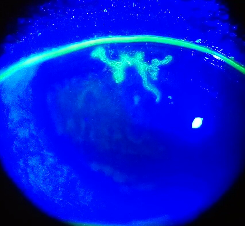
Because bacterial and fungal keratitis in particular can look genuinely similar on clinical appearance alone, a study of experienced cornea specialists reviewing photographs of culture-proven bacterial and smear-proven fungal keratitis found they correctly differentiated the two in fewer than 70% of cases[1] — a strong argument for culture-based diagnosis over visual pattern recognition alone (see Assessment and Diagnosis), and a reminder for a pharmacist not to reassure a patient based on how "typical" a presentation looks.
Risk Factors
Contact lens wear runs through the risk-factor profile of bacterial, fungal, and Acanthamoeba keratitis alike, and is the single most important — and most preventable — risk factor a pharmacist can meaningfully address. See Non-Pharmacologic Management for specific counseling points.
Contact-lens-related risk factors
- Overnight or extended wear, including overnight orthokeratology — the single most consistently identified risk factor for contact-lens-associated microbial keratitis.[1]
- Wearing lenses beyond the manufacturer- or practitioner-recommended replacement schedule.[1]
- Inadequate lens disinfection, including "topping off" old solution rather than replacing it, and contamination of the lens case, including rinsing the case with tap water.[1][5]
- Storing or rinsing lenses in tap water, or using homemade or ineffective/contaminated saline rather than proper contact lens disinfecting solution.[1][5]
- Poor contact lens hygiene generally — inadequate hand washing before lens handling, poor lens case hygiene, and infrequent case replacement.[1][5]
- Swimming, using a hot tub, or showering while wearing contact lenses — water exposure while lenses are worn is specifically associated with Acanthamoeba keratitis.[1][4]
- Using a damaged or poor-fitting lens, sharing lenses, or using unregulated lenses (decorative, internet-purchased, or otherwise obtained without a prescription).[1]
- Lack of routine follow-up: as many as 50% of asymptomatic contact lens wearers presenting for a routine visit have been found to have signs of a lens-wear complication.[1]
Other local and systemic risk factors
- Ocular trauma, including chemical, thermal, and foreign-body injury — particularly vegetable matter trauma (a branch, thorn, or plant material), a specific risk factor for fungal keratitis, along with agricultural work and warm climates more broadly.[3]
- Previous ocular or eyelid surgery, including refractive surgery, cataract surgery, and keratoplasty.[1]
- Ocular surface disease — tear-film deficiency (dry eye), eyelid malposition (entropion, ectropion, lagophthalmos), misdirected lashes (trichiasis), and neurotrophic keratopathy (reduced corneal sensation, including after previous HSV or zoster disease).[1]
- Prior herpes simplex or varicella zoster keratitis — a risk factor for bacterial superinfection, and, for HSV specifically, a strongly recurrence-prone condition in its own right (see Special Populations).[1]
- Topical medication factors — contaminated ocular medications, and chronic use of topical anesthetics, corticosteroids, or preserved products, which can compromise the ocular surface.[1]
- Systemic immunosuppression (medication-induced or from disease), diabetes mellitus, vitamin A deficiency, and severe malnourishment or critical illness.[1]
Red Flags & Refer
Any suspected microbial keratitis needs prompt ophthalmology evaluation — this is not a self-treatable or primary-care-only condition. Unlike many of the eye conditions covered elsewhere on this platform, keratitis is never appropriate for OTC recommendation with reassessment in a few days. The findings below make the situation more urgent still:
- Any red, painful eye in a contact lens wearer — have the patient stop lens wear immediately and switch to glasses, and arrange same-day-to-next-day ophthalmology (or emergency) assessment. An apparently mild-looking presentation can still be early keratitis, and contact lens wear is the leading risk factor for sight-threatening corneal infection.[1]
- Severe pain out of proportion to the visible findings, especially with radial keratoneuritis or in a contact lens wearer — raises specific suspicion for Acanthamoeba keratitis, which carries a genuinely poor prognosis when diagnosis and treatment are delayed.[4]
- A visible corneal opacity, infiltrate, or ulcer of any kind, or significant fluorescein staining.
- Hypopyon — a visible layer of white cells (pus) layering in the anterior chamber, seen as a blunting of the inferior angle or presenting inferiorly if the patient has recently been lying down — a sign of severe, sight-threatening infection.[1]
- Any sign of corneal thinning, melting, or impending or frank perforation.
- Worsening symptoms, or no improvement, despite treatment — including apparent worsening in the first 24-48 hours of appropriate antibiotic therapy, which can genuinely occur with Pseudomonas and other gram-negative organisms and does not necessarily mean the regimen has failed[1] (see Monitoring and Follow-Up), but which still needs specialist reassessment rather than pharmacist reassurance.
- Sudden or significant vision change.
- A patient already using a topical corticosteroid who develops new eye pain, redness, or photophobia — corticosteroids can mask progression of an unrecognized infection and must not be continued or self-adjusted without specialist input.
Do not recommend an OTC lubricant, antihistamine/decongestant eye drop, or "redness reliever," and do not advise watchful waiting, for a patient with signs or symptoms suggestive of keratitis. The appropriate pharmacist action is referral, not symptomatic self-care.
Assessment & Diagnosis
The central diagnostic principle: identify the organism before, or very early alongside, starting therapy
Because bacterial, fungal, and Acanthamoeba keratitis can be genuinely difficult to distinguish by appearance alone (see Recognize the Condition), and because the treatments for each are not interchangeable and starting the wrong one can worsen the outcome, corneal scraping for smear and culture before starting antimicrobial therapy is recommended whenever feasible in specific higher-risk presentations.[1] That said, the majority of small, non-central, community-acquired bacterial ulcers do resolve with topical empiric therapy alone and are managed without smears or cultures[1] — culture is targeted at presentations where the diagnosis or causative organism genuinely changes management.
When smears and cultures are specifically indicated
- A central, large corneal infiltrate (within 3 mm of the corneal center and at least 2 mm in size), and/or two or more adjacent lesions, and/or significant stromal involvement or melting.[1]
- One or more cells in the anterior chamber.[1]
- A history of corneal surgery.[1]
- Atypical clinical features suggestive of fungal, amoebic, or mycobacterial keratitis.[1]
- Multiple corneal infiltrates.[1]
- Poor response to broad-spectrum antibiotic therapy already started.[1]
Corneal material is obtained after a topical anesthetic — preservative-free proparacaine is preferred; tetracaine is avoided for its own antimicrobial effect — using a sterile blade or spatula to scrape the base and periphery of the lesion. If Rose Bengal dye has been used on the eye, note that it is virucidal under light exposure and can produce falsely negative viral cultures.[1][2] Polymerase chain reaction (PCR) and other immunodiagnostic techniques are increasingly available and useful, particularly for HSV, though PCR cannot always distinguish pathogenic viral shedding from normal viral shedding in the tear film.[1][2]
Etiology-specific diagnostic tools
- HSV epithelial (dendritic) keratitis is usually diagnosed clinically by slit-lamp appearance alone, and laboratory testing is not typically needed for a classic presentation. Where testing is needed, PCR of a corneal or tear-film sample is more sensitive than viral culture (which can take up to 10 days) and is considered comparably specific for confirming HSV epithelial keratitis; direct fluorescent antibody testing offers a faster (minutes) but somewhat less sensitive alternative.[2]
- Fungal keratitis is diagnosed by microscopy (potassium hydroxide or Gram stain of corneal scrapings) and culture, classically on Sabouraud agar; confocal microscopy can also help visualize fungal elements directly.[3][1]
- Acanthamoeba keratitis is diagnosed by confocal microscopy, which can directly visualize trophozoites or cysts, culture on non-nutrient agar seeded with E. coli, and increasingly by PCR.[4]
Differential diagnosis
The differential includes both infectious mimics — viral (HSV, VZV, Epstein-Barr) keratitis producing immunologically mediated infiltrates that can resemble bacterial, fungal, or Acanthamoeba disease, and viral epithelial defects that are themselves prone to secondary bacterial superinfection, particularly in older, larger, or immunosuppressed patients — and non-infectious causes: contact-lens-associated sterile infiltrates, staphylococcal marginal keratitis, peripheral ulcerative keratitis from autoimmune disease, and infiltrative keratitis from systemic connective tissue or vasculitic disease.[1] When there is genuine clinical uncertainty about etiology, initial management of a presumed infectious ulcer should still include empiric antibacterial coverage while the workup proceeds.[1]
Treatment Targets
- Eradicate the causative organism from the cornea.
- Minimize corneal scarring, thinning, and neovascularization — particularly in the central or paracentral cornea, where scarring most directly threatens vision.
- Relieve pain and photophobia.
- Resolve associated discharge and anterior chamber inflammation.
- Prevent complications: corneal perforation, endophthalmitis, secondary cataract, glaucoma from prolonged corticosteroid use, and permanent vision loss.
- Restore corneal integrity and optimize long-term visual function, including timely referral for visual rehabilitation where permanent scarring has occurred.
- Identify and correct modifiable risk factors — above all, unsafe contact lens practices — to prevent recurrence.
- Recognize that etiology-specific antimicrobial selection is an ophthalmology-specialist decision; the pharmacist's role is recognition, urgent referral, patient education, and supporting adherence to a specialist-directed regimen, not initiating or adjusting antimicrobial therapy for suspected keratitis.
Choose Initial Treatment
Antimicrobial selection for keratitis is an ophthalmology-specialist decision — but pharmacists should understand the classes involved
Because etiology genuinely changes the correct treatment class, and because misdirected therapy — most importantly, a corticosteroid started before the organism is identified — can worsen outcomes, specific antimicrobial selection, dosing, and any corticosteroid decision for keratitis should be made by the treating ophthalmologist, not initiated or substituted by a pharmacist. What follows is a pharmacist-level overview of treatment principles by etiology, intended to support counseling, dispensing accuracy, and adherence, not independent prescribing.
Bacterial keratitis
Topical antibiotic eye drops, which achieve high corneal tissue levels, are the preferred treatment for the great majority of bacterial keratitis.[1] Two general strategies are used:
- Single-drug fluoroquinolone monotherapy — for example moxifloxacin, ofloxacin, gatifloxacin, besifloxacin, ciprofloxacin, or levofloxacin — has been shown to be as effective as combination fortified-antibiotic therapy for most cases, and is the usual choice for small, non-central, community-acquired ulcers.[1]
- Fortified (compounded) antibiotics — most often fortified cefazolin combined with fortified tobramycin, or a fluoroquinolone — should be considered for large and/or visually significant infiltrates, especially when a hypopyon is present, and are prepared by a specialized compounding pharmacy.[1] For central or severe keratitis, an initial loading dose (for example every 5-15 minutes) followed by frequent maintenance dosing (for example hourly) is typical.[1]
Topical corticosteroids may have a role in some cases of bacterial keratitis, but only once the organism has been identified and/or the infection has shown a clinical response to antibiotic therapy — generally not before 48 hours of treatment, and never in cases with suspected Acanthamoeba, Nocardia, or fungal infection, where corticosteroids should be avoided.[1] This timing, and the decision itself, is made by the treating ophthalmologist with close intraocular pressure follow-up, not initiated empirically.
Herpes simplex virus (HSV) keratitis
Treatment differs by which corneal layer is involved, and this distinction matters enormously for corticosteroid safety:
Epithelial vs. stromal/endothelial HSV keratitis
Epithelial (dendritic) keratitis
Antiviral alone is the treatment of choice. Topical corticosteroids should be avoided in the initial management of active HSV epithelial keratitis — a strong recommendation based on good-quality evidence, because corticosteroid use during active epithelial viral replication can worsen and prolong the disease.[2]
Stromal or endothelial keratitis
A topical corticosteroid combined with an oral antiviral is the preferred treatment — the corticosteroid treats the immune-mediated stromal inflammation, while the antiviral provides essential cover against viral reactivation during corticosteroid use.[2] A typical stromal-keratitis course is at least 10 weeks of topical corticosteroid, tapered under specialist supervision, with a prophylactic oral antiviral continued throughout.[2]
Both topical antiviral agents (trifluridine solution and ganciclovir ophthalmic gel) and oral antiviral agents (acyclovir, valacyclovir, famciclovir) are effective for HSV epithelial keratitis; oral agents are generally preferred for stromal or endothelial disease given their superior corneal penetration and more favourable long-term safety profile — topical trifluridine, in particular, causes toxic keratoconjunctivitis with prolonged use.[2] Long-term, low-dose oral antiviral prophylaxis (at least one year) is recommended for patients with a history of recurrent HSV stromal keratitis, given the disproportionate share of ocular HSV morbidity that stromal disease causes — see Special Populations and Monitoring and Follow-Up.[2]
Fungal keratitis
Topical natamycin 5% is the usual first-line antifungal, historically shown to outperform other agents for filamentous (mould) fungal keratitis in large clinical trials, applied intensively (for example hourly) initially with a gradual taper over what is typically a considerably longer treatment course than bacterial keratitis — often several weeks.[3] Topical or oral voriconazole is used as an alternative or adjunct, particularly for Fusarium species or when the response to natamycin is inadequate; oral antifungal therapy is added for deep or severe infections.[3] Fungal keratitis has a generally worse prognosis than bacterial keratitis — larger infiltrates/scars, slower re-epithelialization, and a higher perforation rate at comparable time points in comparative trials.[1]
Acanthamoeba keratitis
First-line therapy is a topical biguanide — polyhexamethylene biguanide (PHMB) and/or chlorhexidine — often combined with a diamidine (propamidine or hexamidine), applied intensively and continued for a prolonged course that typically extends over months rather than weeks, reflecting the organism's resistant cyst form and the disease's characteristically slow response to treatment.[4] A single-agent topical PHMB 0.08% product has become available as a licensed treatment option in some jurisdictions. Delayed diagnosis is strongly associated with a worse visual outcome, which is precisely why the pain-out-of-proportion and radial keratoneuritis clues in Recognize the Condition matter so much — a missed or delayed Acanthamoeba diagnosis, often because the presentation was initially treated only as HSV or bacterial keratitis, is one of the most important preventable contributors to permanent vision loss from keratitis.[4]
Non-Pharmacologic Treatment
Contact lens hygiene — the single highest-value pharmacist counseling point in this chapter
Because contact lens wear is the leading modifiable risk factor across bacterial, fungal, and Acanthamoeba keratitis, and because most of the individual practices below are genuinely within a pharmacist's counseling scope, this is the section with the most direct preventive impact.
- Never sleep in contact lenses unless a specific extended-wear lens has been prescribed for that purpose by an eye care provider, and even then, understand that overnight wear substantially raises infection risk. The incidence of microbial keratitis is as high as 20 per 10,000 wearers per year with extended wear, compared with 1-2 per 10,000 wearers per year with daily wear.[1]
- Avoid all water contact while wearing lenses — remove lenses before swimming, using a hot tub, showering, bathing, or washing the hair, and never rinse lenses or the lens case in tap water or store lenses in homemade saline.[1][5] Water exposure while wearing lenses is specifically associated with Acanthamoeba keratitis.[1][4]
- Use fresh disinfecting solution every time — never "top off" old solution in the case, and never reuse solution.[5]
- Wash and dry hands thoroughly before handling lenses, replace the lens case regularly, and never share lenses with anyone else.[5][1]
- Follow the prescribed replacement schedule — do not wear lenses beyond their intended disposal interval, and do not use a damaged or poorly fitting lens.[1]
- Avoid decorative, novelty, or internet-purchased lenses obtained without a valid prescription and fitting — these carry a disproportionate infection risk.[1]
- Stop lens wear immediately at the first sign of any eye symptom — redness, pain, photophobia, discharge, or blurred vision — switch to glasses, and seek prompt assessment; do not simply "wait and see" while continuing to wear lenses.
- Keep scheduled contact lens follow-up visits even when asymptomatic — as many as half of asymptomatic wearers presenting for a routine visit have signs of a lens-related complication on examination.[1]
- Encourage patients and prescribers to report contact-lens-related corneal infections through the relevant medical-device adverse-event reporting program, since extended-wear contact lenses are a higher-risk medical device category and adverse-event reporting helps inform future labelling and safety communication.[1]
Other non-pharmacologic measures
- Do not patch a contact-lens-associated or traumatic corneal abrasion. Patching, or using a therapeutic bandage contact lens, in these settings is specifically advised against, because it increases the risk of secondary bacterial keratitis rather than protecting the eye.[1]
- Prompt treatment of any corneal abrasion — a broad-spectrum topical antibiotic started within 24 hours of a traumatic corneal abrasion has been shown to reduce the risk of subsequent corneal ulceration.[1]
- Address correctable ocular surface risk factors — eyelid malposition, severe tear-film deficiency, exposure — as part of preventing both first episodes and recurrence.[1]
- For patients with recurrent HSV keratitis, education about the nature of the condition, its recurrence risk, and the early warning signs of a flare supports both adherence to long-term prophylaxis and prompt self-recognition of a recurrence.[2]
Special Populations
Contact lens wearers
This population deserves dedicated emphasis given how central contact lens wear is to this chapter (see Risk Factors and Non-Pharmacologic Management). Any contact lens wearer with a red, painful eye should stop lens wear immediately and be assessed urgently. After an episode of bacterial keratitis, the risks and timing of resuming contact lens wear should be discussed with the treating ophthalmologist, and switching to daily disposable lenses — which have a lower complication rate than reusable lenses — is often recommended.[1]
Pediatrics
Keratitis in children requires the same urgency as in adults, though presentation and history-taking can be harder — a young child may not reliably localize pain or report photophobia. Teenage contact lens wearers are a relevant at-risk group for the same reasons as adults. Children and atopic individuals with HSV keratitis may require relatively higher doses of oral antivirals and can be more difficult to manage.[2] Doxycycline is avoided under 8 years (permanent tooth staining) if an oral tetracycline adjunct for corneal thinning is being considered.
Pregnancy and breastfeeding
Keratitis is not a condition to manage more conservatively because a patient is pregnant or breastfeeding — untreated sight-threatening infection is the greater risk, and the treating ophthalmologist selects therapy accordingly. Oral acyclovir and valacyclovir have extensive safety data in pregnancy from their use in other HSV indications and are generally considered the antivirals of choice when systemic therapy is needed. Oral tetracyclines (doxycycline) are avoided in pregnancy given the risk of fetal tooth and bone effects; this is relevant only to the adjunctive oral-tetracycline role for corneal melt, not to primary antimicrobial therapy for the keratitis itself.
Immunocompromised patients
Immunosuppression — from systemic disease, medication (including topical or systemic corticosteroids), diabetes, or critical illness — is both a risk factor for keratitis and a reason to expect a more severe course, an atypical organism, or a poorer response to standard therapy.[1] HSV keratitis in immunocompromised patients may be more severe and require higher oral antiviral doses.[2] A lower threshold for culture and specialist involvement is appropriate throughout this population.
Older adults
Age itself is a component of several of the systemic risk factors in this chapter — diabetes, polypharmacy, immunosenescence — and older patients may have more difficulty instilling frequent eye drops accurately, a genuine barrier to the intensive dosing schedules (sometimes hourly) that severe keratitis requires. Confirm a patient's or caregiver's ability to administer the prescribed regimen, and flag any difficulty to the prescriber; hospitalization is sometimes needed specifically to ensure adequate dosing frequency and adherence.[1]
Renal impairment
Topical ocular antimicrobials (antibiotics, antivirals, antifungals, biguanides) have minimal systemic absorption and generally require no renal dose adjustment. Oral antivirals used for HSV keratitis (acyclovir, valacyclovir, famciclovir) do require renal dose adjustment, and dosing in significant renal insufficiency should be coordinated with the prescriber or a nephrologist.[2]
Monitoring & Follow-Up
Follow-up frequency for keratitis is set by disease severity rather than being routine. Severe cases — deep stromal involvement, infiltrates larger than 2 mm with extensive suppuration, or any hypopyon — are typically followed daily, at least until stable or clear clinical improvement is confirmed; a mild, small, non-central bacterial ulcer may be reassessed less frequently, but always by the treating ophthalmology service, not by community pharmacy alone.[1]
Signs of a favourable response
- Reduced pain and reduced amount of discharge.
- Reduced eyelid edema or conjunctival injection.
- Consolidation and sharper demarcation of the perimeter of the stromal infiltrate, with decreasing density in the absence of progressive stromal loss.
- Reduced stromal edema and endothelial inflammatory plaque.
- Reduced anterior chamber cells, fibrin, or hypopyon.
- Initial re-epithelialization and cessation of progressive corneal thinning.[1]
Important caveat: in Pseudomonas and other gram-negative bacterial keratitis, increased inflammatory signs during the first 24-48 hours despite genuinely appropriate therapy can occur and does not automatically indicate treatment failure[1] — a genuine clinical nuance worth knowing so a pharmacist does not inadvertently alarm a patient, or contribute to premature regimen-switching, based on apparent early worsening alone. The treating ophthalmologist is best placed to distinguish expected early inflammation from true non-response.
Corticosteroid-specific monitoring
Any patient started on a topical corticosteroid for keratitis needs a defined follow-up plan for intraocular pressure and lens clarity; the patient is typically examined within 1-2 days of starting a corticosteroid.[1] Warn patients that inflammation and symptoms can temporarily increase as a corticosteroid is tapered or withdrawn — this reflects the loss of local immune suppression and is not necessarily worsening infection, though any new or increasing symptoms still warrant reassessment.[1]
HSV-specific follow-up
Patients on long-term oral antiviral prophylaxis after HSV stromal keratitis should have their ongoing need for prophylaxis, adherence, and any breakthrough recurrence reviewed periodically; the protective effect of prophylaxis does not appear to persist once the medication is stopped, so the decision to eventually discontinue is a specialist one made in the context of ongoing recurrence risk.[2]
Common Errors to Avoid
- Treating a red, painful eye in a contact lens wearer as ordinary conjunctivitis or dry eye, recommending an OTC product, and reassessing in a few days, rather than referring urgently.
- Patching a contact-lens-associated or traumatic corneal abrasion — this increases, rather than reduces, the risk of secondary bacterial keratitis.
- Assuming a "typical-looking" corneal ulcer confirms the etiology by appearance alone — even experienced cornea specialists correctly distinguish bacterial from fungal keratitis by photograph alone in fewer than 70% of cases.
- Missing severe pain out of proportion to findings, or radial keratoneuritis, as clues to Acanthamoeba keratitis, and treating a contact-lens-associated pseudodendrite as HSV without considering Acanthamoeba in the differential.
- Recommending or continuing a topical corticosteroid for a red eye before the causative organism has been identified, or in a case with suspected fungal, Acanthamoeba, or Nocardia infection.
- Starting a topical corticosteroid in active HSV epithelial (dendritic) keratitis without antiviral cover — a genuinely dangerous error that can worsen and prolong the disease.
- Stopping the antiviral when a corticosteroid is added for HSV stromal or endothelial keratitis, rather than continuing both together.
- Interpreting increased inflammation in the first 24-48 hours of antibiotic therapy for Pseudomonas or other gram-negative keratitis as treatment failure and unnecessarily switching or escalating therapy.
- Recommending water contact (swimming, hot tub, showering) while wearing contact lenses, or the use of tap water or homemade saline for lens storage or rinsing.
- Not asking about contact lens wear, hygiene practices, and water exposure as a routine part of assessing any red or painful eye.
- Treating fungal keratitis with a treatment course of the same duration used for bacterial keratitis and concluding treatment has failed prematurely.
- Recommending oral doxycycline in a pregnant patient or a child under 8 years without considering the timing and necessity of that adjunct.
- Failing to set a follow-up plan for intraocular pressure and lens clarity when a topical corticosteroid is part of the regimen.
Clinical Decision Algorithm
Drug Tables
How to use this table: Doses are adult doses and require patient-specific confirmation (renal/hepatic function, weight, allergy history). Pediatric dosing is weight-based per the current product monograph unless stated. Drug pricing is intentionally omitted. Select any drug name to open its complete Pharmasist Drug Database page.
Topical Antibacterial Therapy — Empiric and Fortified
Fluoroquinolone monotherapy is appropriate for most small, non-central, community-acquired ulcers. Fortified (compounded) antibiotics are reserved for large, central, or severe infiltrates, especially with hypopyon, and are prepared by a specialized compounding pharmacy — not commercially available at these concentrations. All dosing below is specialist-directed and individualized to severity.
| Drug | Dosage | Adverse effects | Drug interactions | Comments |
|---|---|---|---|---|
| Moxifloxacin (ophthalmic) 0.5% | Adults: 1 drop hourly while awake initially, tapered per clinical response | Local irritation, punctate keratopathy with prolonged use | None significant at standard ophthalmic doses | Fourth-generation fluoroquinolone; broad gram-positive and gram-negative coverage. |
| Ofloxacin 0.3% | Adults: 1 drop hourly while awake initially, tapered per clinical response | Local irritation, punctate keratopathy with prolonged use | None significant at standard ophthalmic doses | FDA-approved for bacterial keratitis; comparable efficacy to fortified therapy in trials. |
| Gatifloxacin 0.3% | Adults: 1 drop hourly while awake initially, tapered per clinical response | Local irritation, punctate keratopathy with prolonged use | None significant at standard ophthalmic doses | Better gram-positive coverage than earlier-generation fluoroquinolones in vitro. |
| Besifloxacin 0.6% | Adults: 1 drop hourly while awake initially, tapered per clinical response | Local irritation, punctate keratopathy with prolonged use | None significant at standard ophthalmic doses | Not available as an oral agent, which may reduce selection pressure for systemic fluoroquinolone resistance. |
| Ciprofloxacin 0.3% (ophthalmic) | Adults: 1 drop hourly while awake initially, tapered per clinical response | Local irritation, corneal precipitate deposition reported with frequent use | None significant at standard ophthalmic doses | FDA-approved for bacterial keratitis; an earlier-generation fluoroquinolone with somewhat weaker gram-positive coverage than newer agents. |
| Fortified cefazolin 50 mg/mL | Adults: Loading dose every 5-15 minutes, then hourly, alternating with the paired agent | Local toxicity with prolonged use | None significant at standard ophthalmic doses | Compounded from an injectable vial; combined with fortified tobramycin for broad-spectrum coverage of severe ulcers. |
| Fortified tobramycin 9-14 mg/mL | Adults: Loading dose every 5-15 minutes, then hourly, alternating with the paired agent | Local toxicity with prolonged use | None significant at standard ophthalmic doses | Compounded from an injectable vial; paired with fortified cefazolin for gram-negative coverage, including Pseudomonas. |
| Fortified vancomycin 25-50 mg/mL | Adults: Loading dose every 5-15 minutes, then hourly, per culture/sensitivity results | Local toxicity with prolonged use | None significant at standard ophthalmic doses | Reserved for confirmed or strongly suspected methicillin-resistant Staphylococcus aureus or other resistant gram-positive organisms. |
Topical Corticosteroids — Adjunctive, Specialist-Directed Only
Never started empirically before the organism is identified and clinical response confirmed (generally not before 48 hours for bacterial keratitis). Contraindicated in suspected Acanthamoeba, Nocardia, or fungal infection, and in active HSV epithelial (dendritic) keratitis without antiviral cover. Every course needs a defined follow-up plan for intraocular pressure and lens clarity.
| Drug | Dosage | Adverse effects | Drug interactions | Comments |
|---|---|---|---|---|
| Prednisolone acetate 1% | Adults: Specialist-directed dose and taper | Elevated intraocular pressure, cataract with prolonged use, delayed corneal healing, worsened infection if used prematurely | None significant at standard ophthalmic doses | Used as an adjunct in selected bacterial keratitis once the organism is identified, and as part of the HSV stromal/endothelial regimen with concurrent oral antiviral cover. |
| Loteprednol etabonate 0.5% | Adults: Specialist-directed dose and taper | Elevated intraocular pressure (less likely than with ketone-based steroids), cataract with prolonged use, delayed corneal healing | None significant at standard ophthalmic doses | A site-specific corticosteroid with a more favourable ocular safety profile; a reasonable specialist-directed alternative to prednisolone acetate. |
Antiviral Therapy for HSV Keratitis
Antiviral therapy alone is the treatment of choice for HSV epithelial (dendritic) keratitis; corticosteroids are avoided in this setting. For stromal or endothelial keratitis, an oral antiviral is combined with a topical corticosteroid — oral agents are generally preferred over topical antivirals for stromal/endothelial disease given superior corneal penetration and a more favourable long-term safety profile.
| Drug | Dosage | Adverse effects | Drug interactions | Comments |
|---|---|---|---|---|
| Trifluridine 1% ophthalmic solution | Adults: 1 drop up to 9 times daily initially, tapered as the epithelium heals | Toxic keratoconjunctivitis, allergic conjunctivitis, and punctal stenosis with long-term use | None significant at standard ophthalmic doses | FDA-approved topical antiviral for HSV epithelial keratitis; not recommended for stromal/endothelial disease given inadequate corneal stromal penetration. |
| Ganciclovir 0.15% ophthalmic gel | Adults: 1 drop 5 times daily until the ulcer heals, then 3 times daily for 7 days | Local irritation, blurred vision immediately after instillation | None significant at standard ophthalmic doses | FDA-approved topical antiviral for HSV epithelial keratitis; better tolerated than trifluridine with long-term use, though long-term safety data are more limited. |
| Acyclovir | Treatment: 400 mg 5 times daily Prophylaxis: 400 mg twice daily for at least 1 year | Generally well tolerated; renal dose adjustment needed | None significant at standard oral doses | Treatment of HSV epithelial, stromal, and endothelial keratitis (with topical corticosteroid for stromal/endothelial disease); long-term prophylaxis after recurrent stromal keratitis. |
| Valacyclovir | Treatment: 500 mg 3 times daily Prophylaxis: 500 mg once daily for at least 1 year | Generally well tolerated; renal dose adjustment needed | None significant at standard oral doses | Acyclovir prodrug with improved oral bioavailability; as effective as acyclovir for prophylaxis in comparative trials. |
| Famciclovir | Prophylaxis: 250 mg twice daily | Generally well tolerated; renal dose adjustment needed | None significant at standard oral doses | No dedicated HSV keratitis trial data; a reasonable extrapolated alternative to acyclovir/valacyclovir based on genital herpes and ocular penetration data. |
Antifungal Therapy for Fungal Keratitis
Treatment courses are substantially longer than for bacterial keratitis, often extending over several weeks given a slower fungal response to therapy.
| Drug | Dosage | Adverse effects | Drug interactions | Comments |
|---|---|---|---|---|
| Natamycin 5% ophthalmic suspension | Adults: 1 drop hourly initially, tapered over a prolonged course of several weeks | Local irritation | None significant at standard ophthalmic doses | First-line antifungal, particularly for filamentous (mould) organisms; the only topical antifungal specifically FDA-approved for fungal keratitis. |
| Voriconazole | Adults: Specialist-directed dose and route | GI upset; hepatic enzyme elevation, visual disturbances (oral) | Numerous CYP-mediated interactions with oral use — review the full drug interaction profile before oral dosing | Alternative or adjunct, particularly for Fusarium species or an inadequate natamycin response; used topically, intrastromally, or orally for deep/severe infection. |
| Amphotericin B | Adults: Specialist-directed dose and route | Local irritation (topical); nephrotoxicity, infusion reactions (systemic) | Additive nephrotoxicity with other nephrotoxic drugs (systemic use) | Used for yeast (e.g., Candida) keratitis or refractory filamentous disease; systemic use reserved for severe or deep infection. |
Anti-Amoebic Therapy for Acanthamoeba Keratitis
Treatment courses typically extend over months rather than weeks. Delayed diagnosis is strongly associated with a worse visual outcome, so any suspicion (severe pain out of proportion to findings, radial keratoneuritis, a pseudodendrite in a contact lens wearer) warrants urgent specialist referral rather than empiric treatment as HSV or bacterial keratitis.
| Drug | Dosage | Adverse effects | Drug interactions | Comments |
|---|---|---|---|---|
| Polyhexamethylene biguanide (PHMB) 0.02-0.08% | Adults: Intensive initial dosing (e.g., hourly), tapered over a prolonged course of months | Local toxicity with prolonged intensive use | None significant at standard ophthalmic doses | First-line biguanide; a single-agent 0.08% product has become available as a licensed treatment option in some jurisdictions. |
| Chlorhexidine 0.02% (ophthalmic) | Adults: Intensive initial dosing (e.g., hourly), tapered over a prolonged course of months | Local toxicity with prolonged intensive use | None significant at standard ophthalmic doses | Used alone or in combination with PHMB and/or a diamidine. |
| Propamidine isethionate 0.1% | Adults: Intensive initial dosing, tapered over a prolonged course of months | Local irritation | None significant at standard ophthalmic doses | A diamidine typically combined with a biguanide rather than used alone. |
Adjunctive Oral Therapy for Corneal Thinning/Melt
An adjunct to, never a substitute for, etiology-specific antimicrobial therapy. Considered when progressive stromal thinning or melting occurs despite appropriate antimicrobial treatment.
| Drug | Dosage | Adverse effects | Drug interactions | Comments |
|---|---|---|---|---|
| Doxycycline | Adults: 100 mg twice daily, specialist-directed duration | Photosensitivity, GI upset | Reduced absorption with calcium/iron/antacids | Inhibits matrix metalloproteinases and bacterial collagenases; avoid in pregnancy and in children under 8 years (tooth staining). |
References
- Bacterial Keratitis Preferred Practice Pattern — American Academy of Ophthalmology Preferred Practice Pattern Cornea/External Disease Panel (Rhee MK, Ahmad S, Amescua G, Cheung AY, Choi DS, Jhanji V, Lin A, Mian SI, Viriya ET, Mah FS, Varu DM). Ophthalmology 2024;131(4):P87-P133
- Herpes Simplex Virus Keratitis: A Treatment Guideline — Massachusetts Eye and Ear Infirmary, Department of Ophthalmology, Harvard Medical School (White ML, Chodosh J); reviewed and accepted by the American Academy of Ophthalmology Hoskins Center for Quality Eye Care. Approved by the Ocular Microbiology and Immunology Group, May 2014
- Management of Filamentous Fungal Keratitis: A Pragmatic Approach — Journal of Fungi (Hoffman JJ, Arunga S, Mohamed Ahmed AHA, Hu VH, Burton MJ). J Fungi (Basel) 2022;8(10):1067
- New Frontiers in Acanthamoeba Keratitis Diagnosis and Management — Biology (Shareef O, Shareef S, Saeed HN). Biology (Basel) 2023;12(12):1489
- Preventing Eye Infections When Wearing Contacts — Centers for Disease Control and Prevention, Healthy Contact Lens Wear and Care. Accessed 2026
Professional reference only. Apply clinical judgment and verify patient-specific decisions against current authoritative guidance and product information.