Lichen Planus
Clinical Snapshot
Lichen planus (LP) is a chronic, T-cell-mediated inflammatory disease that can involve the skin, oral and genital mucosa, nails, and hair-bearing scalp, alone or in combination.[1] Cytotoxic CD8+ T cells, activated against an unidentified antigenic change on basal keratinocytes, mount a band-like attack along the dermo-epidermal junction that destroys the basal cell layer and produces the disease's signature "saw-tooth" histology.[1] Cutaneous LP affects roughly 0.2-1% of adults worldwide, while oral LP is more common still, affecting an estimated 1-4% of the population - mucosal disease frequently outlasts skin disease by years.[1] A modest female predominance (about 1.5:1) and a peak incidence between 30 and 60 years are typical; up to 10% of patients have an affected first-degree relative, and incidence is reported higher in patients of African American, Indian, and Arab descent.[1]
The classic cutaneous lesion is captured by the mnemonic the "6 P's" - purple, polygonal, planar (flat-topped), pruritic papules and plaques - but LP's actual clinical range is far broader, spanning hypertrophic, atrophic, bullous, actinic, inverse, and pigmented cutaneous variants; reticular, erosive, atrophic, papular, plaque-like, and bullous oral disease; vulvar, vaginal, and penile involvement; a scarring, permanently hair-destroying scalp variant (lichen planopilaris); and a nail dystrophy that can end in irreversible pterygium formation.[1] Three threads run through this entire chapter. First, LP has a genuine, well-quantified association with hepatitis C infection, with a pooled odds ratio around 4.5 for HCV seropositivity in LP patients.[2] Second, a recognized list of medications produces a near-identical "lichenoid" eruption that must be actively distinguished from idiopathic LP, since the fix is simply identifying and stopping the drug.[1] Third, erosive oral (and, less often, genital) LP carries a small but real malignant transformation risk - on the order of 1% in the best pooled estimates - that turns this disease from a purely symptomatic problem into one needing lifelong surveillance.[3]
Recognize the Condition

Classic cutaneous lichen planus - the "6 P's"
The hallmark lesion is purple (violaceous; yellow-brown on palms and soles), polygonal, planar (flat-topped rather than dome-shaped), pruritic papules and plaques, classically a few millimetres wide and favouring the flexor wrists, dorsal hands, lower back, ankles, and shins.[1] Close inspection or dermoscopy of an individual papule often reveals Wickham striae - a fine, lace-like network of white lines on the surface, sometimes bordered by red globules on dermoscopy - one of the most useful bedside clues to LP.[1] New lesions frequently arise in sites of skin trauma or scratching, the Koebner (isomorphic) phenomenon, and established plaques often resolve into grey-brown post-inflammatory hyperpigmentation that can outlast the active disease by months to years.[1]
Cutaneous variants
| Variant | Key features |
|---|---|
| Hypertrophic | Thick, verrucous, intensely pruritic red-brown-to-grey plaques, classically on the shins and ankles; slowest to resolve and the cutaneous variant most associated with malignant transformation, so a persistent, thickening plaque deserves a low threshold for biopsy.[1] |
| Atrophic | Uncommon; annular plaques with an atrophic, thinned centre.[1] |
| Bullous / lichen planus pemphigoides | Tense blisters arising on established LP plaques (bullous LP) or on both LP lesions and normal-appearing skin with true anti-BP180 autoantibodies (lichen planus pemphigoides, a genuine overlap with bullous pemphigoid); biopsy with direct immunofluorescence clarifies which is present.[1] |
| Actinic (LP actinicus) | Red-brown annular plaques or hyperpigmented patches confined to sun-exposed skin (face, dorsal hands, forearms); flares in summer and improves in winter; more common in younger patients with darker skin tones and in Middle Eastern/North African populations. Because sun exposure is the trigger, phototherapy is specifically the wrong treatment for this variant (see Common Errors to Avoid).[1] |
| Pigmentosus | Ill-defined grey-brown macules without a preceding inflammatory papule/plaque phase, in linear, follicular, or Blaschkoid patterns; provoked or worsened by sun exposure.[1] |
| Inverse | Erythematous, lichenified plaques with indistinct borders confined to skin folds - axillae, groin, inframammary skin.[1] |
| Annular / linear / ulcerative | Annular plaques with central clearing (often genital/intertriginous); linear lesions following a line of trauma (a Koebner variant); painful erosive/ulcerative lesions on the soles and interdigital spaces that can impair walking.[1] |
Oral lichen planus
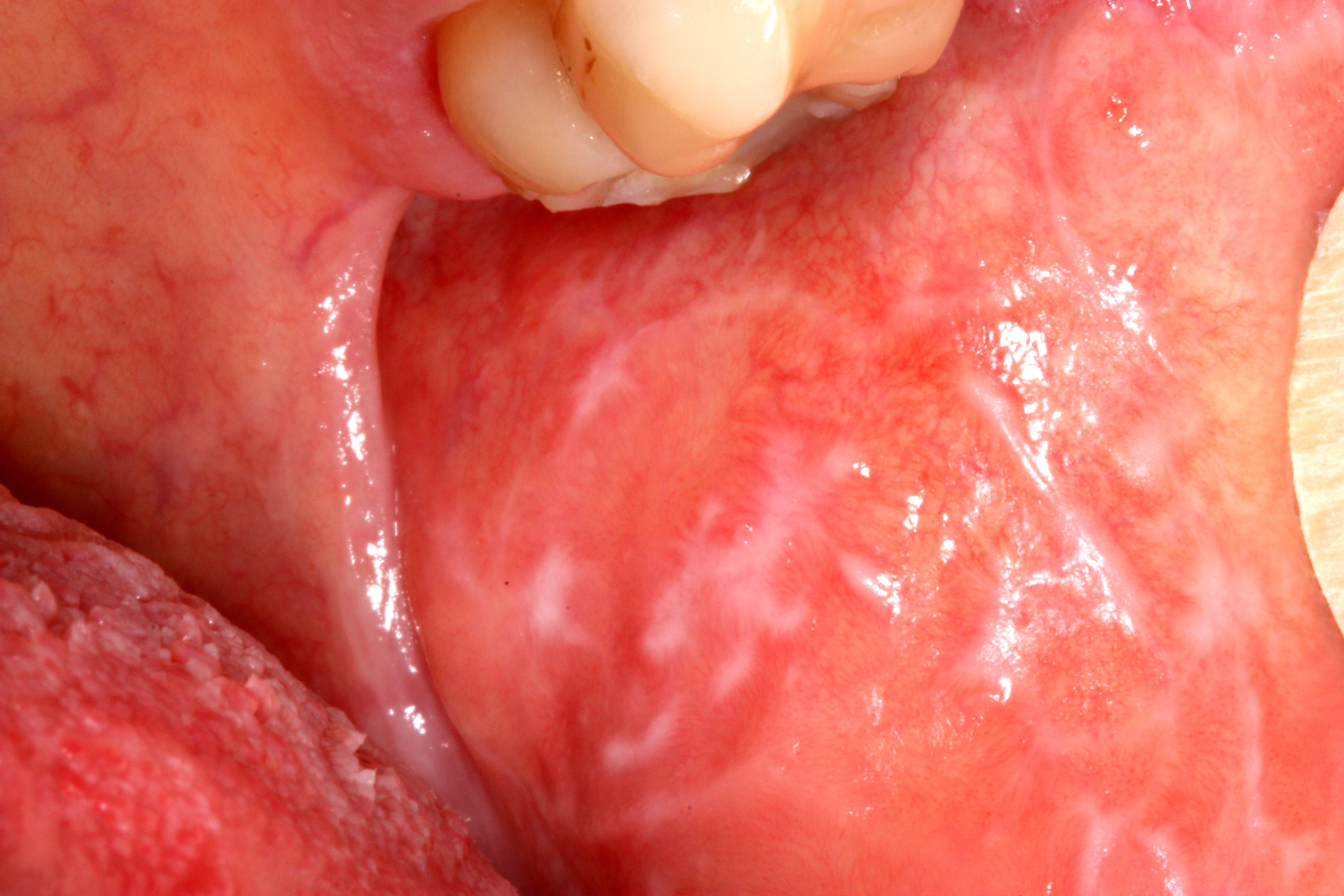
Mucosal involvement affects over half of all LP patients and is frequently the sole presenting sign, with no skin disease at all.[1] 6 recognized oral patterns exist, and more than 1 often coexists in the same patient:[1]
- Reticular - the most common pattern: asymptomatic, lacy white Wickham striae, typically bilateral and symmetric on the buccal mucosa, sometimes extending to the tongue, gingiva, or lips. Easily mistaken for leukoplakia or chronic candidiasis if the classic lacy pattern is not specifically recognized.[1]
- Erosive - painful, irregular erosions and ulcers, often ringed by radiating white striae, worsened by hot, spicy, or acidic food; the pattern most often responsible for symptoms driving patients to seek care, and the pattern carrying the highest malignant transformation risk.[1][3]
- Atrophic - diffuse redness and thinning of the mucosa, often painful; when confined to the gingiva it produces desquamative gingivitis - diffuse red, peeling gingiva that is also a shared endpoint of pemphigus vulgaris and mucous membrane pemphigoid, so biopsy with direct immunofluorescence is often needed to tell these apart.[1]
- Papular, plaque-like, and bullous - less common patterns; plaque-like lesions in particular can resemble leukoplakia clinically.[1]
A localized oral lichenoid lesion in direct topographic contact with a dental amalgam restoration suggests a contact reaction to mercury or another dental metal rather than idiopathic OLP - patch testing and, if positive, amalgam replacement can be diagnostic and curative for this specific presentation (see Assessment and Diagnosis).[1]
Genital lichen planus
On the glans penis, LP classically forms an annular ring of papules around the glans; white reticular streaks and, less often, erosive disease also occur.[1] Vulvar disease ranges from asymptomatic lacy white streaks to painful, persistent erosions and ulcers, and - particularly with the erosive subtype - can progress to genuinely disabling scarring: labial fusion, resorption of the labia minora, clitoral hood adhesions, and introital stenosis narrowing the vaginal opening. Concurrent vaginal involvement produces a painful desquamative vaginitis that can itself prevent intercourse.[1] The combination of erosive vulvovaginal LP, erosive oral LP, and (often) gingival involvement defines the vulvovaginal-gingival syndrome, a severe, scarring, difficult-to-treat presentation with a reported association to the HLA-DQB1*02:01 allele.[1] Because scarring is progressive and only partially reversible once established, genital LP - especially the erosive pattern - deserves early, adequately potent treatment rather than a "wait and see" approach (see Red Flags and Choose Initial Treatment).
Nail lichen planus
Affects roughly 10% of LP patients, usually several nails at once, and can occur with or without skin/mucosal disease.[1] The typical sequence is nail-plate thinning, longitudinal ridging and fissuring, then scarring of the nail matrix producing a pterygium - permanent adhesion of the proximal nail fold to the nail bed - progressing in severe, untreated cases to a rough, sandpaper-like texture (trachonychia) or complete, irreversible nail loss (anonychia).[1] When all 20 nails are affected with no skin or mucosal disease elsewhere, this is termed 20-nail dystrophy, a pattern seen more often in children.[1] Because pterygium formation is not reversible once it has occurred, nail LP - like lichen planopilaris below - is a genuine "treat early" situation rather than one where a watch-and-wait approach is low-risk.
Lichen planopilaris and other hair-bearing scalp involvement
Lichen planopilaris (LPP) presents as tiny, red, spiny follicular papules with perifollicular erythema and scale, extending into smooth, shiny, permanently bald patches punctuated by isolated surviving hairs ("lonely hairs") - a scarring alopecia in which lost hair follicles do not regrow.[1] Frontal fibrosing alopecia is a distinct clinical pattern of LPP - a band of scarring hair loss along the frontal and temporal hairline, often with eyebrow loss, seen predominantly in postmenopausal women.[1] Graham-Little-Piccardi-Lasseur syndrome is a rare familial variant combining scalp LPP, typical cutaneous/mucosal LP elsewhere, and non-scarring loss of axillary and pubic hair with follicular papules.[1] Pseudopelade of Brocq describes late-stage, "burnt-out" scarring alopecia patches - sometimes an LPP end-stage, sometimes idiopathic - classically likened to "footprints in the snow."
Esophageal lichen planus
An underrecognized site, predominantly in women, that can cause dysphagia, odynophagia, and esophageal strictures, and - like erosive oral and genital disease - carries a risk of malignant transformation to squamous cell carcinoma.[1] Consider it specifically in an LP patient (especially one with erosive oral disease) reporting new or worsening swallowing difficulty (see Red Flags).
Risk Factors
- Age 30-60 years is the typical window at onset; LP is uncommon in children (under 5% of all cases), though 20-nail dystrophy and actinic LP are both disproportionately pediatric presentations.[1]
- Female sex - a modest overall predominance (about 1.5:1); frontal fibrosing alopecia in particular is overwhelmingly a disease of postmenopausal women.[1]
- Family history - up to 10% of patients have an affected first-degree relative, suggesting a genetic contribution.[1]
- Hepatitis C infection - a genuine, well-quantified bidirectional association. A 2023 systematic review/meta-analysis found LP patients have significantly higher odds of being HCV-seropositive than controls (pooled odds ratio 4.48, 95% CI 3.48-5.77), with global HCV prevalence of 9.42% among LP patients overall and LP prevalence of 7.05% among HCV-positive patients; the association was stronger for cutaneous LP (odds ratio 5.95) than for oral LP (odds ratio 3.49), and stronger still in Mediterranean-basin countries.[2] HCV-positive status independently raises the odds of malignant transformation in oral lichenoid disease as well (see Assessment and Diagnosis).[3]
- Dental metal contact allergy (mercury from amalgam restorations, less often other metals) - relevant specifically to an oral lichenoid lesion in direct contact with a restoration, and confirmed by patch testing (see Assessment and Diagnosis).[1]
- Graft-versus-host disease - chronic cutaneous and oral GVHD can produce lichenoid lesions that are histologically and clinically similar to idiopathic LP; always consider this in a patient with a history of allogeneic hematopoietic stem cell transplant.[1]
- An emerging, still-developing literature has reported LP flares temporally associated with COVID-19 infection and, less often, COVID-19 vaccination; causality is not established, and this should not change routine vaccination counselling.[1]
Drug-induced lichenoid reactions - a critical mimic
A recognized list of medications can trigger an eruption that looks and, on biopsy, largely behaves like idiopathic LP - a lichenoid drug eruption. Distribution is often more photodistributed and symmetric/generalized than idiopathic LP, Wickham striae are typically absent, and the latency from starting the drug to eruption can run from several months to over a year, which is exactly why the connection is so often missed.[1] A thorough medication review is a genuinely essential part of assessing any new LP presentation.
| Drug class | Examples |
|---|---|
| Antihypertensives | ACE inhibitors, beta-blockers, methyldopa, thiazide diuretics (classically photodistributed) |
| Antimalarials | Hydroxychloroquine, quinine, quinidine (quinine/quinidine classically photodistributed) - notable because antimalarials are also used to treat LP itself (see Choose Initial Treatment), so a worsening eruption on hydroxychloroquine needs re-examination rather than an automatic dose increase |
| NSAIDs | Most agents in the class have been implicated |
| Gold and penicillamine | Historic but well-documented causes, particularly relevant in patients with longstanding rheumatoid arthritis on older disease-modifying therapy |
| TNF-alpha inhibitors and other biologics | Etanercept, adalimumab, infliximab and others |
| Other reported culprits | Sulfonylureas, proton pump inhibitors, statins, imatinib and other tyrosine kinase inhibitors, interferon |
Red Flags & Refer
Any of the following changes the urgency of assessment and, often, the treatment plan itself:
- An enlarging nodule, an ulcer with rolled or indurated edges, or a persistently non-healing erosion within longstanding erosive oral, genital, or hypertrophic cutaneous LP - biopsy urgently to exclude malignant transformation rather than assuming it is simply an unresponsive LP lesion; the tongue margin and floor of mouth are particularly high-risk oral sites.[3][4]
- Active, spreading lichen planopilaris (perifollicular erythema and scale at the advancing edge of a bald patch) - refer promptly for aggressive treatment, since hair loss in LPP is permanent once scarring is established; this is not a "try a mild topical and see" situation.[1]
- Progressive nail LP with early pterygium formation - similarly time-sensitive, since matrix scarring is irreversible.[1]
- Progressive genital scarring - labial fusion, introital stenosis, clitoral hood adhesions, or painful desquamative vaginitis preventing intercourse - needs coordinated dermatology/gynecology (or urology, for penile disease) management; surgical intervention may eventually be needed for established adhesions.[1]
- New or worsening dysphagia or odynophagia in any LP patient, especially with erosive oral disease - evaluate for esophageal LP, which can stricture and, rarely, transform malignantly.[1]
- Tense blisters arising on LP plaques or on both lesional and normal skin - biopsy with direct immunofluorescence to distinguish bullous LP from lichen planus pemphigoides (a true bullous pemphigoid overlap), since management differs.[1]
- A newly started, temporally plausible culprit medication (see Risk Factors) - pursue a structured medication review and, where a reasonable alternative exists, discontinuation, rather than treating the eruption as idiopathic LP indefinitely.[1]
- Erosive oral or genital disease severe enough to limit oral intake, hydration, or basic hygiene - needs prompt, adequately potent treatment, and nutritional/hydration support where needed, rather than a slow step-up.
- Unknown hepatitis C status in a patient with a new LP diagnosis, particularly erosive or widespread disease - arrange HCV screening given the well-documented bidirectional association.[2]
Assessment & Diagnosis
Diagnostic approach
A classic cutaneous presentation - violaceous, flat-topped, polygonal, pruritic papules with Wickham striae in a typical distribution - can often be diagnosed clinically.[1] Biopsy is genuinely useful, and often necessary, whenever the presentation is atypical, mucosal-only or scalp-only (where LP and lupus erythematosus can look very similar), before starting a systemic immunosuppressant, or whenever malignant transformation is a concern (see Red Flags).[1]
Histopathology
The classic findings are hyperkeratosis without parakeratosis, wedge-shaped hypergranulosis, irregular "saw-tooth" rete ridges, liquefactive degeneration of the basal cell layer, a dense, band-like lymphocytic infiltrate hugging the dermo-epidermal junction, and colloid (Civatte) bodies - apoptotic keratinocytes - in the lower epidermis and upper dermis.[1] Eosinophils are typically absent in idiopathic LP; their presence should raise suspicion for a drug-induced lichenoid reaction instead.[1]
Direct immunofluorescence and dermoscopy
Direct immunofluorescence (DIF) is not needed routinely but is genuinely useful when the differential includes lupus erythematosus (particularly scalp-only or mucosal-only disease) or an immunobullous disease (bullous LP vs. lichen planus pemphigoides, or desquamative gingivitis vs. pemphigus/mucous membrane pemphigoid) - DIF in LP characteristically shows colloid bodies staining for IgM (and, less consistently, IgG, IgA, and C3) and "shaggy" fibrinogen deposition along the basement membrane zone.[1] Dermoscopy of a cutaneous or mucosal lesion shows Wickham striae as a network of white lines, sometimes with red globules or dots at the periphery reflecting the underlying vascular pattern - a fast, non-invasive way to support the diagnosis at the bedside.[1]
Patch testing for suspected dental-metal contact reaction
For an oral lichenoid lesion in direct topographic contact with a dental amalgam restoration, patch testing for mercury and other dental metals helps distinguish a genuine contact reaction from idiopathic OLP; if positive and the lesion has not responded to standard therapy, replacing the amalgam restoration with a composite material (coordinated with dentistry) can be diagnostic and, in confirmed cases, curative.[1]
Distinguishing idiopathic LP from a drug-induced lichenoid reaction
Favour a drug cause when the eruption is more photodistributed or symmetric/generalized than typical idiopathic LP, Wickham striae are absent, onset followed a new medication by several months to over a year, and/or eosinophils are seen on biopsy.[1] The diagnosis is ultimately confirmed by response to withdrawal - lesions gradually resolve once the culprit drug is stopped, though this can take weeks to several months, so counsel patients accordingly rather than expecting rapid clearing.[1]
Hepatitis C screening
Given the well-documented bidirectional association, arrange HCV screening for newly diagnosed LP patients, particularly with erosive or widespread disease or in a higher HCV-prevalence population/region - screening both identifies a treatable infection and flags a patient at somewhat higher malignant-transformation risk if oral disease is present.[2][3]
Differential diagnosis
| Condition | Features that point away from lichen planus |
|---|---|
| Lupus erythematosus (cutaneous/discoid) | Follicular plugging, atrophy, and scale more prominent than the flat-topped LP papule; scalp-only or oral-only cases can closely mimic LP and often need DIF to distinguish. |
| Lichen simplex chronicus | Thickened, lichenified plaque from chronic scratching without the violaceous colour or Wickham striae of hypertrophic LP. |
| Lichen sclerosus | For vulvar/genital disease specifically: porcelain-white, atrophic (rather than violaceous, flat-topped) plaques, often figure-of-eight around the vulva/perianal skin; a genuinely different, primarily anogenital chronic sclerosing condition covered in its own separate chapter, not this one. |
| Psoriasis | Thicker plaques with a silvery, easily detached scale (vs. LP's thin, fine Wickham striae); nail pitting rather than LP's ridging/pterygium pattern. |
| Secondary syphilis | Papulosquamous eruption often involving the palms/soles with systemic symptoms and a positive serology; consider and test for in an atypical or sexually active patient. |
| Pityriasis rosea | An initial herald patch, a "Christmas-tree" truncal distribution, and a self-limited several-week course without the chronicity or Wickham striae of LP. |
| Lichen nitidus | Numerous tiny, skin-coloured, pinpoint papules without the violaceous colour or Wickham striae of LP. |
| Graft-versus-host disease | History of allogeneic stem cell transplant; otherwise can be clinically and histologically near-identical to LP. |
| Leukoplakia / chronic candidiasis (oral) | Leukoplakia is a solitary, non-lacy white patch that does not wipe off; candidiasis classically wipes off, leaving raw mucosa, and improves with antifungal therapy - neither shows the bilateral, symmetric lacy pattern of reticular OLP. |
| Pemphigus vulgaris / mucous membrane pemphigoid | Both can produce desquamative gingivitis indistinguishable from atrophic OLP on inspection alone; DIF is often needed to separate them. |
| Drug-induced lichenoid reaction | See above - photodistribution, absent Wickham striae, longer latency, eosinophils on biopsy, and resolution after stopping the culprit drug. |
Treatment Targets
- Control pruritus (cutaneous LP) or pain (erosive oral, genital, and ulcerative disease), which are frequently the dominant drivers of quality-of-life impact and treatment-seeking.
- Induce remission of active lesions and reduce time to resolution, while accepting that post-inflammatory hyperpigmentation can persist long after active disease has cleared.
- Prevent irreversible structural complications specifically where they are time-sensitive: permanent scarring alopecia in lichen planopilaris, nail matrix scarring/pterygium formation, and genital scarring/adhesions - all reasons to treat promptly rather than deferring in these particular presentations.
- Identify and, where feasible, discontinue a causative medication in suspected drug-induced lichenoid disease, rather than treating it as idiopathic LP indefinitely.
- Screen for and, where positive, coordinate management of hepatitis C infection, given the well-documented bidirectional association.
- Maintain lifelong malignant-transformation surveillance for erosive oral (and genital) disease - a genuine, ongoing treatment goal in its own right, not a one-time box to check at diagnosis.
- Minimize treatment-related morbidity, particularly from prolonged high-potency topical or systemic corticosteroid use, which carries real risks - including adrenal suppression from "just a topical steroid" applied in the mouth - that are easy to underestimate.
- Support nutrition, oral hygiene, and psychosocial wellbeing through what is frequently a chronic, relapsing-remitting, and visibly disfiguring or painful disease course.
Choose Initial Treatment
Cutaneous lichen planus - localized disease
1st line a superpotent topical corticosteroid, clobetasol propionate 0.05% ointment, applied twice daily for 2-4 weeks to affected areas.[1] Inadequate response intralesional triamcinolone acetonide 5-10 mg/mL injected directly into resistant or thickened lesions, particularly useful for the slower-to-resolve hypertrophic variant.[1]
Cutaneous lichen planus - widespread/diffuse disease
1st line prednisone 30-60 mg/day PO (roughly 0.5-1 mg/kg/day), tapered over 2-6 weeks.[1] Steroid-sparing alternative phototherapy - narrowband UVB (NB-UVB) and psoralen plus UVA (PUVA) show broadly comparable effectiveness (about 86% vs. 90% achieving a complete or good response in a large retrospective series), so the choice can be guided by access and patient preference rather than one being clearly superior.[5] NB-UVB is typically given 2-3 times weekly, starting at 0.3-0.6 J/cm² depending on skin phototype and increased by 10-20% increments in the absence of treatment-induced erythema, to a maximum of about 3.0 J/cm², with a mean of roughly 29 exposures to reach response. Oral PUVA uses 8-methoxypsoralen 0.6 mg/kg approximately 1 hour before UVA exposure, also 2-3 times weekly, starting at about 70% of the minimal phototoxic dose, with a mean of roughly 25 exposures to response; bath PUVA (8-MOP 5 mg/L, 15-minute immersion before UVA) is a further option where available. Oral PUVA carries a meaningfully higher rate of gastrointestinal adverse effects than NB-UVB.[5]
2nd line acitretin 30 mg/day PO for 8 weeks has among the best trial-level evidence of any systemic option for cutaneous LP, along with sulfasalazine and griseofulvin, all associated with an increased overall response rate versus placebo in meta-analysis - though overall evidence quality across cutaneous LP treatments ranges from very low to only moderate, and trials are generally small.[6] Methotrexate 15 mg PO or SC once weekly in adults (0.25 mg/kg/week in children), with folic acid per usual practice, showed high efficacy with a good safety profile in extensive disease and no significant difference in complete response compared with oral betamethasone in trial data.[6][1] Sulfasalazine, started at 500 mg PO twice daily and titrated by 500 mg every 3 days to a target of 2.5 g/day over 3-6 weeks, and isotretinoin 10 mg PO twice daily for about 2 months are further 2nd-line options.[1]
3rd line / refractory disease cyclosporine, mycophenolate mofetil, azathioprine, hydroxychloroquine, tetracyclines, TNF-alpha inhibitors (etanercept, adalimumab), trimethoprim-sulfamethoxazole, griseofulvin, terbinafine, and low-molecular-weight heparin have all been reported effective, generally on weaker (case-series-level) evidence; reserve for disease that has genuinely failed the options above.[1]
Oral lichen planus
Do not treat asymptomatic reticular OLP - the burden of adverse effects from topical or systemic treatment is not justified when the patient has no symptoms; monitor instead (see Monitoring and Follow-Up).[1]
1st line, symptomatic (erosive/atrophic) disease a very-high-potency topical corticosteroid applied directly to lesions 3 times daily until remission - clobetasol propionate, typically as a 0.025% oral gel formulation (a lower concentration than the 0.05% used on skin, reflecting the mouth's more absorptive mucosa) or an ointment used under an occlusive tray - alongside dietary counselling to avoid spicy, acidic, or very hot foods, alcohol, and tobacco, all of which aggravate erosive lesions.[1][7] Equally reasonable 1st-line alternative topical tacrolimus 0.1% ointment applied to lesions - a systematic review/meta-analysis found a markedly higher odds of clinical improvement with tacrolimus (odds ratio approximately 8.0) than with clobetasol (odds ratio approximately 1.2) versus placebo/comparator, supporting tacrolimus as a genuine first-line option rather than only a steroid-refractory fallback, particularly useful where prolonged topical steroid exposure is a concern.[8]
A real, easily missed risk prolonged or frequent topical corticosteroid use in the mouth is not risk-free: a cohort study of patients on long-term topical clobetasol propionate 0.025% gel for OLP found roughly 1 in 5 had biochemical evidence of glucocorticoid-induced adrenal insufficiency, some severe enough to need ongoing hydrocortisone replacement. Counsel patients on long-term topical mouth steroids about this risk and the need for stress-dose coverage during intercurrent illness, and do not treat this route as inherently safer than it is (see Monitoring and Follow-Up).[7]
2nd line, no improvement after 6 weeks a course of oral corticosteroid, or switching to/adding a topical calcineurin inhibitor if not already used.[1] 3rd line, refractory disease cyclosporine (topical swish-and-spit or systemic), azathioprine, mycophenolate mofetil, or methotrexate.[1]
Genital lichen planus
1st line a potent-to-superpotent topical corticosteroid ointment (an ointment base is generally better tolerated on genital mucosa than a cream, which can sting from its preservatives/vehicle) applied to affected vulvar or penile skin.[1] Alternative or steroid-sparing option topical tacrolimus, following the same rationale as oral disease. Treat erosive vulvovaginal or vulvovaginal-gingival syndrome disease promptly and adequately rather than conservatively, given the real risk of progressive, only partially reversible scarring; established adhesions or introital stenosis may eventually need surgical management, coordinated with gynecology.[1]
Nail lichen planus
1st line, active/progressive disease intralesional or systemic corticosteroid therapy, following the same rationale as lichen planopilaris below - because matrix scarring and pterygium formation are irreversible once established, treat multi-nail or rapidly progressive disease promptly rather than waiting to see if it resolves on its own.[1]
Lichen planopilaris and scalp involvement
Treat early - hair loss is permanent 1st line a high-potency topical or intralesional corticosteroid to the active, inflamed margin of disease, combined with hydroxychloroquine 200-400 mg/day PO as the first-choice systemic steroid-sparing agent.[1] Adjuncts doxycycline (an anti-inflammatory, sub-antimicrobial-style adjunct) and pioglitazone have both been used, generally on weaker evidence.[1] Refractory disease mycophenolate mofetil, methotrexate, or cyclosporine.[1] Set expectations early: treatment aims to halt further hair loss and inflammation, not to regrow hair in already-scarred, permanently bald areas.
Drug-induced lichenoid reaction
Definitive management identify and, wherever a reasonable alternative exists, discontinue the causative medication (see Risk Factors for the culprit-drug list); a potent topical corticosteroid can be used for symptomatic relief in the meantime. Counsel that resolution after stopping the drug can take weeks to several months, and that this delayed timeline is expected, not a sign the wrong drug was implicated.[1]
Pruritus control (adjunct across all cutaneous presentations)
A sedating antihistamine at bedtime (for example, hydroxyzine) can meaningfully improve sleep and comfort in itchy cutaneous LP; this is symptomatic relief only and does not substitute for adequately potent lesion-directed therapy.
Non-Pharmacologic Treatment
Skin and mucosal care
- Use gentle, fragrance-free cleansers and regular emollients on affected skin; avoid harsh soaps, hot water, and vigorous scrubbing, all of which can Koebnerize new lesions.[1]
- Avoid unnecessary skin trauma, tight clothing over active plaques, and picking or scratching, given the Koebner phenomenon.
- For oral disease: a soft-bristled toothbrush, gentle oral hygiene technique, and avoidance of spicy, acidic, very hot, or coarse/crunchy foods, alcohol, and tobacco during flares of erosive disease.[1]
Photoprotection - critical, and different by variant
Routine sun protection (broad-spectrum sunscreen, protective clothing, shade-seeking) is reasonable general advice, but it is a genuine treatment requirement - not just good practice - for actinic LP and LP pigmentosus, both of which are directly triggered or worsened by ultraviolet exposure; phototherapy, which is a legitimate treatment for other cutaneous LP, should specifically be avoided in these 2 variants for exactly this reason (see Common Errors to Avoid).[1]
Dental and periodontal care
Maintain regular dental hygiene and periodontal care in patients with oral LP - untreated periodontal disease and dental plaque can independently aggravate gingival LP. Coordinate with dentistry for patch testing and possible amalgam replacement when a lesion is directly apposed to a metal restoration and standard OLP therapy has not helped (see Assessment and Diagnosis).[1]
Smoking and alcohol cessation
Counsel smoking and alcohol cessation specifically in patients with erosive oral LP - both are independently associated with a higher odds of malignant transformation (smoking approximately 1.8-fold, alcohol approximately 3.3-fold, in pooled analysis) on top of their well-established, unrelated risks for oral cancer generally.[3]
Genital and sexual health counselling
For genital LP, counsel on gentle hygiene, avoidance of irritant soaps/wipes, and the use of a bland emollient or barrier ointment before intercourse where dyspareunia from erosive disease or early scarring is a concern; involve a pelvic floor physiotherapist or sex therapist where relevant to quality of life, and coordinate early with gynecology or urology if adhesions or stenosis are developing (see Red Flags).
Nutrition and hydration support
For painful erosive oral disease significant enough to limit intake, consider a temporary softer/blended diet and nutritional supplementation while pharmacologic treatment takes effect, rather than allowing dehydration or weight loss to go unaddressed.
Psychosocial support and expectation-setting
- Explain the expected natural history up front: cutaneous LP often resolves within 1-2 years even without treatment, though post-inflammatory hyperpigmentation can persist much longer; mucosal (oral/genital) disease and lichen planopilaris are typically more chronic, relapsing-remitting conditions that can persist for a decade or more.[1]
- Be explicit that hair loss in established lichen planopilaris and nail-matrix scarring in advanced nail LP are permanent - this shapes both the urgency of early treatment and realistic goals once scarring has occurred.[1]
- For visible or genital disease, acknowledge the real psychosocial and intimacy impact directly rather than focusing only on lesion counts, and offer camouflage makeup counselling or a mental health/sexual health referral where appropriate.
- Reinforce that LP is not contagious - a common and reasonable patient concern worth addressing explicitly, especially with visible facial or genital lesions.
Special Populations
Pregnancy
Low-to-mid potency topical corticosteroids are preferred where practical, given minimal systemic absorption at recommended use. Avoid acitretin - a potent teratogen contraindicated in pregnancy and for at least 3 years after discontinuation given prolonged tissue retention - and methotrexate and mycophenolate mofetil, both established teratogens. A short course of oral prednisone is reasonable for severe disease when the benefit outweighs the risk, ideally decided together with the treating obstetric provider. Hydroxychloroquine is generally considered compatible with pregnancy at LP-relevant doses.
Breastfeeding
Topical corticosteroids at any potency are generally compatible with breastfeeding; wash the area before the next feed if applied near the breast/nipple. Avoid acitretin, methotrexate, and mycophenolate mofetil as above. A short oral prednisone course is also generally compatible.
Children
LP is uncommon in children (under 5% of cases) but when it occurs, 20-nail dystrophy and actinic LP are both disproportionately represented.[1] Children have a higher body-surface-area-to-weight ratio and thinner skin, increasing systemic absorption from topical corticosteroids - favour the lowest effective potency and shortest effective course, and dose systemic agents (methotrexate 0.25 mg/kg/week, and others) by weight per the current product monograph.[1]
Darker skin tones
Post-inflammatory hyperpigmentation is common after LP of any type and can persist for months to years after the active lesion itself has resolved - counsel on this expected course explicitly so it is not mistaken for ongoing active disease or treatment failure. LP pigmentosus and actinic LP are both reported more often in patients with darker skin tones and in Middle Eastern/North African/South Asian populations specifically.[1]
Hepatitis C-positive patients
Coordinate LP management with the patient's hepatology/infectious disease team managing HCV treatment; treating the underlying HCV infection does not reliably clear coexisting LP, so continue LP-directed therapy in parallel rather than assuming antiviral treatment alone will resolve the skin/mucosal disease. HCV-positive status is also an independent risk factor for malignant transformation in oral lichenoid disease, reinforcing the case for surveillance in this group (see Monitoring and Follow-Up).[3]
Renal impairment
Cyclosporine is nephrotoxic in its own right and needs particularly careful renal and blood-pressure monitoring in any degree of pre-existing renal impairment; adjust or avoid methotrexate with reduced eGFR per current Canadian labelling conventions used throughout this content library (avoid below 50 mL/min/1.73 m²).
Hepatic impairment
Acitretin, methotrexate, and azathioprine all carry hepatotoxicity risk and need closer monitoring, or an alternative agent, in significant hepatic impairment; mycophenolate mofetil's comparatively lower hepatotoxicity profile can make it a preferable alternative in this situation.
Monitoring & Follow-Up
Common Errors to Avoid
- Treating asymptomatic reticular oral LP with topical or systemic therapy "just in case," rather than monitoring it - the treatment's adverse-effect burden is not justified without symptoms.[1]
- Using phototherapy for actinic LP or LP pigmentosus - both variants are directly triggered or worsened by ultraviolet exposure, so phototherapy is specifically the wrong treatment here even though it is a legitimate option for other cutaneous LP.[1]
- Treating a longstanding hypertrophic, erosive oral, or erosive genital plaque as "just unresponsive LP" rather than biopsying a new ulcer, enlarging nodule, or indurated area to exclude malignant transformation.
- Missing a drug-induced lichenoid reaction by not performing a structured medication review, especially when the eruption is photodistributed, generalized, or lacks Wickham striae, or the patient recently started a recognized culprit medication.
- Treating "just a topical steroid in the mouth" as inherently risk-free - prolonged, frequent use carries a real, biochemically demonstrable risk of adrenal suppression.
- Deferring treatment of active lichen planopilaris or progressive nail LP with a watch-and-wait approach - unlike most cutaneous LP, the resulting scarring alopecia and nail pterygium are permanent once established.
- Missing early genital scarring (labial fusion, introital stenosis) because genital sites were not specifically examined in a patient presenting with cutaneous or oral LP - actively ask about and, with consent, examine genital involvement rather than waiting for the patient to raise it.
- Attributing new dysphagia in an LP patient to reflux or simple oral discomfort without considering esophageal LP.
- Assuming a worsening rash on hydroxychloroquine, an antimalarial also used to treat LP itself, must reflect undertreated disease rather than considering a hydroxychloroquine-induced lichenoid reaction.
- Not screening for hepatitis C in a newly diagnosed LP patient, particularly with erosive or widespread disease, given the well-documented bidirectional association.
- Prescribing acitretin without confirming pregnancy status and providing pregnancy-prevention counselling, given its prolonged teratogenic risk after discontinuation.
- Stopping a prolonged systemic or high-potency topical corticosteroid course abruptly rather than tapering, risking rebound flare and, for systemic courses, adrenal insufficiency.
Clinical Decision Algorithm
Drug Tables
How to use this table: Doses are adult doses and require patient-specific confirmation (renal/hepatic function, weight, allergy history). Pediatric dosing is weight-based per the current product monograph unless stated. Drug pricing is intentionally omitted. Select any drug name to open its complete Pharmasist Drug Database page.
Topical and Intralesional Corticosteroids
First-line for localized cutaneous, oral, and genital disease. Use an ointment base (not cream) on genital skin for better tolerability; use a lower-concentration gel formulation in the mouth.
| Drug | Dosage | Adverse effects | Drug interactions | Comments |
|---|---|---|---|---|
| Clobetasol propionate, topical | Cutaneous: 0.05% ointment, apply BID for 2-4 weeks Oral (erosive/atrophic): 0.025% gel/ointment, apply TID until remission Genital: 0.05% ointment, apply to affected skin | Skin atrophy, striae, telangiectasia with prolonged use; in the mouth, absorbed doses carry a real risk of adrenal suppression with frequent long-term use | None clinically significant with topical use at recommended doses | About 1 in 5 patients on long-term topical mouth steroid had biochemical adrenal suppression in cohort data; counsel on stress-dose coverage during illness and step down once controlled. |
| Triamcinolone acetonide, intralesional | Dosing: 5-10 mg/mL injected intralesionally | Local skin/mucosal atrophy and hypopigmentation at the injection site | None clinically significant | Reserve for resistant or thickened (hypertrophic) lesions not adequately responding to topical therapy alone; also used for progressive nail-matrix disease. |
Topical Calcineurin Inhibitors
A genuine first-line alternative to topical corticosteroids for oral and genital LP, particularly where prolonged steroid exposure is a concern; also used as a cutaneous adjunct.
| Drug | Dosage | Adverse effects | Drug interactions | Comments |
|---|---|---|---|---|
| Tacrolimus, topical | Application: 0.1% ointment applied to lesions | Local burning/stinging on application; rare lymphoma/malignancy signal with long-term calcineurin inhibitor use (class boxed warning) | Avoid concurrent use with other significant local immunosuppressants without specialist guidance | Meta-analysis found markedly higher odds of clinical improvement in oral LP than clobetasol (odds ratio approximately 8 vs approximately 1.2, both vs placebo/comparator). |
| Pimecrolimus, topical | Application: 1% cream applied to lesions | Local burning/stinging on application; same class boxed warning as tacrolimus | Avoid concurrent use with other significant local immunosuppressants without specialist guidance | A reasonable alternative calcineurin inhibitor where tacrolimus is not tolerated or available. |
Systemic Corticosteroid Induction
First-line for widespread/diffuse cutaneous LP and for severe, rapidly symptomatic erosive oral or genital disease needing faster control than topical therapy alone can provide.
| Drug | Dosage | Adverse effects | Drug interactions | Comments |
|---|---|---|---|---|
| Prednisone, oral | Dosing: 30-60 mg/day PO (approximately 0.5-1 mg/kg/day), tapered over 2-6 weeks | Hyperglycemia, hypertension, osteoporosis, GI upset, mood changes, infection risk, adrenal suppression with prolonged use | Additive hyperglycemia with other diabetogenic drugs; reduced vaccine response during immunosuppressive dosing | Taper rather than stop abruptly. Arrange bone, glucose, blood pressure, and GI monitoring for courses beyond a few weeks. |
Retinoids
Among the better trial-evidenced second-line systemic options for cutaneous LP, though absolute contraindication in pregnancy limits use in patients who could become pregnant.
| Drug | Dosage | Adverse effects | Drug interactions | Comments |
|---|---|---|---|---|
| Acitretin | Dosing: 30 mg/day PO for about 8 weeks | Mucocutaneous dryness and cheilitis, hepatotoxicity, hyperlipidemia, teratogenicity | Increased hepatotoxicity with methotrexate; avoid alcohol (converts acitretin to the longer-lived teratogen etretinate) | Confirm pregnancy status and provide pregnancy-prevention counselling before starting; contraindicated in pregnancy and for at least 3 years after discontinuation. |
| Isotretinoin | Dosing: 10 mg PO BID for about 2 months | Mucocutaneous dryness, teratogenicity, mood changes (monitor), hepatotoxicity, hyperlipidemia | Avoid concurrent tetracyclines (pseudotumor cerebri risk) | An alternative retinoid option for cutaneous LP; same pregnancy-prevention requirements as acitretin apply. |
Steroid-Sparing Immunosuppressive Agents
Used for refractory cutaneous, oral, or genital LP, and (hydroxychloroquine specifically) as first-choice therapy for lichen planopilaris.
| Drug | Dosage | Adverse effects | Drug interactions | Comments |
|---|---|---|---|---|
| Methotrexate | Adults: 15 mg PO or SC once weekly Children: 0.25 mg/kg/week | Hepatotoxicity, myelosuppression, GI upset, mucositis, rare pulmonary toxicity | Increased toxicity with NSAIDs, trimethoprim-sulfamethoxazole, and other folate antagonists | Comparable complete-response rate to oral betamethasone in trial data for extensive cutaneous LP; give with folic acid per usual practice. |
| Hydroxychloroquine | Adults: 200-400 mg/day PO | Retinopathy with long-term use, GI upset, rare cardiomyopathy/QTc effects | Additive QTc prolongation with other QTc-prolonging drugs | First-choice steroid-sparing agent for lichen planopilaris; itself a recognized cause of drug-induced lichenoid eruption, so a worsening rash needs reassessment rather than an automatic dose increase. |
| Cyclosporine | Dosing: Per specialist-directed, weight-based protocol | Nephrotoxicity, hypertension, gingival hyperplasia, hirsutism | Numerous CYP3A4 interactions; avoid with other nephrotoxic agents where possible | Reserve for refractory disease given renal-toxicity/hypertension risk with prolonged use; monitor blood pressure and renal function closely. |
| Mycophenolate mofetil | Adults: 2-3 g/day PO in divided doses | GI upset (diarrhea common), myelosuppression, increased infection risk | Reduced absorption with antacids or cholestyramine; avoid live vaccines during treatment | Reasonable alternative when methotrexate or azathioprine is contraindicated or not tolerated. |
| Azathioprine | Adults: 1-2.5 mg/kg/day PO | Myelosuppression, hepatotoxicity, GI upset | Severe myelosuppression with allopurinol (reduce azathioprine dose substantially if co-administered); reduced efficacy of warfarin | Dose according to thiopurine methyltransferase (TPMT) activity where available. |
| Sulfasalazine | Dosing: 500 mg PO BID, titrated by 500 mg every 3 days to 2.5 g/day over 3-6 weeks | GI upset, hypersensitivity reactions, rare myelosuppression | Reduced absorption of folic acid and digoxin; caution with other sulfonamide-sensitive patients | Second-line option for cutaneous LP; evidence quality low-to-moderate. Titrate slowly to limit GI intolerance. |
| Doxycycline | Adults: 100 mg PO once or twice daily | GI upset, photosensitivity, esophageal irritation | Chelation with antacids, calcium, iron, or dairy reduces absorption | Anti-inflammatory, sub-antimicrobial-style adjunct for lichen planopilaris; weaker evidence base than hydroxychloroquine. |
Pruritus Adjunct
Symptomatic relief only for itchy cutaneous LP; does not substitute for adequately potent lesion-directed therapy.
| Drug | Dosage | Adverse effects | Drug interactions | Comments |
|---|---|---|---|---|
| Hydroxyzine | Adults: 25-50 mg PO at bedtime PRN | Sedation, dry mouth, blurred vision, dizziness | Additive CNS depression with alcohol, opioids, or other sedatives | Reserve for bedtime use when itching disrupts sleep; use caution in older adults given anticholinergic burden and fall risk. |
References
- Lichen Planus — StatPearls [Internet] (Arnold DL, Syed HA, Krishnamurthy K). StatPearls Publishing, updated Oct 29, 2024
- Bidirectional Association between Lichen Planus and Hepatitis C—An Update Systematic Review and Meta-Analysis — Journal of Clinical Medicine (García-Pola M, Rodríguez-Fonseca L, Suárez-Fernández C, Sanjuán-Pardavila R, Seoane-Romero J, Rodríguez-López S). J Clin Med 2023;12(18):5777
- Rate of Malignant Transformation Differs Based on Diagnostic Criteria for Oral Lichenoid Conditions: A Systematic Review and Meta-Analysis of 24,277 Patients — Cancers (Basel) (Li JW, Li KY, Chan BWA, McGrath CP, Zheng LW). Cancers (Basel) 2023;15(9):2537
- Oral Lichen Planus: risk factors of malignant transformation and follow up. Ten years retrospective study — Journal of Clinical and Experimental Dentistry (Zotti F, Nocini R, Capocasale G, Bertossi D, Fior A, Peretti M, Manfrin E, Albanese M). J Clin Exp Dent 2021;13(7):e630-e636
- Effectiveness of narrowband UVB phototherapy and psoralen plus UVA photochemotherapy in the treatment of generalized lichen planus: Results from a large retrospective analysis and an update of the literature — Photodermatology, Photoimmunology & Photomedicine (Weber B, Marquart E, Radakovic S, Tanew A). Photodermatol Photoimmunol Photomed 2022;38(2):104-111
- Treatments for Cutaneous Lichen Planus: A Systematic Review and Meta-Analysis — American Journal of Clinical Dermatology (Atzmony L, Reiter O, Hodak E, Gdalevich M, Mimouni D). Am J Clin Dermatol 2016;17(1):11-22
- Topical clobetasol treatment for oral lichen planus can cause adrenal insufficiency — Oral Diseases (Einarsdottir MJ, Bankvall M, Robledo-Sierra J, Rödström P-O, Bergthorsdottir R, Trimpou P, Hasséus B, Ragnarsson O). Oral Dis 2024;30(3):1304-1312
- Efficacy of tacrolimus and clobetasol in the treatment of oral lichen planus: a systematic review and meta-analysis — International Journal of Dermatology (Chamani G, Rad M, Zarei MR, Lotfi S, Sadeghi M, Ahmadi Z). Int J Dermatol 2015;54(9):996-1004
- European S1 guidelines on the management of lichen planus: a cooperation of the European Dermatology Forum with the European Academy of Dermatology and Venereology — Journal of the European Academy of Dermatology and Venereology (Ioannides D, Vakirlis E, Kemeny L, et al.). J Eur Acad Dermatol Venereol 2020;34(7):1403-1414
Professional reference only. Apply clinical judgment and verify patient-specific decisions against current authoritative guidance and product information.