Myasthenia Gravis
Clinical Snapshot
Myasthenia gravis (MG) is an autoimmune disease of the neuromuscular junction, most often driven by antibodies against the postsynaptic acetylcholine receptor (AChR), with antibodies against muscle-specific kinase (MuSK) and lipoprotein-related protein 4 (LRP4) accounting for most of the antibody-defined remainder.[1] The hallmark clinical feature is fatigable, fluctuating weakness — weakness that worsens with sustained or repeated muscle use and characteristically improves with rest — most classically affecting the eyelids and extraocular muscles first.[2]
MG is genuinely uncommon but not rare: a Canadian population-based study found Ontario's age- and sex-standardized prevalence rose steadily from 16.3 per 100,000 in 1996 to 26.3 per 100,000 in 2013, with a crude prevalence of 32.0 per 100,000 (3,611 diagnosed cases) by the end of that period, while the standardized incidence remained essentially stable at 2.3 to 2.7 per 100,000 per year throughout — a pattern the authors interpreted as reflecting patients living longer, likely because of improved treatment rather than more new disease.[3] Onset follows a bimodal age distribution: younger women in their 20s to 40s, and older men, who also carry a higher rate of the thymoma sometimes found alongside MG.[2]
Two themes run through this entire chapter and deserve equal weight to the symptomatic and immunosuppressive treatment ladder. First, an unusually long list of otherwise routine medications — aminoglycoside, fluoroquinolone, and macrolide antibiotics; intravenous magnesium; beta-blockers, including ophthalmic timolol; certain antiarrhythmics and calcium channel blockers; botulinum toxin in any formulation; and the neuromuscular blocking agents and inhalational anesthetics used in surgery — can worsen or unmask MG, sometimes precipitating a genuine myasthenic crisis, making careful medication selection and reconciliation one of the highest-yield things a pharmacist does for a patient with MG.[4] Second, myasthenic crisis — respiratory failure requiring ventilatory support — affects an estimated 15 to 20% of MG patients at some point and is, in about one-fifth of cases, the very presentation that first brings MG to medical attention; recognizing it promptly, and distinguishing it from the much rarer but genuinely different cholinergic crisis caused by acetylcholinesterase-inhibitor excess, is this chapter's other central safety message.[5]
Recognize the Condition
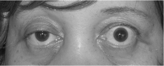
The core clinical pattern: fatigable, fluctuating weakness
At the neuromuscular junction, autoantibodies deplete or functionally block postsynaptic AChR directly (the majority pattern), or — in MuSK- and LRP4-antibody disease — disrupt the agrin-LRP4-MuSK signalling pathway that normally clusters AChR at the postsynaptic membrane, producing the same net effect of unreliable neuromuscular transmission through a mechanistically distinct route.[1] Clinically, this produces localized muscle weakness in a characteristic distribution that is exacerbated by repeated activity and typically worsens later in the day.[2] Asymmetric ptosis and variable diplopia are the presenting symptoms in about 60% of patients; in most, weakness goes on to spread, causing fluctuating dysarthria, dysphagia, difficulty chewing, and dyspnea. Limb involvement is less common as a presenting feature and, when present, is typically proximal.[2]
Simple bedside tests for fatigable weakness
Three simple bedside maneuvers can demonstrate the fatigable pattern directly: asking the patient to look upward for one minute to watch for worsening ptosis, asking the patient to sustain lateral gaze for 30 to 60 seconds and report new double vision, and asking the patient to perform five consecutive resisted shoulder abductions to observe gradual weakening of the deltoid.[2]
Myasthenia Gravis Foundation of America (MGFA) clinical classification
The MGFA clinical classification is the standard framework for describing MG severity and distribution at a point in time, and is used throughout the literature (including the trials in Pharmacologic Treatment Options) to describe study populations. It is presented here as a teaching reference.
| Class | Description |
|---|---|
| I | Any ocular muscle weakness, possibly including weakness of eye closure; all other muscle strength normal |
| II (IIa / IIb) | Mild weakness affecting muscles other than the ocular muscles (IIa: predominantly limb/axial; IIb: predominantly oropharyngeal/respiratory); ocular weakness of any severity may also be present |
| III (IIIa / IIIb) | Moderate weakness affecting muscles other than the ocular muscles, with the same limb/axial versus oropharyngeal/respiratory split |
| IV (IVa / IVb) | Severe weakness affecting muscles other than the ocular muscles, with the same split |
| V | Intubation needed to maintain the airway (with or without mechanical ventilation), other than during routine postoperative management — the definition of myasthenic crisis |
Ocular versus generalized disease
Roughly half of patients present with weakness confined to the eyes (ocular MG, MGFA Class I). Left untreated, a substantial majority go on to generalize — most within the first two years after onset — and current consensus guidance favours early treatment specifically to reduce this risk, generally preferring low-dose over high-dose corticosteroids when an acetylcholinesterase inhibitor alone is insufficient.[6]
Antibody subtypes carry real prognostic and treatment weight
AChR antibodies are highly specific and positive in about 85% of generalized MG but only about 50% of pure ocular disease — a normal AChR antibody does not exclude ocular MG.[2] MuSK-antibody-positive MG is a clinically distinct entity: it more often produces prominent bulbar, facial, neck, and respiratory weakness, sometimes with visible facial and tongue muscle atrophy; it often responds poorly to — and can even be worsened by — pyridostigmine; thymectomy has no established benefit because the thymic pathology of AChR-positive disease is absent; and rituximab, rather than the standard steroid-sparing agents, is the treatment of choice for refractory disease (see Special Populations and Choose Initial Treatment).[4][6] A smaller remaining group is LRP4-antibody-positive or fully seronegative, generally following a course closer to AChR-positive disease.[1]
Risk Factors
- Sex and age — a bimodal pattern: women in their 20s to 40s, and older men, who also carry a higher rate of associated thymoma.[2]
- Thymoma — found on chest imaging in 10 to 15% of MG patients overall, more often in older-onset disease; every newly diagnosed patient needs chest computed tomography to look for it (see Assessment and Diagnosis).[2]
- Thymic hyperplasia — common in early-onset, AChR-antibody-positive generalized MG, and the rationale for thymectomy in that specific subgroup (see Choose Initial Treatment).[1]
- Other autoimmune disease — autoimmune thyroid disease in particular, but also rheumatoid arthritis, systemic lupus erythematosus, and others, occur at higher-than-expected rates alongside MG.[1]
- Certain drugs and immunotherapies — D-penicillamine and the immune checkpoint inhibitors are established causes of genuine, de novo drug-induced MG, distinct from simply worsening pre-existing disease (see Drug Tables).[4]
Red Flags & Refer
MG is a chronic condition punctuated by genuinely emergent exacerbations. Escalate urgently for:
- New or worsening dyspnea, orthopnea, a weak or ineffective cough, or difficulty clearing secretions — treat as possible impending myasthenic crisis until proven otherwise; do not wait for overt respiratory failure (see Assessment and Diagnosis and the Clinical Decision Algorithm for bedside respiratory-monitoring thresholds).[5]
- New or worsening dysphagia, or difficulty handling secretions — a genuine aspiration risk, and a marker of the same bulbar-predominant deterioration that can precede respiratory crisis.
- Rapid symptom worsening shortly after starting any new medication — reflexively suspect a drug on the avoidance list (see Drug Tables) rather than assuming spontaneous disease progression, and report/discontinue the likely culprit while arranging urgent reassessment.[4]
- The first two weeks after starting a high-dose corticosteroid — a well-documented, genuinely paradoxical window in which weakness can worsen, occasionally into crisis, in a substantial proportion of patients; this is a reason for a cautious induction strategy, not a reason to avoid corticosteroids altogether (see Choose Initial Treatment).[4][5]
- Any pregnant patient with MG being considered for intravenous magnesium sulfate (for pre-eclampsia or eclampsia, or for tocolysis) — flag this urgently to the obstetric team before it is given; magnesium is a well-documented precipitant of myasthenic crisis (see Drug Tables and Special Populations).[4]
- New, unexplained weakness in a patient recently started on an immune checkpoint inhibitor — consider de novo, checkpoint-inhibitor-associated MG, which can be severe and carries a real mortality risk; this needs urgent oncology and neurology co-management, not routine outpatient follow-up.[4]
Assessment & Diagnosis
Diagnosis combines the clinical pattern with serologic and electrodiagnostic testing
A neurologist typically performs most of the confirmatory work-up.[2] AChR antibodies are highly specific and positive in about 85% of generalized MG, but only about 50% of pure ocular disease, so a negative result does not exclude ocular MG.[2] When AChR antibodies are negative, testing for MuSK and LRP4 antibodies helps subtype the remainder (see Recognize the Condition).[1]
Single-fibre electromyography has the highest sensitivity of any confirmatory test, at about 90%, but requires a neuromuscular specialist to perform. Repetitive nerve stimulation testing (looking for a decremental response with repeated stimulation) is more widely available but has relatively poor sensitivity.[2]
The edrophonium (Tensilon) test — intravenous edrophonium producing transient improvement of weakness — is now considered obsolete in North America because of drug shortages and concerns about cardiorespiratory compromise during the test itself; do not expect it to be used or available.[2]
Chest imaging to rule out thymoma
Computed tomography of the chest is usually ordered once the diagnosis of MG has been confirmed, specifically to identify thymoma, which occurs in 10 to 15% of patients.[2] Its presence changes management directly: surgical removal is indicated for a thymoma regardless of the patient's age or MG severity, both as MG treatment and as oncologic treatment (see Choose Initial Treatment).
Differential diagnosis
In a young patient with diplopia, consider multiple sclerosis — additional sensory symptoms are common in MS but essentially never occur in MG. In an older patient presenting with dyspnea or limb weakness, consider amyotrophic lateral sclerosis (ALS) — ALS spares eye movements (which MG frequently affects) and typically produces visible fasciculations.[2] Against the other classic causes of acute neuromuscular weakness, MG's fluctuating, fatigable pattern is the key distinguishing feature, in contrast to Guillain-Barré syndrome's steadily ascending weakness or botulism's steadily descending weakness — see the differential table in this platform's Guillain-Barré Syndrome chapter for the full three-way comparison.
Treatment Targets
- Control fatigable weakness symptomatically with pyridostigmine, recognizing that it does not modify the underlying disease.
- Achieve minimal symptom expression, or remission, with immunosuppression appropriately tailored to disease severity, distribution, and antibody status.
- Identify and treat thymoma, and appropriately select nonthymomatous patients for thymectomy.
- Avoid iatrogenic worsening — the genuinely practical centerpiece of pharmacist involvement in MG care — by screening every new prescription, including over-the-counter and as-needed items, against the drug-avoidance list.
- Recognize and rapidly escalate myasthenic crisis, and correctly distinguish it from cholinergic crisis.
- Minimize corticosteroid-related morbidity through an early steroid-sparing strategy.
- Support safe medication use and family planning across pregnancy, breastfeeding, and the surgical/perioperative setting.
Choose Initial Treatment
Step 1 — Symptomatic therapy with pyridostigmine
First-line, all subtypes except often MuSK-positive disease start pyridostigmine 30 mg PO three to four times daily and titrate to the patient's own symptom timing (typically before meals and before anticipated exertion), usually reaching 60 mg every four to six hours while awake.[6] It is purely symptomatic and does not alter the disease course; it is often inadequate — and can even worsen neuromuscular transmission — in MuSK-antibody-positive MG (see Special Populations).[4]
Step 2 — Corticosteroid induction plus an early steroid-sparing agent
For inadequate symptom control or generalized disease add prednisone, generally started low and titrated up rather than started at full dose in an unmonitored outpatient: high starting doses can paradoxically worsen weakness — sometimes into crisis — in a substantial proportion of patients within the first two weeks of treatment.[4][5] If a rapid, high starting dose cannot be avoided (for example, significant bulbar or early respiratory involvement), do so as an inpatient or with IVIG/plasma exchange pretreatment (see Drug Tables).[4]
Start concomitantly, not sequentially add a non-steroid immunosuppressant — azathioprine or mycophenolate mofetil are the standard first choices — alongside induction, rather than waiting to see whether corticosteroid monotherapy fails; both are formally endorsed by international consensus, alongside cyclosporine, tacrolimus, and methotrexate as further options.[6]
Thymectomy
Nonthymomatous, AChR-antibody-positive, generalized MG current international consensus encourages thymectomy for patients aged 18 to 50 with generalized MG and AChR antibodies, even without a thymoma.[6] This rests on the randomized MGTX trial (extended transsternal thymectomy plus prednisone versus prednisone alone, ages 18 to 65): at 3 years, thymectomy produced a lower time-weighted average quantitative MG (QMG) score (2.85 points lower, p less than 0.001), a lower time-weighted average prednisone dose (16 mg/day lower, p less than 0.001), less need for rescue azathioprine (17% versus 48%, p less than 0.001), and fewer hospitalizations for exacerbations (9% versus 37%, p less than 0.001); benefit was durable through a 5-year open-label extension.[7]
Not beneficial in MuSK-positive MG the thymic pathology that makes thymectomy effective in AChR-positive disease is absent in MuSK-antibody-positive MG, and thymectomy has no established role there (see Special Populations).[4]
Thymoma present surgical removal is indicated regardless of age or MG severity, as both oncologic and MG treatment (see Assessment and Diagnosis).
Refractory disease
Refractory, AChR-antibody-positive generalized MG a complement C5 inhibitor (eculizumab, ravulizumab, or zilucoplan) or a neonatal Fc receptor (FcRn) antagonist (efgartigimod alfa, rozanolixizumab, or nipocalimab) for disease not adequately controlled despite corticosteroids and a non-steroid immunosuppressant (see Pharmacologic Treatment Options and Drug Tables for the individual trial evidence and monitoring requirements).[6]
MuSK-positive, or refractory to the above rituximab, per specialist-directed protocol — the preferred escalation specifically in MuSK-positive disease.[6]
Non-Pharmacologic Treatment
Patient and caregiver education
- Explain the fatigable pattern explicitly — symptoms genuinely worsen with sustained activity and heat (including hot showers or baths) and improve with rest — so patients can pace activity and time pyridostigmine doses before anticipated exertion or meals rather than reactively after symptoms appear.
- Set expectations for the delayed onset of steroid-sparing agents (often many months to full effect) so a patient does not lose confidence in a new medication before it has had time to work.
- Review the specific symptoms that warrant urgent contact rather than waiting for the next visit (see Red Flags and Refer) — new breathlessness, difficulty swallowing or handling secretions, and any rapid worsening after a new medication chief among them.
The pharmacist's central role: medication reconciliation against the avoidance list
Screen every new prescription, and every over-the-counter or as-needed item, against the drug-avoidance list in Drug Tables at every encounter — not only at diagnosis. This is genuinely one of the highest-value, lowest-cost interventions available for an MG patient, given how many routinely prescribed drugs can worsen the disease or precipitate crisis.
Aspiration precautions and nutrition
For patients with bulbar involvement, arrange a formal swallowing assessment, time meals to coincide with peak pyridostigmine effect, and consider texture modification; escalate to enteral nutrition support if oral intake cannot safely meet needs.
Vaccination planning
Confirm age-appropriate vaccination status, including meningococcal vaccination specifically before any complement inhibitor is started (see Drug Tables), proactively and early — ideally before significant immunosuppression begins, since live vaccines should generally be avoided during substantial immunosuppressive therapy.
Perioperative and surgical planning
Coordinate any planned surgery — including thymectomy — with anesthesia in advance; MG patients are more sensitive to neuromuscular blocking agents and inhalational anesthetics, with a block that can be prolonged and unpredictable (see Drug Tables).
Psychosocial support
MG is a chronic, often relapsing-remitting disease with a genuine fatigue burden even outside overt flares; screen for and address the psychosocial impact, and connect patients with peer-support and neuromuscular-disease resources where available.
Special Populations
Pregnancy
MG can flare during pregnancy, particularly in the first trimester and postpartum period, and disease activity should be optimized before conception whenever the clinical situation allows planning.[5] Critical avoidance intravenous magnesium sulfate — commonly used for pre-eclampsia, eclampsia, or tocolysis — is a well-documented precipitant of myasthenic crisis; flag any pregnant patient with MG to the obstetric team before magnesium is given, and ensure the flag is visible in the chart (see Red Flags and Drug Tables).[4] Pyridostigmine, prednisone, azathioprine, and IVIG are generally considered compatible with pregnancy; mycophenolate mofetil and methotrexate are teratogenic and should be discontinued with an adequate washout before a planned conception. Roughly 10 to 20% of infants born to mothers with MG develop transient neonatal myasthenia from transplacental antibody transfer — a self-limited weakness (poor feeding, hypotonia, occasionally respiratory difficulty) typically resolving within weeks.[1] Alert the neonatal team in advance so this can be anticipated and supported rather than misattributed to another cause.
Breastfeeding
Pyridostigmine, prednisone, azathioprine, and IVIG are generally considered compatible with breastfeeding; avoid mycophenolate mofetil and methotrexate as with pregnancy.
MuSK-antibody-positive MG
Treat differently from AChR-positive disease MuSK-positive MG more often produces prominent bulbar, facial, neck, and respiratory weakness, sometimes with visible facial and tongue atrophy; it frequently responds poorly to — and can even be worsened by — pyridostigmine.[4] Thymectomy has no established benefit, since the thymic hyperplasia that justifies it in AChR-positive disease is absent.[6] Rituximab, rather than the standard first-line steroid-sparing agents, is the preferred escalation for refractory disease in this subgroup.[6]
Juvenile and pediatric MG
MG in children and adolescents is genuinely rarer than adult-onset disease and more often presents as ocular-predominant disease; distinguish it clearly from transient neonatal myasthenia (above), which is passive antibody transfer rather than the child's own autoimmune disease. Management principles otherwise follow the same treatment ladder, adjusted for age and weight, with pediatric neurology involvement.
Older adults and thymoma
Late-onset MG in older men carries a higher rate of associated thymoma and warrants the same mandatory chest imaging as any newly diagnosed patient (see Assessment and Diagnosis).[2] Polypharmacy compounds the drug-avoidance risk in this group — reconcile medication lists against the avoidance list (see Drug Tables) with particular vigilance, since older adults are more likely to be prescribed several of the higher-risk classes (beta-blockers, calcium channel blockers, fluoroquinolones) for unrelated indications.
Perioperative and anesthesia considerations
MG patients are more sensitive than unaffected individuals to nondepolarizing neuromuscular blocking agents, with a block that can be prolonged and unpredictable; nondepolarizing agents and inhalational anesthetics are best avoided where a suitable alternative exists, and when a nondepolarizing agent is required, close postoperative monitoring and sugammadex reversal reduce the risk of postoperative myasthenic crisis (see Drug Tables).[4]
Renal impairment
Adjust azathioprine and methotrexate cautiously in reduced renal function, and note that renal impairment independently raises the risk of aminoglycoside- and magnesium-associated neuromuscular blockade (see Drug Tables) — a genuine double reason for caution when these agents cannot be avoided.[4]
Hepatic impairment
Azathioprine and methotrexate both carry hepatotoxicity risk and need closer monitoring, or an alternative agent, in significant hepatic impairment.
Monitoring & Follow-Up
Disease-activity monitoring
Track functional status at every visit using a validated instrument such as the MG Activities of Daily Living (MG-ADL) scale or the Quantitative Myasthenia Gravis (QMG) score — the same instruments used in the pivotal trials referenced in Pharmacologic Treatment Options and Drug Tables — rather than relying on antibody titres, which correlate poorly with clinical activity in individual patients.
Steroid-sparing agent monitoring
- Azathioprine: dose according to TPMT activity where available; monitor CBC and liver enzymes regularly.
- Mycophenolate mofetil: monitor CBC and for signs of infection.
- Methotrexate: routine CBC, liver enzymes, and renal function; give with folic acid.
- Cyclosporine and tacrolimus: monitor renal function, blood pressure, and (for tacrolimus) trough levels.
Corticosteroid monitoring
Standard bone- and GI-protective bundle for a prolonged corticosteroid course: vitamin D and adequate calcium intake with bone-density monitoring, regular blood pressure and glucose checks, and a proton pump inhibitor for corticosteroid-related reflux where indicated (see Drug Tables).
Biologic-specific monitoring
- Complement C5 inhibitors (eculizumab, ravulizumab, zilucoplan): confirm meningococcal vaccination status is complete and current before starting, and maintain a low threshold to investigate fever given the encapsulated-organism infection risk throughout treatment.
- FcRn antagonists (efgartigimod alfa, rozanolixizumab, nipocalimab): monitor for infection given the on-target reduction in total IgG; no meningococcal-specific requirement.
- Rituximab: monitor B-cell counts, immunoglobulin levels, and for infection per specialist-directed protocol.
Ongoing medication reconciliation
Re-screen the full medication list against the drug-avoidance list (see Drug Tables) at every follow-up, not only at diagnosis — new prescriptions from other providers are a recurring, preventable source of exacerbation.
Post-thymectomy follow-up
Continue routine disease-activity and steroid-sparing-agent monitoring after thymectomy; benefit accrues gradually over months to years rather than immediately post-surgery, matching the MGTX trial's 3- and 5-year time horizons.[7]
Common Errors to Avoid
- Prescribing an aminoglycoside, fluoroquinolone, or macrolide antibiotic for an MG patient without checking for a safer alternative first, or without at least counseling closer monitoring if no alternative exists.[4]
- Starting a high dose of corticosteroid in an unmonitored outpatient with generalized MG, rather than titrating up gradually or arranging inpatient/IVIG-plasma-exchange bridging for patients who need rapid disease control.[4][5]
- Giving or allowing intravenous magnesium sulfate for pre-eclampsia, eclampsia, or tocolysis in a pregnant patient with MG without flagging the risk to the obstetric team first.[4]
- Confusing myasthenic crisis with cholinergic crisis and empirically increasing pyridostigmine when a patient's weakness worsens — this can make a true cholinergic crisis significantly worse (see Clinical Decision Algorithm).[5]
- Offering thymectomy to a MuSK-antibody-positive patient expecting the same benefit seen in AChR-positive disease — the underlying thymic pathology, and the trial evidence, simply do not apply.[4][6]
- Starting a complement C5 inhibitor (eculizumab, ravulizumab, or zilucoplan) without first confirming meningococcal vaccination is complete.
- Skipping chest computed tomography for thymoma once MG is diagnosed, or assuming a young patient's age makes thymoma unlikely enough to defer imaging.[2]
- Using a nondepolarizing neuromuscular blocking agent or inhalational anesthetic for a surgical MG patient without coordinating the anesthetic plan with anesthesia and neurology in advance.[4]
- Assuming pyridostigmine is disease-modifying, or that a patient whose symptoms are well controlled on pyridostigmine alone therefore has mild, self-limited disease rather than a condition still requiring monitoring for progression.
- Reassuring a patient with new bulbar or respiratory symptoms as "just fatigue" and deferring escalation, rather than obtaining bedside respiratory assessment promptly.[5]
- Injecting botulinum toxin, for a cosmetic or unrelated therapeutic indication, in a patient with known MG without explicit caution and slow dose titration.[4]
- Prescribing methotrexate for MG on the assumption it carries the same steroid-sparing evidence as azathioprine or mycophenolate mofetil — a dedicated randomized trial found no steroid-sparing benefit.[8]
Clinical Decision Algorithm
Drug Tables
How to use this table: Doses are adult doses and require patient-specific confirmation (renal/hepatic function, weight, allergy history). Pediatric dosing is weight-based per the current product monograph unless stated. Drug pricing is intentionally omitted. Select any drug name to open its complete Pharmasist Drug Database page.
Symptomatic Acetylcholinesterase Inhibitor Therapy
First-line for essentially all newly diagnosed patients except often MuSK-antibody-positive MG; purely symptomatic and not disease-modifying.
| Drug | Dosage | Adverse effects | Drug interactions | Comments |
|---|---|---|---|---|
| Pyridostigmine | Adults: Start 30 mg PO 3-4 times daily; typical maintenance 60 mg every 4-6 hours while awake (maximum about 600 mg/day) | Muscarinic effects: abdominal cramping, diarrhea, salivation, sweating; overdose can itself cause a cholinergic crisis | Additive cholinergic effects with other cholinesterase inhibitors; muscarinic side effects can be managed with glycopyrrolate without blunting the therapeutic nicotinic effect | Titrate to symptom timing (before meals and anticipated exertion); often inadequate, or even counterproductive, in MuSK-positive MG. |
| Glycopyrrolate | Adults: Per product labelling, titrated to symptom relief | Dry mouth, constipation, urinary retention | Additive anticholinergic burden with other agents | Does not cross the blood-brain barrier and does not blunt pyridostigmine's therapeutic nicotinic effect; a reasonable adjunct for bothersome muscarinic side effects. |
Corticosteroid Induction Therapy
First-line immunosuppression for inadequate symptomatic control or generalized disease; start a steroid-sparing agent concomitantly rather than after steroid failure.
| Drug | Dosage | Adverse effects | Drug interactions | Comments |
|---|---|---|---|---|
| Prednisone | Initial: 10-20 mg/day PO, titrated to about 1 mg/kg/day or equivalent alternate-day dosing | Can paradoxically worsen weakness, sometimes into crisis, in the first 2 weeks of high-dose therapy in a substantial proportion of patients; hyperglycemia, hypertension, osteoporosis, mood changes, infection risk | Additive hyperglycemia with other diabetogenic drugs | Start low (about 10-20 mg/day or 0.25-0.5 mg/kg/day) and titrate up over 1-2 weeks; if rapid high-dose therapy is needed, bridge with IVIG or plasma exchange rather than starting at full dose unmonitored. |
| Methylprednisolone (IV pulse) | Pulse dose: Per specialist-directed protocol, then transition to oral maintenance | Hyperglycemia, mood changes, rarely arrhythmia with rapid infusion | Additive hyperglycemia with other diabetogenic drugs | Reserved for crisis or near-crisis presentations, started together with IVIG or plasma exchange rather than alone, given the same early-worsening risk as oral induction. |
Steroid-Sparing / Non-Steroid Immunosuppressants
Onset of effect is delayed by months — start alongside induction corticosteroid, not after it fails.
| Drug | Dosage | Adverse effects | Drug interactions | Comments |
|---|---|---|---|---|
| Azathioprine | Adults: 2-3 mg/kg/day PO | Myelosuppression, hepatotoxicity, GI upset | Severe myelosuppression with allopurinol (reduce azathioprine dose substantially if co-administered); reduced efficacy of warfarin | First choice; dose according to TPMT activity where available; delayed onset (often 12 months or more) needs explicit counseling. |
| Mycophenolate mofetil | Adults: 1-1.5 g PO twice daily (2-3 g/day) | GI upset (diarrhea common), myelosuppression, increased infection risk | Reduced absorption with antacids or cholestyramine; avoid live vaccines during treatment | First-choice alternative to azathioprine; teratogenic, avoid in pregnancy. |
| Methotrexate | Adults: 15-20 mg PO or SC once weekly, with folic acid | Hepatotoxicity, myelosuppression, GI upset, mucositis | Increased toxicity with NSAIDs, trimethoprim-sulfamethoxazole, and other folate antagonists | A randomized, placebo-controlled trial found no steroid-sparing benefit at 12 months; consensus still allows it as a later option, but evidence is weaker than for azathioprine or mycophenolate. |
| Cyclosporine | Adults: Divided doses, titrated to trough level and response | Nephrotoxicity, hypertension, tremor, hyperglycemia, gingival hyperplasia | Numerous CYP3A4 interactions; avoid with other nephrotoxic agents where possible | An alternative when azathioprine and mycophenolate are contraindicated or not tolerated; monitor renal function and blood pressure closely. |
| Tacrolimus | Adults: Per trough-level-guided dosing | Nephrotoxicity, hypertension, tremor, hyperglycemia | Numerous CYP3A4 interactions; avoid with other nephrotoxic agents where possible | An alternative steroid-sparing option, dosed to a target trough level. |
| Cyclophosphamide | Adults: Per specialist-directed protocol | Myelosuppression, hemorrhagic cystitis, infertility risk, infection risk | Additive myelosuppression with other cytotoxic/immunosuppressive agents | Reserved for the most severe, refractory disease; coordinate with a tertiary neuromuscular centre. |
Rapid Immunomodulation — Myasthenic Crisis and Refractory Exacerbation
Onset within days; used for crisis, rapid pre-operative optimization, or disease refractory to maintenance immunosuppression.
| Drug | Dosage | Adverse effects | Drug interactions | Comments |
|---|---|---|---|---|
| Immunoglobulin (human), IV (IVIG) | Adults: 0.4 g/kg IV once daily for 5 consecutive days (total 2 g/kg) | Headache, aseptic meningitis, renal impairment, thromboembolism, anaphylaxis in IgA deficiency | May blunt live-vaccine response for a period afterward | Onset in 4-5 days, effect lasts 4-8 weeks; does not require apheresis resources. |
| Plasma exchange (plasmapheresis) | Adults: Five exchanges (about one plasma volume each), every other day over about 10 days | Hypotension, catheter-related infection or thrombosis, citrate-related hypocalcemia, coagulopathy | Removes recently administered IV drugs and some coagulation factors — coordinate timing of other IV medications with the apheresis team | Onset within about 2 days, effect lasts 3-4 weeks; requires apheresis resources and central venous access. If initial therapy fails, sequential therapy (the alternative modality) may be used. |
Complement C5 Inhibitors — Refractory AChR-Antibody-Positive Generalized MG
All three require confirmed, current meningococcal vaccination before starting because of the encapsulated-organism infection risk inherent to terminal complement blockade.
| Drug | Dosage | Adverse effects | Drug interactions | Comments |
|---|---|---|---|---|
| Eculizumab | Adults: 900 mg IV weekly for 4 doses, then 1200 mg IV every 2 weeks | Meningococcal infection risk, infusion reactions, headache | None specific beyond the shared infection-risk precaution | Complete meningococcal vaccination at least 2 weeks before the first dose wherever possible; REGAIN trial showed a significant improvement in QMG total score at 26 weeks versus placebo. |
| Ravulizumab | Adults: Weight-based IV loading dose, then maintenance infusion every 8 weeks | Meningococcal infection risk, infusion reactions, headache | None specific beyond the shared infection-risk precaution | Longer-acting than eculizumab, allowing 8-weekly maintenance dosing; CHAMPION MG trial showed significantly greater improvement in QMG total score versus placebo at 26 weeks. |
| Zilucoplan | Adults: Weight-based subcutaneous injection once daily | Meningococcal infection risk, injection-site reactions | None specific beyond the shared infection-risk precaution | Self-administered subcutaneous injection once trained; RAISE trial showed significantly greater MG-ADL improvement versus placebo at 12 weeks. |
Neonatal Fc Receptor (FcRn) Antagonists — Refractory Generalized MG
Accelerate degradation of circulating IgG, including the pathogenic autoantibody; infection risk relates to the resulting drop in total IgG rather than to complement blockade, so there is no meningococcal-specific requirement.
| Drug | Dosage | Adverse effects | Drug interactions | Comments |
|---|---|---|---|---|
| Efgartigimod alfa | Adults: IV infusion once weekly for 4 doses per cycle, cycles repeated based on clinical response | Headache, infection risk from lowered IgG | None specific | ADAPT trial: 68% of AChR-antibody-positive patients were MG-ADL responders in the first treatment cycle versus 30% on placebo; a subcutaneous formulation also exists. |
| Rozanolixizumab | Adults: Weekly subcutaneous injection, weight-based, for a defined treatment cycle | Headache, infection risk from lowered IgG, hypersensitivity reactions | None specific | MycarinG trial studied both AChR- and MuSK-antibody-positive patients, with clinically meaningful improvement and good tolerability at both studied doses. |
| Nipocalimab | Adults: Per specialist-directed protocol | Headache, infection risk from lowered IgG | None specific | A further FcRn antagonist option; confirm current antibody-status eligibility criteria before selecting over the other agents in this class. |
B-Cell Depletion
The preferred escalation specifically for MuSK-antibody-positive disease, where thymectomy has no established role.
| Drug | Dosage | Adverse effects | Drug interactions | Comments |
|---|---|---|---|---|
| Rituximab | Dosing: Per specialist-directed protocol | Infusion reactions, infection risk, hypogammaglobulinemia | Avoid live vaccines during and after treatment per usual practice | Preferred for refractory MuSK-positive MG; also used for refractory AChR-positive disease. |
Cholinergic Crisis and Muscarinic Symptom Management
Atropine treats the muscarinic symptoms of acetylcholinesterase-inhibitor excess; it does not reverse the underlying weakness, which requires holding the offending inhibitor and, if needed, ventilatory support.
| Drug | Dosage | Adverse effects | Drug interactions | Comments |
|---|---|---|---|---|
| Atropine | Adults: 0.5-1 mg IV, repeated as needed for muscarinic symptom control | Tachycardia, dry mouth, urinary retention, blurred vision | Additive anticholinergic burden with other agents | For cholinergic crisis or severe muscarinic side effects from an acetylcholinesterase inhibitor; hold pyridostigmine and reassess before resuming. |
Drugs That Can Worsen or Unmask MG, or Precipitate Myasthenic Crisis
This is the single highest-yield table in this chapter for day-to-day pharmacist practice. Evidence levels reflect the strength of the published association; "Avoid" means avoid if any reasonable alternative exists and monitor closely if not, not an absolute contraindication in a genuine emergency.
| Drug class / agents | Mechanism | Practical guidance |
|---|---|---|
| gentamicin, tobramycin, amikacin Definite | Impairs both pre- and postsynaptic neuromuscular transmission | Avoid if any alternative antibiotic exists; well documented to cause exacerbation and, at high doses or in renal failure, myasthenia-like symptoms even in patients without MG |
| ciprofloxacin, levofloxacin, moxifloxacin, norfloxacin, ofloxacin Probable | Impairs pre- and postsynaptic neuromuscular transmission | Avoid if a safer alternative exists; exacerbation has been reported from 15 minutes to 4 days after the first dose |
| azithromycin, clarithromycin, erythromycin, telithromycin Definite | Presynaptic impairment of acetylcholine release | Azithromycin is the antibiotic most frequently implicated in real-world MG exacerbations; telithromycin carries a specific severe-exacerbation warning and is no longer marketed in Canada or the US |
| amoxicillin, ampicillin, cephalexin, sulfamethoxazole-trimethoprim, clindamycin, doxycycline Generally safe | No meaningful signal for most; a small number of case reports exist for penicillins | Reasonable first-choice antibiotics when an alternative to the classes above is needed |
| magnesium sulfate (IV) Definite | Blocks presynaptic acetylcholine release and impairs the postsynaptic response | Use with extreme caution; myasthenic crisis has been reported after standard obstetric (pre-eclampsia/eclampsia) and general replacement dosing — flag any MG patient started on IV magnesium urgently |
| onabotulinumtoxinA, abobotulinumtoxinA, incobotulinumtoxinA Definite | Presynaptic reduction of acetylcholine release — the same neuromuscular-junction target as MG itself | Avoid where possible for any indication (cosmetic or therapeutic); may be offered with caution and slow dose titration for mild, stable disease with a compelling indication such as blepharospasm |
| procainamide, quinidine, disopyramide Definite | Impairs pre- and postsynaptic neuromuscular transmission | Avoid, or use only with extreme caution and close monitoring; procainamide has caused respiratory failure in MG patients and myasthenia-like symptoms in non-MG patients with renal failure |
| chloroquine, hydroxychloroquine Probable to definite | Direct effect on pre- and postsynaptic neuromuscular transmission; chloroquine can also induce AChR antibodies | Use only when clearly indicated, with close monitoring for new or worsening weakness after starting |
| penicillamine Definite (causes de novo MG) | Alters antigen-presenting cell surface molecules, breaking immune tolerance | MG develops in about 1-7% of patients on long-term therapy, usually within a year, and typically remits within 6-10 months of stopping — this is a genuinely distinct drug-induced form of MG, not simply an exacerbation |
| pembrolizumab, nivolumab, ipilimumab Definite (causes de novo MG) | T-cell activation and loss of peripheral immune tolerance | Immune checkpoint inhibitor-associated MG causes respiratory failure in close to half of affected patients and carries a real mortality risk; needs early, aggressive combined immunosuppression and joint oncology/neurology management, and generally precludes re-challenge after a severe episode |
| propranolol, timolol (including ophthalmic drops) Possible | Unclear effect on neuromuscular transmission | Can generally be used in stable, well-controlled MG at the lowest effective dose with close monitoring, especially when starting; do not overlook the ophthalmic route as a source of systemic exposure |
| verapamil Possible | Unclear effect on neuromuscular transmission | Respiratory failure has been reported after starting verapamil in severe generalized MG; use the lowest effective dose with monitoring, especially at initiation |
| simvastatin, rosuvastatin, atorvastatin Probable | Proposed T-cell polarization shift, superimposed myopathy, or mitochondrial effects | An infrequent cause of exacerbation and a rare cause of de novo MG; not a reason to withhold an indicated statin, but worth considering if new weakness develops after starting one |
| succinylcholine, rocuronium and other nondepolarizing agents Definite | Postsynaptic neuromuscular block, exaggerated by the already-reduced receptor reserve in MG | MG patients are more sensitive, and the block can be prolonged and unpredictable; avoid nondepolarizing agents and inhalational anesthetics where a suitable alternative exists, and use sugammadex reversal with close postoperative monitoring when a nondepolarizing agent is required |
| lithium carbonate Probable | Reduced presynaptic acetylcholine synthesis/release and reduced postsynaptic AChR density | Exacerbation is rarely reported; can generally be used in MG with monitoring |
| phenytoin, gabapentin, carbamazepine Case reports / possible | Unclear; proposed effects on neuromuscular transmission at the presynaptic terminal | Generally usable; be alert to new or worsening weakness after starting, particularly with phenytoin |
Supportive Therapy Alongside Long-Term Corticosteroids
Standard bone- and GI-protective bundle for any patient on a prolonged corticosteroid course.
| Drug | Dosage | Adverse effects | Drug interactions | Comments |
|---|---|---|---|---|
| Calcium carbonate + vitamin D3 | Adults: Combo: calcium carbonate 500-1000 mg elemental PO daily + vitamin D3 800-2000 IU PO daily | GI upset, constipation, hypercalcemia with excessive intake | Reduced absorption of concurrently administered levothyroxine, tetracyclines, or fluoroquinolones if not spaced apart | Combine with usual bone-density monitoring. |
| Pantoprazole | Adults: 40 mg PO once daily | Headache, GI upset, rare hypomagnesemia with long-term use | May reduce absorption of drugs requiring an acidic environment | Reserve for patients with corticosteroid-related reflux or another clear GI-protective indication. |
References
- Myasthenia Gravis — New England Journal of Medicine (Gilhus NE). N Engl J Med 2016;375(26):2570-2581
- Myasthenia gravis — CMAJ, Canadian Medical Association Journal (Bourque PR, Breiner A). CMAJ 2018;190(38):E1141
- Epidemiology of myasthenia gravis in Ontario, Canada — Neuromuscular Disorders (Breiner A, Widdifield J, Katzberg HD). Neuromuscul Disord 2016;26(1):41-46
- Drugs That Induce or Cause Deterioration of Myasthenia Gravis: An Update — Journal of Clinical Medicine (Sheikh S, Alvi U, Soliven B, Rezania K). J Clin Med 2021;10(7):1537
- Myasthenic Crisis — The Neurohospitalist (Wendell LC, Levine JM). Neurohospitalist 2011;1(1):16-22
- International Consensus Guidance for Management of Myasthenia Gravis: 2020 Update — Neurology, Myasthenia Gravis Foundation of America Task Force (Narayanaswami P, Sanders DB, Wolfe G, et al.). Neurology 2021;96(3):114-122
- Randomized Trial of Thymectomy in Myasthenia Gravis — New England Journal of Medicine, MGTX Study Group (Wolfe GI, Kaminski HJ, Aban IB, et al.). N Engl J Med 2016;375(6):511-522
- A randomized controlled trial of methotrexate for patients with generalized myasthenia gravis — Neurology, Methotrexate in MG Investigators of the Muscle Study Group (Pasnoor M, He J, Herbelin L, et al.). Neurology 2016;87(1):57-64
- Safety and efficacy of eculizumab in anti-acetylcholine receptor antibody-positive refractory generalised myasthenia gravis (REGAIN): a phase 3, randomised, double-blind, placebo-controlled, multicentre study — The Lancet Neurology, REGAIN Study Group (Howard JF Jr, Utsugisawa K, Benatar M, et al.). Lancet Neurol 2017;16(12):976-986
- Safety, efficacy, and tolerability of efgartigimod in patients with generalised myasthenia gravis (ADAPT): a multicentre, randomised, placebo-controlled, phase 3 trial — The Lancet Neurology, ADAPT Investigator Study Group (Howard JF Jr, Bril V, Vu T, et al.). Lancet Neurol 2021;20(7):526-536
- Safety and efficacy of rozanolixizumab in patients with generalised myasthenia gravis (MycarinG): a randomised, double-blind, placebo-controlled, adaptive phase 3 study — The Lancet Neurology (Bril V, Drużdż A, Grosskreutz J, et al.). Lancet Neurol 2023;22(5):383-394
- Safety and efficacy of zilucoplan in patients with generalised myasthenia gravis (RAISE): a randomised, double-blind, placebo-controlled, phase 3 study — The Lancet Neurology, RAISE Study Group (Howard JF Jr, et al.). Lancet Neurol 2023;22(5):395-406
- Terminal Complement Inhibitor Ravulizumab in Generalized Myasthenia Gravis — NEJM Evidence, CHAMPION MG Study Group (Vu T, Meisel A, Mantegazza R, et al.). NEJM Evid 2022;1(5):EVIDoa2100066
Professional reference only. Apply clinical judgment and verify patient-specific decisions against current authoritative guidance and product information.