Myocarditis
Myocarditis
References
- 2024 ACC Expert Consensus Decision Pathway on Strategies and Criteria for the Diagnosis and Management of Myocarditis: A Report of the American College of Cardiology Solution Set Oversight Committee — Journal of the American College of Cardiology, American College of Cardiology Solution Set Oversight Committee (Drazner MH, Bozkurt B, Cooper LT, Aggarwal NR, Basso C, Bhave NM, Caforio ALP, Ferreira VM, Heidecker B, Kontorovich AR, Martín P, Roth GA, Van Eyk JE). J Am Coll Cardiol, published online Dec 10, 2024
- Management of Patients With Giant Cell Myocarditis: JACC Review Topic of the Week — Journal of the American College of Cardiology (Bang V, Ganatra S, Shah SP, Dani SS, Neilan TG, Thavendiranathan P, Resnic FS, Barac A, Yang EH, Lenihan DJ, Cheng RK). J Am Coll Cardiol 2021;77(8):1122-1134
- Management of Immune-Related Adverse Events in Patients Treated With Immune Checkpoint Inhibitor Therapy: ASCO Guideline Update — Journal of Clinical Oncology, American Society of Clinical Oncology (Schneider BJ, Naidoo J, Santomasso BD, et al.). J Clin Oncol 2021;39(36):4073-4126
- Myocarditis and Pericarditis After mRNA COVID-19 Vaccination: 2024 Status and Management Update — Canadian Journal of Cardiology, Canadian Cardiovascular Society (McDonald MA, Kafil TS, Khoury M, Luk AC, Wright MK, Hawkins NM). Can J Cardiol 2024;40(9):1536-1540
- Clinical Considerations for Competitive Sports Participation for Athletes With Cardiovascular Abnormalities: A Scientific Statement From the American Heart Association and American College of Cardiology — Journal of the American College of Cardiology, American Heart Association/American College of Cardiology (Kim JH, Baggish AL, Levine BD, et al.). J Am Coll Cardiol 2025;85(10):1059-1108
- The Role of Endomyocardial Biopsy in the Management of Cardiovascular Disease: A Scientific Statement From the American Heart Association, the American College of Cardiology, and the European Society of Cardiology — Circulation, American Heart Association/American College of Cardiology/European Society of Cardiology (Cooper LT, Baughman KL, Feldman AM, Frustaci A, Jessup M, Kuhl U, Levine GN, Narula J, Starling RC, Towbin J, Virmani R). Circulation 2007;116(19):2216-2233
- Clinical guidance for youth with myocarditis and pericarditis following mRNA COVID-19 vaccination — Canadian Paediatric Society (Wong PD, McCrindle BW, Wong K, Khoury M, Chan K, Salvadori M). Canadian Paediatric Society Practice Point, 2021
- CCS/CHFS Heart Failure Guidelines Update: Defining a New Pharmacologic Standard of Care for Heart Failure With Reduced Ejection Fraction — Canadian Journal of Cardiology, Canadian Cardiovascular Society/Canadian Heart Failure Society (McDonald M, Virani S, Chan M, et al.). Can J Cardiol 2021;37(4):531-546
- Long-Term Outcome of Fulminant Myocarditis as Compared with Acute (Nonfulminant) Myocarditis — New England Journal of Medicine (McCarthy RE 3rd, Boehmer JP, Hruban RH, Hutchins GM, Kasper EK, Hare JM, Baughman KL). N Engl J Med 2000;342(10):690-695
Clinical Snapshot
Myocarditis is inflammation of the myocardium, most commonly triggered by a viral infection or by the immune response that follows one, though the list of recognized triggers is genuinely broad — autoimmune and hypersensitivity reactions, specific drugs, and, of real current clinical importance, both mRNA COVID-19 vaccination and immune checkpoint inhibitor (ICI) cancer therapy.[1] Clinical severity spans an enormous range: from an entirely subclinical finding, through mild chest pain and transient ECG changes that resolve without specific treatment, to fulminant cardiogenic shock, life-threatening ventricular arrhythmia, or sudden cardiac death as the very first presentation — sometimes in a previously healthy young person only days removed from what looked like an ordinary viral illness.[1] This is precisely why this chapter leads with emergency recognition rather than treatment: the central clinical skill this condition demands is recognizing when apparent viral symptoms, chest pain, or fatigue are not "just a viral illness" but the leading edge of a process that can deteriorate within days.
The 2024 ACC Expert Consensus Decision Pathway organizes myocarditis into four stages that parallel the familiar heart-failure staging framework, and this chapter uses that same structure throughout: Stage A (exposure to a known myocarditis trigger — a viral illness, an mRNA vaccine dose, an ICI — without cardiac symptoms or abnormal testing), Stage B (asymptomatic but with objective evidence of myocardial injury or inflammation on testing), Stage C (symptomatic myocarditis — chest pain, dyspnea, palpitations, or overt heart failure), and Stage D (hemodynamic or electrical instability — cardiogenic shock, sustained ventricular arrhythmia, or high-grade heart block; this is fulminant myocarditis).[1] Roughly half of patients have complete resolution within 2-4 weeks, but about a quarter go on to persistent cardiac dysfunction and 12-25% progress to end-stage heart failure or death — outcomes that are meaningfully worse with biventricular dysfunction, Stage D presentation at diagnosis, and the presence of late gadolinium enhancement on cardiac MRI.[1]
There is no specific antiviral treatment for most viral myocarditis, which is why heart-failure-directed pharmacotherapy is the genuine backbone of management for the majority of patients with reduced left ventricular function. That backbone, however, is not the whole story: giant cell myocarditis and eosinophilic myocarditis require immunosuppression as first-line, disease-specific therapy rather than supportive care alone, and recognizing which pathway a patient is on — supportive versus immunosuppressive — is one of this chapter's central organizing decisions.[2] A parallel decision applies to the two triggered forms this chapter specifically emphasizes: ICI-associated myocarditis, which carries a strikingly high mortality rate and demands immediately holding the ICI and starting corticosteroids at first suspicion, and mRNA-vaccine-associated myocarditis, which is usually mild and self-limited but still requires prompt recognition, supportive care, and specific guidance on future vaccine dosing.[3][4] Finally, activity restriction — genuinely avoiding competitive sport and vigorous exercise for a defined period after diagnosis — is a specific, concrete counseling point running through this entire chapter, because vigorous exercise during active myocardial inflammation is a recognized trigger for malignant ventricular arrhythmia.[5]
Recognize the Condition
Epidemiology and etiology
The true incidence of myocarditis is difficult to pin down because so many cases are subclinical or never biopsied; it is likely substantially underdiagnosed. Viral infection is the most common identified trigger worldwide — historically coxsackievirus and other enteroviruses, with adenovirus, parvovirus B19, human herpesvirus 6, and, more recently, SARS-CoV-2 itself all well described. SARS-CoV-2 infection is, in fact, a meaningfully more common and more severe trigger of myocarditis than the mRNA vaccines used to prevent it — an important, genuinely reassuring point for patient counseling, and one addressed directly in Special Populations and Common Errors to Avoid.[4] Beyond infection, recognized triggers include autoimmune and connective tissue disease, drug hypersensitivity reactions, hypereosinophilic states, and, of specific and growing current importance, mRNA COVID-19 vaccination and immune checkpoint inhibitor cancer therapy — see Risk Factors for the full list.[1]
Typical presentation
- A preceding viral prodrome — fever, myalgias, upper respiratory or gastrointestinal symptoms — in the 1-3 weeks before cardiac symptoms begin, though this history is not always present or elicited.
- Chest pain, which can be pleuritic/positional (suggesting an accompanying pericardial component) or can closely mimic acute coronary syndrome, including troponin elevation and ST-segment changes — a genuine diagnostic trap addressed in Assessment & Diagnosis.
- Dyspnea, fatigue, and reduced exercise tolerance, from evolving left ventricular dysfunction.
- Palpitations, from atrial or ventricular ectopy, and, less commonly, sustained arrhythmia.
- Presyncope or syncope, which can reflect a life-threatening arrhythmia or high-grade AV block and is a genuine red flag (see Red Flags/Refer).
- In children, presentation is often less specific — vomiting, lethargy, poor feeding, or abdominal pain rather than classic chest pain — which is exactly why a high index of suspicion matters in this age group (see Special Populations).
- Tragically, sudden cardiac death or cardiogenic shock can be the first presentation, particularly in young people and especially during or immediately after vigorous exertion — the central reason this chapter treats activity restriction as a genuine safety issue rather than a routine convalescence recommendation.[5]
The core distinction this chapter is organized around: supportive versus immunosuppression-first management
Typical (lymphocytic/viral) myocarditis vs. the immunosuppression-requiring forms
Typical viral/lymphocytic myocarditis
The most common pattern. There is no specific antiviral treatment; management is heart-failure-directed supportive pharmacotherapy plus activity restriction, with most patients recovering fully within weeks.[1]
Giant cell and eosinophilic myocarditis
Rarer, but genuinely different in both course and treatment: these are immune-mediated forms that often progress rapidly — heart failure, ventricular arrhythmia, and high-grade AV block within days to weeks — and require immunosuppression as first-line, disease-specific therapy, not simply supportive care. Recognizing this pathway early, through the specific endomyocardial biopsy indications in Assessment & Diagnosis, materially changes outcomes.[2][6]
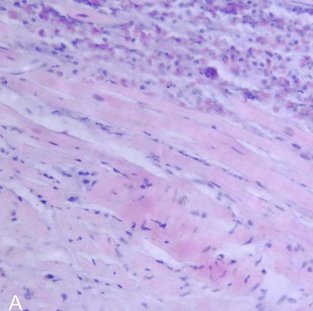
Two current, specifically important triggered forms
mRNA COVID-19 vaccine-associated myocarditis is uncommon (roughly 1.2 cases per 100,000 vaccine doses in Canada as of January 2024) but has a recognizable pattern: most often young males under 30, more common after the second dose, with symptom onset typically within 1-7 days (peaking days 1-3) of vaccination, and the great majority of cases mild and self-limited.[4][7] ICI-associated myocarditis is rarer still but disproportionately dangerous, with reported mortality in the range of 25-50% despite its low overall incidence — early recognition and immediately holding the causative agent are genuinely time-critical.[3] Both are covered in full in their own parts of Choose Initial Treatment and Special Populations, reflecting their real, current clinical importance.
Risk Factors
Infectious and post-infectious triggers
- Viral infection — historically coxsackievirus B and other enteroviruses, adenovirus, parvovirus B19, human herpesvirus 6, influenza, and SARS-CoV-2, among others; a preceding viral illness in the 1-3 weeks before cardiac symptoms is the most commonly identifiable trigger.[1]
- Less commonly, bacterial (including Lyme carditis and diphtheria), fungal, or parasitic infection (e.g., Chagas disease, more relevant with a relevant travel/exposure history), and, in immunocompromised patients, opportunistic organisms such as cytomegalovirus (see Special Populations).
Immune-mediated and hypersensitivity triggers
- Autoimmune and connective tissue disease — systemic lupus erythematosus, systemic sclerosis, sarcoidosis (cardiac sarcoidosis is a distinct, granulomatous form with its own specialist-directed pathway), and inflammatory myopathy.
- Drug hypersensitivity reactions, including DRESS (drug reaction with eosinophilia and systemic symptoms) — a recognized trigger specifically for eosinophilic myocarditis; identifying and stopping the causative drug is itself a core part of treatment (see Choose Initial Treatment).
- Hypereosinophilic syndrome and eosinophilic granulomatosis with polyangiitis (EGPA/Churg-Strauss) as underlying drivers of eosinophilic myocarditis when no drug trigger is identified.
- An association with thymoma, myasthenia gravis, and inflammatory bowel disease is recognized in giant cell myocarditis, though many cases have no identified associated condition.[2]
Drug- and therapy-triggered forms — the two current forms this chapter emphasizes
- Immune checkpoint inhibitor (ICI) therapy — anti-PD-1/PD-L1 and anti-CTLA-4 agents used in cancer treatment. Risk is substantially higher with combination ICI therapy (dual checkpoint blockade) than with monotherapy, onset is typically early — a median of around 30 days after starting therapy and often within the first few doses — and concurrent immune-related myositis and/or myasthenia gravis (an overlap syndrome) carries a markedly higher mortality than isolated myocarditis.[3]
- mRNA COVID-19 vaccination — age under 30, male sex, and the second dose of the primary series are the most consistent risk factors identified; onset is typically within 1-7 days of vaccination.[4][7]
Other recognized risk factors
- Cardiotoxin exposure — cocaine, excessive alcohol use, and certain other drugs.
- A family history of cardiomyopathy or unexplained sudden cardiac death, reflecting a genetic predisposition component in some patients that is increasingly recognized as clinically relevant — genetic counselling and testing is now a specific 2024 ACC recommendation where feasible (see Non-Pharmacologic Treatment).[1]
- Young age and male sex are epidemiologic associations across several of this chapter's triggered forms (viral myocarditis generally, vaccine-associated myocarditis, and the sudden-cardiac-death-in-athletes literature specifically).
Red Flags & Refer
This is not simply a viral illness if any of the following are present — escalate or refer urgently:
- Hemodynamic instability — hypotension, poor perfusion, cardiogenic shock, or rapidly worsening heart failure — is fulminant myocarditis (Stage D) until proven otherwise; arrange urgent transfer to a center capable of mechanical circulatory support without delaying for an outpatient work-up.[1]
- Sustained ventricular arrhythmia or high-grade (Mobitz type II or third-degree) AV block in a patient with suspected or confirmed myocarditis — a life-threatening electrical presentation that needs the same urgency as hemodynamic instability.[1]
- Syncope or presyncope in a patient with suspected myocarditis — treat as a potential marker of a life-threatening arrhythmia or high-grade block, not a benign vasovagal event, until specialist evaluation says otherwise.
- Chest pain with troponin elevation mimicking acute coronary syndrome — this needs the same urgent evaluation pathway as suspected ACS; myocarditis is a diagnosis reached partly by excluding obstructive coronary disease, not a reason to defer that evaluation (see Assessment & Diagnosis).
- Suspected giant cell or eosinophilic myocarditis — rapid clinical deterioration, new ventricular arrhythmia, new high-grade AV block, or failure to respond to usual supportive care within 1-2 weeks — warrants urgent cardiology referral for endomyocardial biopsy; this is a genuinely different, immunosuppression-requiring pathway and delay has a real cost (see Assessment & Diagnosis, Choose Initial Treatment).[6][2]
- Any new cardiac symptom, ECG change, or troponin elevation in a patient currently receiving an immune checkpoint inhibitor — hold the ICI immediately and refer urgently to cardio-oncology; ICI myocarditis carries a mortality of roughly 25-50% and this is one of the most time-critical referrals in this entire chapter.[3]
- New chest pain, dyspnea, or palpitations within 1-7 days of an mRNA COVID-19 vaccine dose, particularly in a young male — evaluate promptly with ECG and troponin; most cases are mild, but admission or close monitoring is appropriate until the clinical course is established.[7]
- Sudden cardiac arrest or death as a first presentation, especially in a young athlete during or shortly after vigorous exertion — tragic, but the entire rationale for this chapter's emphasis on activity restriction during active myocardial inflammation.[5]
Assessment & Diagnosis
Initial testing — every patient with suspected myocarditis
ECG, cardiac troponin, and transthoracic echocardiography are the core initial tests and carry real prognostic value, but a normal result on any of them does not exclude myocarditis when clinical suspicion remains — do not let reassuring initial testing close the diagnostic question prematurely.[1] ECG findings are nonspecific and varied: diffuse ST-segment and T-wave changes (which can mimic pericarditis when a pericardial component coexists), sinus tachycardia, ectopy, or frank conduction block. Troponin is elevated in most patients with active myocarditis and can be markedly elevated — occasionally above 10,000 ng/L in mRNA-vaccine-associated cases — though the degree of elevation does not reliably predict severity on its own.[7] BNP or NT-proBNP tracks the degree of hemodynamic/heart-failure involvement. Echocardiography assesses left and right ventricular function (wall-motion abnormalities in myocarditis are often more global than the territorial pattern of an acute coronary occlusion, though regional patterns do occur and are not reassuring on their own) and any pericardial effusion.
Cardiac MRI — the cornerstone noninvasive diagnostic tool
Cardiac MRI is central to modern myocarditis diagnosis, characterizing myocardial edema, hyperemia, necrosis/fibrosis (via late gadolinium enhancement), pericardial inflammation, and LV function in a single noninvasive study.[1] The updated (2018) Lake Louise diagnostic criteria require at least one T2-based marker of myocardial edema (T2-weighted imaging or T2 mapping) and at least one T1-based marker of non-edematous injury (late gadolinium enhancement, T1 mapping, or extracellular volume), with pericardial effusion and systolic dysfunction as supportive additional findings. Late gadolinium enhancement in particular carries independent prognostic weight — its presence is associated with a worse long-term trajectory — which is why CMR findings feed directly into the follow-up risk-stratification described in Monitoring & Follow-Up.[1]
Endomyocardial biopsy — reserved, not routine, but genuinely decision-changing in specific scenarios
Endomyocardial biopsy (EMB) remains the reference standard for identifying a specific, etiology-directed diagnosis (most importantly, distinguishing giant cell and eosinophilic myocarditis from the far more common lymphocytic/viral pattern) and for excluding mimics, but it is not recommended routinely given its procedural risk and imperfect sensitivity from sampling error.[1] The classic case-definition indications, still in current use, are:
| Clinical scenario | Recommendation |
|---|---|
| New-onset heart failure of less than 2 weeks' duration, associated with a normal-to-mildly-dilated left ventricle and hemodynamic compromise | Urgent EMB — specifically to identify or exclude giant cell myocarditis or fulminant necrotizing eosinophilic myocarditis, both of which need immunosuppression started without delay |
| New heart failure of 2 weeks to 3 months' duration, associated with a dilated left ventricle, and new ventricular arrhythmias, Mobitz type II or third-degree AV block, or failure to respond to usual heart-failure care within 1-2 weeks | EMB recommended — the combination of a subacute course with one of these specific red-flag features is what distinguishes a patient worth biopsying from routine subacute heart failure |
[6] Outside these and a small number of other specific scenarios (e.g., suspected cardiac sarcoidosis, or unexplained cardiomyopathy where the result would change management), EMB adds procedural risk without changing the treatment plan for the large majority of hemodynamically stable, non-progressive presentations — see Common Errors to Avoid.
Additional testing and differential diagnosis
- Coronary angiography (or CT coronary angiography in appropriate lower-risk patients) to exclude obstructive coronary artery disease when the presentation mimics acute coronary syndrome — myocarditis is a diagnosis reached partly by exclusion in this scenario, and this should not be skipped out of an assumption that a young patient "can't have a heart attack."
- Inflammatory markers (CRP, ESR) are nonspecific but can support the overall clinical picture.
- Routine viral serology is generally low-yield and rarely changes management; targeted testing (e.g., HIV, Lyme serology, specific testing suggested by epidemiologic exposure or travel history) is more useful than a broad, undirected viral panel.
- A peripheral eosinophil count can support suspected eosinophilic myocarditis, though a normal count does not exclude it.
- Genetic counselling and testing, per the 2024 ACC pathway, for patients where a hereditary cardiomyopathy component is plausible (see Non-Pharmacologic Treatment).[1]
- Differential diagnosis: acute coronary syndrome/myocardial infarction, Takotsubo (stress) cardiomyopathy, other forms of acute or chronic cardiomyopathy, and, in the ICI-therapy population specifically, other immune-related adverse events (myositis, pericarditis) that can coexist with or mimic myocarditis.
Treatment Targets
- Recognize the fulminant (Stage D) presentation early and escalate to a mechanical-circulatory-support-capable center without delay — this single decision has the largest effect on outcome of anything in this chapter.[1]
- For the large majority of patients with reduced LV function: initiate and up-titrate heart-failure-directed pharmacotherapy, since no specific antiviral treatment exists for most viral myocarditis.[8]
- Distinguish, through history, course, and — where indicated — endomyocardial biopsy, the patients who need immunosuppression as first-line therapy (giant cell myocarditis, eosinophilic myocarditis) from the majority who do not.[2][6]
- For ICI-associated myocarditis: hold the causative agent immediately at first suspicion, regardless of severity grade, and start corticosteroids promptly given the disease's genuinely high mortality.[3]
- For mRNA-vaccine-associated myocarditis: confirm the diagnosis, provide supportive care, and give clear guidance on future vaccine dosing per current national recommendations.[4][7]
- Restrict competitive sport and vigorous exercise for a defined period during active inflammation, and reassess against objective criteria — not a calendar alone — before clearing return to play.[5]
- Identify and manage life-threatening arrhythmia and high-grade conduction block promptly, including temporary or permanent pacing and defibrillator support where indicated.
- Where a hereditary component is plausible, offer genetic counselling/testing and consider first-degree-relative implications.[1]
Choose Initial Treatment
Reduced LVEF or overt heart failure, hemodynamically stable initiate heart-failure-directed pharmacotherapy as the backbone of treatment — there is no specific antiviral therapy for most viral myocarditis, so this is genuinely first-line, not a fallback. Start an ACE inhibitor or, where appropriate and tolerated, an angiotensin receptor-neprilysin inhibitor, together with a beta-blocker once hemodynamically stable, a mineralocorticoid receptor antagonist, and an SGLT2 inhibitor, following standard heart-failure-with-reduced-ejection-fraction principles and titration.[8] Add a loop diuretic for congestive symptoms, titrated to volume status.
Avoid routine NSAID use when LV dysfunction or heart failure is present — animal and clinical data raise concern that NSAIDs can worsen myocardial inflammation and outcomes in this setting; NSAIDs remain a reasonable option specifically for pericarditic-type chest pain without LV dysfunction (see the anti-inflammatory-analgesia directive below).[1]
Cardiogenic shock, refractory arrhythmia, or high-grade AV block urgent transfer to a center capable of mechanical circulatory support (intra-aortic balloon pump, percutaneous ventricular assist device, or VA-ECMO); inotropic/vasopressor support (dobutamine, milrinone, or norepinephrine) is a reasonable bridge while arranging transfer and support, but should not substitute for early escalation to mechanical support — prolonged high-dose catecholamine exposure in a myocardium that may well recover is itself a real concern.[1] Arrange early endomyocardial biopsy in parallel, since fulminant presentation is exactly the scenario in which giant cell or eosinophilic myocarditis needs to be identified without delay.[6]
A genuinely counterintuitive but well-established point. Despite its dramatic presentation, fulminant myocarditis carries a better long-term prognosis than the more indolent acute (nonfulminant) form, provided the patient survives the acute phase with appropriate support — a landmark biopsy-based cohort found 93% 11-year survival in fulminant myocarditis versus 45% in acute (nonfulminant) myocarditis.[9] The clinical message is not to be falsely reassured by a milder-looking presentation, and not to be falsely pessimistic about a dramatic one — aggressive acute support genuinely pays off in this specific disease.
Biopsy-confirmed or strongly suspected giant cell myocarditis start combination immunosuppression without delay: IV methylprednisolone 10 mg/kg/day for the first 3 days, transitioning to an oral corticosteroid taper over several months, combined with cyclosporine (titrated to a specialist-directed trough level), historically with the addition of muromonab-CD3 (no longer available) and now more often anti-thymocyte globulin or alemtuzumab in more severe or refractory presentations.[2] Combination therapy including cyclosporine has been shown to improve median transplant-free survival roughly four-fold compared with corticosteroids alone (from about 3.0 to 12.4 months in the founding registry data), and an early cohort treated with cyclosporine and corticosteroids (with or without an additional T-cell-targeted agent) achieved 91% survival at 1 year.[2] Even with treatment, giant cell myocarditis carries a substantial risk of progressing to transplant or death, so early referral to a transplant-capable center for parallel evaluation is appropriate rather than a "treat and reassess" approach alone.[2]
Biopsy-confirmed eosinophilic myocarditis corticosteroids are the mainstay of treatment — oral prednisone/prednisolone (approximately 1 mg/kg/day) for hemodynamically stable presentations, or IV methylprednisolone for more severe or hemodynamically significant disease, tapered over weeks to months guided by symptoms, peripheral eosinophil count, and imaging response. Where a specific trigger is identified, treating it directly is just as important as the corticosteroid itself: stop the causative drug in hypersensitivity/DRESS-associated eosinophilic myocarditis, and treat any underlying hypereosinophilic syndrome or eosinophilic granulomatosis with polyangiitis in coordination with the relevant specialist.[1]
Any grade of suspected ICI myocarditis hold the ICI immediately — this applies even to mild (grade 1) presentations, given the disease's disproportionately high mortality relative to its low incidence.[3] Start corticosteroids promptly: oral prednisone (approximately 0.5-1 mg/kg/day) is a reasonable starting point for milder presentations, while IV methylprednisolone pulse dosing (around 1000 mg/day for 3 days) is appropriate for hemodynamically significant or life-threatening disease, followed by an oral taper over a minimum of 4-6 weeks.[3]
Steroid-refractory disease (no improvement within 24-48 hours) escalate to additional immunosuppression — options described in the current literature include anti-thymocyte globulin, abatacept, ruxolitinib, tocilizumab, IVIG, and plasmapheresis; the specific choice is a multidisciplinary cardio-oncology decision, since comparative trial data are limited and current guidance is largely expert-consensus-based.[3] A genuine and important caution: infliximab has been described as an option for refractory ICI myocarditis, but TNF-alpha inhibitors are independently associated with worsening heart failure at higher doses, and infliximab is generally avoided when concurrent heart failure or significantly reduced LVEF is present — precisely the situation many patients with more severe ICI myocarditis are already in. This is a real clinical paradox worth knowing rather than reflexively reaching for infliximab as a familiar immunosuppressant.[3]
Most confirmed cases, which are mild with preserved LV function supportive care with NSAIDs (ibuprofen or naproxen) is generally sufficient, mirroring the pericarditis-directed approach; admission or close ambulatory monitoring is appropriate until the clinical course is established, since the early trajectory is not always predictable from the initial presentation.[7] More severe or persistent presentations corticosteroids are not routinely required but may be used in specialist-directed fashion for cases with significant LV dysfunction or a protracted course; hospitalize with critical-care support for hemodynamic instability, significant arrhythmia, or heart-failure complications.[7]
Revaccination current Canadian guidance (NACI) recommends deferring the next mRNA dose in anyone who develops myocarditis or pericarditis after a prior dose, pending further information; individuals with a history of myocarditis/pericarditis unrelated to vaccination should discuss individualized recommendations with their care team, and referral to a Special Immunization Clinic is reasonable for future dosing decisions.[7] Report every suspected or confirmed case to public health as an Adverse Event Following Immunization per local/provincial requirements.[7]
Non-Pharmacologic Treatment
- Activity restriction is a genuine, specific counseling point, not a vague "take it easy" instruction — restrict competitive sport and vigorous/high-intensity exercise during active myocardial inflammation, since exercise in this setting is a recognized trigger for malignant ventricular arrhythmia; see Special Populations/Athletes and the Clinical Decision Algorithm for the specific, EF-stratified duration and clearance criteria.[5]
- Mechanical circulatory support — intra-aortic balloon pump, percutaneous ventricular assist device, or VA-ECMO — for fulminant myocarditis with cardiogenic shock or refractory arrhythmia; this is genuinely first-line supportive therapy in this scenario, not a last resort, and early escalation (rather than prolonged high-dose inotrope/vasopressor use alone) is the safer strategy for a myocardium that may substantially recover.[1]
- A wearable cardioverter-defibrillator is a reasonable bridge for patients with a significantly reduced LVEF early after diagnosis, given the real possibility of meaningful LV recovery over weeks to months — avoiding a premature permanent ICD decision in a cardiomyopathy that, unlike many others, can genuinely reverse.
- Standard heart-failure supportive measures (sodium/fluid moderation as appropriate, structured follow-up) for patients with reduced LV function, alongside the pharmacologic backbone.
- Genetic counselling and testing, where feasible, for patients in whom a hereditary cardiomyopathy component is plausible (a suggestive family history, an atypical or recurrent course) — a specific 2024 ACC recommendation reflecting growing recognition of a genetic-predisposition component in some myocarditis presentations.[1]
- Avoid alcohol during the acute recovery period, consistent with general cardiomyopathy precautions.
- For confirmed or suspected mRNA-vaccine-associated cases: report to public health as an AEFI, and provide clear, written guidance on future dosing before discharge rather than leaving it to be sorted out later (see Choose Initial Treatment).[7]
- For ICI myocarditis: coordinate closely and continuously with the treating oncology team — decisions about future cancer therapy (an alternative regimen, or the rare, carefully considered ICI rechallenge) are made jointly by cardio-oncology and oncology, not by cardiology alone.
Pharmacologic Treatment Options
| Drug / intervention | Role | Dose / use | Important adverse effects | Teaching points |
|---|---|---|---|---|
| Ramipril Preferred | ACE inhibitor — heart-failure-directed backbone for reduced LV function | Initiate 1.25-2.5 mg PO BID, titrate as tolerated toward 5 mg PO BID | Hypotension, hyperkalemia, cough, acute kidney injury | Standard HFrEF-principles titration; switch to an ARNI where appropriate and tolerated. |
| Sacubitril/valsartan Preferred | ARNI — preferred over an ACE inhibitor/ARB where feasible for reduced LV function | Initiate 24/26 mg PO BID (or 49/51 mg BID if ACE inhibitor/ARB-experienced and tolerating), titrate toward 97/103 mg PO BID | Hypotension, hyperkalemia, angioedema (rare) | Requires a washout period after an ACE inhibitor before starting, per standard heart-failure protocol. |
| Bisoprolol Preferred | Beta-blocker — added once hemodynamically stable | Initiate 1.25 mg PO once daily, titrate toward 10 mg once daily | Bradycardia, hypotension, fatigue | Do not initiate during active hemodynamic instability; add once the patient is stable on other GDMT. |
| Carvedilol Preferred | Alternative beta-blocker with additional alpha-blocking/vasodilating activity | Initiate 3.125 mg PO BID, titrate toward 25 mg BID (50 mg BID if body weight over 85 kg) | Bradycardia, hypotension, fatigue, dizziness | Equally reasonable alternative to bisoprolol within standard HFrEF GDMT. |
| Spironolactone Preferred | Mineralocorticoid receptor antagonist — reduced LV function | Initiate 12.5-25 mg PO once daily, titrate toward 25-50 mg once daily | Hyperkalemia, gynecomastia, acute kidney injury | Monitor potassium and renal function closely, especially with concurrent ACE inhibitor/ARNI use. |
| Dapagliflozin Preferred | SGLT2 inhibitor — fourth pillar of heart-failure GDMT for reduced LV function | 10 mg PO once daily (fixed dose) | Genitourinary infection, volume depletion, rare euglycemic DKA | No titration required; add once other GDMT is established or in parallel per current heart-failure sequencing. |
| Furosemide Preferred | Loop diuretic for congestive symptoms | 20-40 mg PO/IV, titrated to volume status and symptoms | Hypotension, electrolyte disturbance, acute kidney injury | Symptom-guided; not disease-modifying — continue GDMT titration alongside it. |
| Ibuprofen Alternative | Pericarditic-type chest pain WITHOUT LV dysfunction; supportive care for mild, preserved-EF vaccine-associated myocarditis | 400-600 mg PO TID-QID (adult), or weight-based in children per specialist direction | GI upset, renal effects, fluid retention | Avoid when LV dysfunction/heart failure is present — see the caution below. |
| NSAIDs in LV dysfunction Avoid | Not appropriate when LV dysfunction or heart failure is present | N/A | Concern for worsened myocardial inflammation and outcomes | Reserve NSAIDs for pericarditic pain specifically in patients without LV dysfunction; use acetaminophen and standard HF-directed therapy otherwise. |
| Methylprednisolone (IV pulse) Special situation | Giant cell myocarditis (with cyclosporine), eosinophilic myocarditis (severe), ICI myocarditis (moderate-severe) | 10 mg/kg/day IV for 3 days (giant cell myocarditis), or approximately 1000 mg/day IV for 3 days (ICI myocarditis), then an oral corticosteroid taper | Hyperglycemia, mood changes, GI bleeding, infection risk, adrenal suppression | Dose and taper length differ by indication — see Choose Initial Treatment for each pathway. |
| Cyclosporine Special situation | Giant cell myocarditis — combined with corticosteroids | Specialist-directed, titrated to a target trough level | Nephrotoxicity, hypertension, hirsutism, numerous drug interactions | The addition of cyclosporine to corticosteroids is what meaningfully improves transplant-free survival in giant cell myocarditis. |
| Infliximab Special situation | Steroid-refractory ICI myocarditis WITHOUT concurrent heart failure | 5 mg/kg IV, specialist-directed | Infusion reactions, infection risk, can worsen heart failure at higher doses | Avoid when concurrent heart failure or significantly reduced LVEF is present — a genuine, important exception. |
| Digoxin Avoid | Not a preferred rate-control or inotropic choice during active myocarditis | N/A | Heightened arrhythmia sensitivity during active myocardial inflammation | Use with real caution, if at all, during the active inflammatory phase; prefer standard GDMT and, where rate control is needed, a beta-blocker. |
Special Populations
Pediatrics
Children with myocarditis often present less specifically than adults — vomiting, lethargy, poor feeding, or abdominal pain rather than classic chest pain — which is exactly why a high index of suspicion matters in this age group; a sick-looking infant or child with unexplained tachycardia, hepatomegaly, or poor perfusion deserves cardiac consideration.[7] For mRNA-vaccine-associated cases specifically, the Canadian Paediatric Society recommends NSAID dosing of ibuprofen 10 mg/kg/dose every 8 hours for a week (maximum 400 mg/dose, maximum 40 mg/kg/day up to 1200 mg/day), stepping down to 5 mg/kg/dose every 8 hours for a further week, with corticosteroids avoided as first-line pericarditis therapy given an association with higher recurrence risk.[7] Activity modification for at least 1 month, or longer per specialist direction, is recommended for pediatric vaccine-associated myocarditis specifically; broader pediatric myocarditis activity restriction follows the same general principles as the adult pathway in the Clinical Decision Algorithm, individualized by a pediatric cardiologist. IVIG has been used empirically in some severe pediatric presentations at individual centers, but evidence supporting routine use is limited and inconsistent; it is not a standard, guideline-endorsed therapy and should be considered only in consultation with pediatric cardiology.
Athletes and the activity-restriction/return-to-play decision
Vigorous exercise during active myocardial inflammation can provoke malignant ventricular arrhythmia, which is why activity restriction is treated as a genuine safety intervention throughout this chapter rather than routine convalescence advice.[5] Practice has genuinely evolved here: the older, still widely used standard calls for a flat 3-6 months of complete abstinence from competitive sport and vigorous exercise, with resting echocardiography, ambulatory ECG monitoring, and exercise testing before clearance.[1] The 2025 AHA/ACC scientific statement on competitive sports participation offers a more nuanced, criteria-based, EF-stratified approach: for myocarditis with preserved LV function, resumption may reasonably occur 4-6 weeks after complete symptom resolution, provided inflammation has resolved (typically confirmed by cardiac MRI) and no relevant arrhythmia is present on monitoring; for myocarditis with reduced LV function, the more conservative at least 3 months symptom-free threshold still applies, again with confirmed resolution of inflammation and normalized function.[5] Both approaches remain defensible in current practice, and the decision should be individualized and specialist-directed rather than mechanically applying whichever number is more familiar — see the Clinical Decision Algorithm for the full pathway.
Pregnancy
Peripartum cardiomyopathy is an important differential/overlapping consideration in a pregnant or recently postpartum patient with new heart failure and should be considered alongside myocarditis. ACE inhibitors, ARBs, ARNIs, and mineralocorticoid receptor antagonists are contraindicated in pregnancy; a beta-blocker and, where needed, hydralazine/nitrates form the mainstay of heart-failure pharmacotherapy in this setting, with delivery planning and management coordinated jointly by cardiology and maternal-fetal medicine.
Immunocompromised patients
Broaden the infectious differential in immunocompromised patients (transplant recipients, patients on significant immunosuppression) — cytomegalovirus and other opportunistic organisms become more relevant causes of myocarditis in this population, and specialist infectious disease input is appropriate.
Renal and hepatic impairment
Adjust ACE inhibitor/ARNI, spironolactone, and SGLT2 inhibitor dosing for renal function per standard heart-failure references, and monitor potassium and creatinine closely, particularly when combining an ACE inhibitor or ARNI with an MRA. Cyclosporine requires trough-level-guided dosing that is further complicated by renal or hepatic impairment — manage this specifically with the transplant/immunosuppression specialist team rather than by extrapolation.
Monitoring & Follow-Up
- Repeat echocardiography at 2-4 weeks for patients who presented with Stage C or D disease, then again at 6 months to distinguish a lower-risk group (normalized LVEF, no late gadolinium enhancement) from a higher-risk group who may need continued cardiac MRI surveillance and closer follow-up.[1]
- Reassess for arrhythmia — ambulatory ECG monitoring and, where relevant, exercise testing — as part of both routine follow-up and specifically before any return-to-play decision (see Special Populations/Athletes).[5]
- For patients on immunosuppression (giant cell myocarditis, eosinophilic myocarditis, ICI myocarditis): monitor CBC, renal and hepatic function, and infection risk per the specific regimen, and confirm cyclosporine trough levels are on target where applicable.
- For ICI myocarditis survivors: cardiology and oncology should jointly review before any consideration of ICI rechallenge, given the disease's high mortality and the limited evidence base for safely resuming ICI therapy after a myocarditis event.
- For mRNA-vaccine-associated myocarditis: follow-up with the primary care provider within about a week if symptoms are improving, with specialist follow-up determined by severity and course; confirm the revaccination/deferral plan is documented and understood before the patient leaves care.[7]
- Patient education: teach patients to recognize and promptly report new or worsening chest pain, dyspnea, palpitations, or presyncope during the recovery period; reinforce the specific activity-restriction plan and the objective criteria (not just a calendar date) that will be used to clear return to exercise or sport; and, for ICI or vaccine-triggered cases, explain clearly why the causative agent is being held or a future dose deferred, since this is often a source of real patient anxiety and confusion.
- Where genetic testing was pursued, ensure results are reviewed and, if a pathogenic variant is identified, that first-degree-relative implications are discussed.
Treatment Failure / Next Step
- Reduced LV function not improving despite optimized heart-failure-directed pharmacotherapy: reassess adherence and titration first, then reconsider whether endomyocardial biopsy is now indicated (a new ventricular arrhythmia, new high-grade AV block, or failure to respond within 1-2 weeks are each, on their own, indications per the Class I criteria) rather than continuing to escalate GDMT alone indefinitely.[6]
- Cardiogenic shock refractory to inotropic/vasopressor support: escalate without delay to mechanical circulatory support and formal transplant evaluation at a capable center — do not persist with escalating catecholamine doses as a substitute for mechanical support.[1]
- Giant cell myocarditis not responding to first-line corticosteroid-plus-cyclosporine therapy: escalate to additional immunosuppression (anti-thymocyte globulin or alemtuzumab) and ensure the patient is already listed or actively being evaluated for transplant, given the disease's substantial residual mortality even with treatment.[2]
- ICI myocarditis not improving within 24-48 hours of corticosteroids: escalate promptly to second-line immunosuppression (anti-thymocyte globulin, abatacept, ruxolitinib, tocilizumab, IVIG, or plasmapheresis) as a multidisciplinary cardio-oncology decision — avoid infliximab specifically if concurrent heart failure or significantly reduced LVEF is present.[3]
- Persistent high-grade AV block or life-threatening arrhythmia despite treatment of the underlying myocarditis: manage per standard pacing/ICD indications, using a wearable cardioverter-defibrillator as a bridge where meaningful LV recovery still seems plausible rather than proceeding immediately to a permanent device.
- Vaccine-associated myocarditis with a course that is more severe or more persistent than the typical mild, self-limited pattern: broaden the work-up (cardiac MRI if not already done, reconsider endomyocardial biopsy) rather than continuing to manage it as routine mild disease.[7]
Common Errors to Avoid
- Dismissing chest pain, fatigue, or dyspnea following a recent viral illness in a young, otherwise healthy patient as "just the virus" without considering myocarditis and screening for red flags.
- Underestimating how quickly a seemingly mild presentation can progress — missing the fulminant trajectory by not reassessing hemodynamic status closely enough in the first hours to days.[1]
- Ordering endomyocardial biopsy routinely in hemodynamically stable, non-progressive myocarditis where it adds procedural risk without changing management — versus the opposite, and arguably more consequential, error of not obtaining an urgent biopsy in the two specific Class I scenarios where the result genuinely changes treatment.[6]
- Using NSAIDs in a patient with myocarditis and LV dysfunction or heart failure, rather than reserving them for pericarditic-type pain specifically in patients without LV dysfunction.[1]
- Failing to hold an immune checkpoint inhibitor immediately at the first suspicion of myocarditis, including mild (grade 1) presentations — this is precisely the scenario where ICI myocarditis's disproportionate mortality makes early action non-negotiable.[3]
- Reaching for infliximab as a familiar second-line immunosuppressant in ICI myocarditis without recognizing that TNF-alpha inhibitors can themselves worsen heart failure — avoid it specifically when concurrent heart failure or significantly reduced LVEF is present.[3]
- Treating giant cell or eosinophilic myocarditis with the same supportive-care-only approach as typical viral myocarditis, rather than recognizing the need for immunosuppression as first-line, disease-specific therapy.[2]
- Applying a rigid, one-size-fits-all activity-restriction rule without individualizing to LV function and objective recovery criteria — both the traditional 3-6-month approach and the newer EF-stratified, criteria-based approach are defensible, but neither should be applied mechanically without reassessment.[5]
- Alarming patients about mRNA COVID-19 vaccination out of proportion to its actual risk — SARS-CoV-2 infection itself is a meaningfully more common and more severe trigger of myocarditis than the vaccines used to prevent it, and this context is worth proactively sharing with a concerned patient or parent.[4]
- Forgetting to document and communicate a clear future-dosing/revaccination plan after a vaccine-associated myocarditis event, leaving the patient and their primary care provider without guidance at the next scheduled dose.[7]
- Assuming a dramatic, fulminant presentation necessarily predicts a worse long-term outcome than a milder-looking one — the opposite is often true once the acute phase is survived with appropriate support.[9]
Clinical Decision Algorithm
Drug Tables
How to use this table: Doses are adult doses and require patient-specific confirmation (renal/hepatic function, weight, allergy history). Pediatric dosing is weight-based per the current product monograph unless stated. Drug pricing is intentionally omitted. Select any drug name to open its complete Pharmasist Drug Database page.
Heart-Failure-Directed Pharmacotherapy — the Backbone of Management
For reduced LV function; there is no specific antiviral treatment for most viral myocarditis, so this is genuinely first-line care, following standard HFrEF titration principles.
| Drug | Dosage | Adverse effects | Drug interactions | Comments |
|---|---|---|---|---|
| Ramipril | Initiate 1.25-2.5 mg PO BID, titrate toward 5 mg PO BID | Hypotension, hyperkalemia, cough, acute kidney injury | Additive hyperkalemia with an MRA or potassium supplements; additive hypotension with other vasodilators | ACE inhibitor; switch to an ARNI where feasible and tolerated.[8] |
| Sacubitril/valsartan | Initiate 24/26 mg PO BID (49/51 mg BID if ACE inhibitor/ARB-experienced), titrate toward 97/103 mg PO BID | Hypotension, hyperkalemia, angioedema (rare) | Requires a 36-hour washout after an ACE inhibitor; additive hyperkalemia with an MRA | Preferred over an ACE inhibitor/ARB where feasible per current HFrEF guidance.[8] |
| Bisoprolol | Initiate 1.25 mg PO once daily, titrate toward 10 mg once daily | Bradycardia, hypotension, fatigue | Additive bradycardia with a non-dihydropyridine calcium channel blocker or digoxin | Add once hemodynamically stable — do not initiate during active instability.[8] |
| Carvedilol | Initiate 3.125 mg PO BID, titrate toward 25 mg BID (50 mg BID if over 85 kg) | Bradycardia, hypotension, fatigue, dizziness | Additive bradycardia/hypotension with other AV-nodal or vasodilating agents | Equally reasonable alternative to bisoprolol.[8] |
| Spironolactone | Initiate 12.5-25 mg PO once daily, titrate toward 25-50 mg once daily | Hyperkalemia, gynecomastia, acute kidney injury | Additive hyperkalemia with an ACE inhibitor/ARNI/ARB or potassium supplements | Monitor potassium and renal function closely.[8] |
| Dapagliflozin | 10 mg PO once daily (fixed dose) | Genitourinary infection, volume depletion, rare euglycemic DKA | Additive volume depletion with diuretics | No titration required — the fourth GDMT pillar.[8] |
| Furosemide | 20-40 mg PO/IV, titrated to volume status and symptoms | Hypotension, electrolyte disturbance, acute kidney injury | Additive hypotension/electrolyte disturbance with other diuretics or vasodilators | Symptom-guided congestion control; not disease-modifying.[8] |
Acute Hemodynamic Support — Fulminant Myocarditis
A bridge to mechanical circulatory support, not a substitute for it — early escalation to MCS is preferred over prolonged high-dose catecholamine exposure in a myocardium that may recover.
| Drug | Dosage | Adverse effects | Drug interactions | Comments |
|---|---|---|---|---|
| Dobutamine | 2.5-10 mcg/kg/min IV infusion, titrated | Tachyarrhythmia, hypotension, increased myocardial oxygen demand | Additive arrhythmia risk with other sympathomimetics | First-line inotropic bridge while arranging mechanical circulatory support.[1] |
| Milrinone | 0.125-0.75 mcg/kg/min IV infusion, titrated (consider avoiding a loading dose given hypotension risk) | Hypotension, ventricular arrhythmia | Additive hypotension with vasodilators | Vasodilating properties can worsen hypotension in cardiogenic shock — use with caution and consider combining with a vasopressor.[1] |
| Norepinephrine | 0.01-0.5 mcg/kg/min IV infusion, titrated to mean arterial pressure | Peripheral vasoconstriction/ischemia, arrhythmia | Additive hypertension with other vasopressors | Preferred vasopressor for concurrent hypotension in cardiogenic shock.[1] |
Anti-Inflammatory Analgesia — Pericarditic-Type Pain Only
Reserve for pericarditic-type chest pain in patients WITHOUT LV dysfunction; avoid when LV dysfunction or heart failure is present.
| Drug | Dosage | Adverse effects | Drug interactions | Comments |
|---|---|---|---|---|
| Ibuprofen | 400-600 mg PO TID-QID (adult); 10 mg/kg/dose PO q8h then 5 mg/kg/dose q8h, each for 1 week, in pediatric vaccine-associated pericarditis (max 400 mg/dose, 40 mg/kg/day up to 1200 mg/day) | GI upset, renal effects, fluid retention | Additive GI/renal risk with other NSAIDs, anticoagulants, or ACE inhibitors/ARBs | Avoid if LV dysfunction/heart failure is present.[7] |
| Naproxen | 500 mg PO BID (adult), specialist-directed in children | GI upset, renal effects, fluid retention | Additive GI/renal risk with other NSAIDs, anticoagulants, or ACE inhibitors/ARBs | Reasonable alternative to ibuprofen for pericarditic pain without LV dysfunction.[1] |
| Colchicine | 0.6 mg PO once or twice daily, specialist-directed dose adjustment for renal/hepatic impairment | GI upset (diarrhea most common), rare myelosuppression at high doses | Numerous CYP3A4/P-glycoprotein interactions — review before use | Second-line if NSAID response is inadequate, mirroring pericarditis management principles.[7] |
Giant Cell Myocarditis — Combination Immunosuppression
Start without delay once confirmed or strongly suspected; combined with parallel early transplant evaluation given substantial residual mortality even with treatment.
| Drug | Dosage | Adverse effects | Drug interactions | Comments |
|---|---|---|---|---|
| Methylprednisolone (IV pulse) | 10 mg/kg/day IV for 3 days, then transition to an oral corticosteroid taper over several months | Hyperglycemia, mood changes, GI bleeding, infection risk, adrenal suppression | Additive hyperglycemia/infection risk with other immunosuppressants | Always combined with cyclosporine — corticosteroids alone are markedly less effective.[2] |
| Cyclosporine | Specialist-directed, titrated to a target trough level | Nephrotoxicity, hypertension, hirsutism, tremor | Extensive CYP3A4-mediated interactions; additive nephrotoxicity with other nephrotoxic drugs | The addition that meaningfully improves transplant-free survival — roughly 3.0 to 12.4 months median in the founding registry data.[2] |
| Anti-thymocyte globulin (rabbit) | Specialist-directed, weight-based | Infusion reactions, cytokine release, profound lymphopenia, infection risk | Additive infection risk with other immunosuppressants | Contemporary substitute for muromonab-CD3 (OKT3), no longer marketed, in more severe/refractory presentations.[2] |
| Azathioprine | Specialist-directed, weight-based, with TPMT/NUDT15 testing where feasible | Myelosuppression, hepatotoxicity, infection risk | Severe myelosuppression with allopurinol; additive myelosuppression with other agents | An alternative maintenance immunosuppressant in some regimens.[2] |
Eosinophilic Myocarditis
Corticosteroids are the mainstay; identifying and removing a causative trigger (e.g., a hypersensitivity/DRESS-associated drug) is just as important as the drug therapy itself.
| Drug | Dosage | Adverse effects | Drug interactions | Comments |
|---|---|---|---|---|
| Prednisone/prednisolone | Approximately 1 mg/kg/day PO, tapered over weeks to months guided by symptoms, eosinophil count, and imaging | Hyperglycemia, mood changes, GI upset, immunosuppression | Additive hyperglycemia with other agents affecting glucose control | Hemodynamically stable presentations; switch to IV methylprednisolone if more severe.[1] |
Immune Checkpoint Inhibitor (ICI) Myocarditis — Corticosteroids and Steroid-Refractory Escalation
Hold the ICI immediately at any suspected grade before starting this pathway; escalate to second-line agents if not improving within 24-48 hours of corticosteroids.
| Drug | Dosage | Adverse effects | Drug interactions | Comments |
|---|---|---|---|---|
| Methylprednisolone (IV pulse) | Approximately 1000 mg/day IV for 3 days for hemodynamically significant/life-threatening disease, then an oral taper over at least 4-6 weeks | Hyperglycemia, mood changes, GI bleeding, infection risk | Additive hyperglycemia/infection risk with other immunosuppressants | Oral prednisone 0.5-1 mg/kg/day is a reasonable starting point for milder presentations instead.[3] |
| Infliximab | 5 mg/kg IV, specialist-directed | Infusion reactions, infection risk, can worsen heart failure at higher doses | Additive infection risk with other immunosuppressants | AVOID when concurrent heart failure or significantly reduced LVEF is present — TNF-alpha inhibitors are independently associated with worsening heart failure.[3] |
| Anti-thymocyte globulin (rabbit) | Specialist-directed, weight-based | Infusion reactions, cytokine release, profound lymphopenia, infection risk | Additive infection risk with other immunosuppressants | A second-line option for steroid-refractory disease, particularly where infliximab is unsuitable.[3] |
| Abatacept | Specialist-directed, weight-based | Infection risk, infusion reactions | Avoid combining with another biologic immunosuppressant | Mechanistically rational (CTLA-4-Ig) given the checkpoint-blockade mechanism of ICI myocarditis.[3] |
| Ruxolitinib | Specialist-directed | Cytopenias, infection risk, hepatotoxicity | CYP3A4 substrate — numerous interactions | A JAK-inhibitor option described in the current literature for refractory disease.[3] |
| Tocilizumab | Specialist-directed, weight-based | Infection risk, hepatotoxicity, GI perforation (rare) | Can alter CYP450-mediated metabolism of co-administered drugs | An IL-6-receptor-blocking option described for steroid-refractory disease.[3] |
| Immunoglobulin (Human) — IVIG | Specialist-directed | Aseptic meningitis, hemolytic anemia, anaphylaxis in IgA-deficient patients | None significant at standard doses | Considered among second-line options for steroid-refractory ICI myocarditis.[3] |
Arrhythmia Management — a Specific Caution
Active myocardial inflammation heightens sensitivity to certain agents; prefer standard heart-failure-directed rate control where possible.
| Drug | Dosage | Adverse effects | Drug interactions | Comments |
|---|---|---|---|---|
| Amiodarone | Specialist-directed, per standard arrhythmia protocols | Thyroid dysfunction, pulmonary toxicity, hepatotoxicity, QT prolongation | Raises digoxin levels; numerous CYP3A4-mediated interactions | Use per standard arrhythmia indications; monitor closely given the inflamed, arrhythmia-prone myocardium.[1] |
| Digoxin | Avoid, or use with real caution, during active myocarditis | Heightened arrhythmia sensitivity during active myocardial inflammation | Levels raised by amiodarone, verapamil, and dronedarone | Prefer a beta-blocker for rate control where feasible during the active inflammatory phase.[1] |
Professional reference only. Apply clinical judgment and verify patient-specific decisions against current authoritative guidance and product information.