Nephrotic Syndrome
Clinical Snapshot
Nephrotic syndrome is a clinical syndrome, not a single disease — it is the final common pathway of podocyte injury from many possible underlying glomerular diseases, defined by a tetrad that should be recognized as a whole rather than by any single feature in isolation: heavy ("nephrotic-range") proteinuria — classically ≥3.5 g/day in an adult, or an equivalent spot urine protein-to-creatinine ratio — hypoalbuminemia (serum albumin generally below 30 g/L, often well below 25 g/L when frankly nephrotic), edema, and hyperlipidemia.[1][2] Each feature follows mechanistically from the one before it: podocyte injury lets albumin cross the glomerular filtration barrier faster than the liver can replace it; hypoalbuminemia lowers plasma oncotic pressure and, together with intrarenal sodium retention, drives edema; and the liver's compensatory increase in lipoprotein synthesis — an attempt to restore oncotic pressure that overshoots on the lipid side — produces the hyperlipidemia.
This chapter is deliberately organized around the syndrome itself: its own diagnostic workup, its own genuinely distinct complications (venous thromboembolism, infection risk, dyslipidemia), its own symptom-targeted and adjunctive pharmacotherapy (diuretics with albumin support where indicated, RAAS blockade, statins, and anticoagulation), and the corticosteroid-response classification that drives next-step decisions specifically for the primary podocytopathies — minimal change disease (MCD) and primary focal segmental glomerulosclerosis (FSGS). It deliberately does not re-derive the full etiology-by-etiology immunosuppression choice for membranous nephropathy, FSGS, or IgA nephropathy — that decision framework, and the nephritic-versus-nephrotic differential more broadly, is covered in this platform's Glomerulonephritis chapter, with IgA nephropathy covered in its own dedicated chapter; both are cross-referenced throughout rather than repeated.
Recognize the Condition
How it presents
Edema is usually what brings a patient to care. In children, it classically starts as periorbital edema noticed on waking — often mistaken at first for allergies — before progressing over days to dependent edema of the legs and, in more severe cases, ascites, pleural effusion, and scrotal or labial edema. In adults the same progression occurs but is more often first noticed as new lower-extremity or dependent edema, unexplained rapid weight gain, or frothy/foamy urine (the high protein content lowers urine surface tension) rather than the classically pediatric periorbital sign.
Etiology differs sharply between children and adults — and that difference should shape the initial workup
Which cause to suspect first
Neither list is exhaustive — it is the relative frequency that should shape the initial approach in Assessment & Diagnosis.
Children
Minimal change disease accounts for the large majority of childhood nephrotic syndrome (roughly 70%-90% of cases, most often between ages 1 and 10), and is highly steroid-responsive — steroid response itself is treated as near-diagnostic in a child with a typical presentation, which is why biopsy is generally deferred for a steroid trial first rather than pursued upfront (see Assessment & Diagnosis).[2] FSGS and other primary podocytopathies account for most of the remainder; post-infectious GN and other nephritic-pattern diseases can occasionally present with a mixed or overlapping nephrotic picture — see the Glomerulonephritis chapter.
Adults
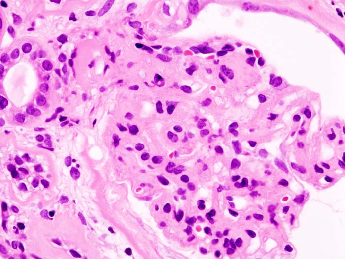
No single cause dominates the way MCD does in children. Diabetic nephropathy is the single most common overall cause of nephrotic syndrome in adults once secondary causes are counted, reflecting the prevalence of diabetes itself rather than any unusual propensity of diabetic kidney disease to become nephrotic. Among the primary (immune-mediated) podocytopathies, FSGS and membranous nephropathy are the most frequently biopsied causes — see the Glomerulonephritis chapter for the complete risk-stratification and immunosuppression framework for each. MCD still occurs in adults (a genuine, if less common, cause), and is a particularly important cause to recognize when it is drug-induced (NSAIDs) or paraneoplastic — a new MCD diagnosis in an adult, especially with lymphadenopathy or B symptoms, should prompt screening for an underlying lymphoma, classically Hodgkin lymphoma.[1]
Other secondary causes worth actively screening for
NSAIDs, lithium, interferon, and anti-VEGF agents (e.g., bevacizumab) are recognized drug-induced causes — bisphosphonates (particularly pamidronate) are specifically associated with a collapsing variant of FSGS.[1] Hepatitis B, hepatitis C, and HIV infection are each associated with specific glomerular lesions (membranous nephropathy, membranoproliferative/cryoglobulinemic patterns, and HIV-associated nephropathy respectively) — see the Glomerulonephritis chapter. In an older adult, amyloidosis and other paraproteinemic diseases are an important and easily missed secondary cause; a serum/urine protein electrophoresis with immunofixation and free light chains is a reasonable addition to the standard workup in this population (see Assessment & Diagnosis and Special Populations).
Risk Factors
- Age under 10 — the age range in which minimal change disease dominates the differential.[2]
- Diabetes mellitus, particularly long-standing or poorly controlled — the leading overall cause of nephrotic syndrome in adults.
- Obesity and reduced nephron mass (a solitary kidney, low birth weight) — drivers of secondary/adaptive FSGS.
- Recent or ongoing NSAID use — a recognized cause of minimal change disease, particularly a new or relapsing presentation in an adult.
- Lymphoma, especially Hodgkin lymphoma — a classic paraneoplastic association with minimal change disease in adults; solid-organ malignancy is a recognized secondary cause of membranous nephropathy, particularly in older adults.[1]
- Lithium, interferon, anti-VEGF agents (e.g., bevacizumab), and bisphosphonates (particularly pamidronate) — recognized drug-induced causes.
- Hepatitis B, hepatitis C, or HIV infection.
- A known plasma cell dyscrasia (multiple myeloma, monoclonal gammopathy) or systemic amyloidosis, or an older adult with unexplained nephrotic syndrome and no clear primary cause on initial workup.
- Systemic lupus erythematosus or another known autoimmune disease.
- A family history of childhood nephrotic syndrome, consanguinity, or onset in the first year of life — raises suspicion for a genetic (congenital/infantile) form; see Special Populations.
Red Flags & Refer
The following change the plan from routine outpatient management to same-day or emergency evaluation.
- Fever with abdominal pain in a nephrotic patient (especially a child) — nephrotic syndrome produces a genuine, elevated risk of spontaneous bacterial peritonitis from urinary loss of immunoglobulins and complement factor B; do not assume this is a benign viral illness or attribute abdominal pain to ascites alone.[2]
- Signs or symptoms of venous thromboembolism — new leg swelling or pain disproportionate to known edema, sudden chest pain or dyspnea, or flank pain with worsening renal function (raising suspicion for renal vein thrombosis) — treat as a possible VTE emergency and reassess anticoagulation strategy urgently (see Choose Initial Treatment).[3][4]
- Signs of intravascular volume depletion despite total-body fluid overload — tachycardia, orthostatic hypotension, oliguria, or an unexpected rise in creatinine, particularly during or after diuresis — nephrotic patients can be genuinely intravascularly depleted from low oncotic pressure even while grossly edematous, and aggressive diuresis carries a disproportionate acute kidney injury risk in this setting (see Choose Initial Treatment and Common Errors to Avoid).
- Severe, symptomatic edema — respiratory compromise from a large pleural effusion or tense ascites, or scrotal/labial edema causing skin breakdown — needs urgent, not routine, management.
- Nephritic-pattern features superimposed on a nephrotic presentation — an active urine sediment (dysmorphic red cells, red cell casts), a rapidly rising creatinine, or new hypertension out of proportion to volume status — raises concern for a mixed nephritic/nephrotic picture or rapidly progressive glomerulonephritis; use the Glomerulonephritis chapter's RPGN algorithm and refer urgently.[1]
- Uncontrolled hypertension or a hypertensive emergency in a patient with known or suspected nephrotic syndrome.
- New-onset nephrotic syndrome in an infant under 3 months of age (congenital nephrotic syndrome) — a distinct, high-risk presentation needing specialized pediatric nephrology management; see Special Populations.
Assessment & Diagnosis
Confirming the tetrad
Quantify proteinuria with a first-morning or spot urine protein-to-creatinine ratio (24-hour collection is an acceptable alternative but adds collection burden without materially improving accuracy for most purposes). The KDIGO 2025 pediatric guideline's complete-remission threshold — a urine protein-to-creatinine ratio ≤200 mg/g (≤20 mg/mmol), or a negative/trace dipstick, or under 100 mg/m²/day, on 3 or more consecutive days — is also a practical, current reference point for what "proteinuria has resolved" means when tracking treatment response in either a child or an adult.[2] Pair this with serum albumin, a fasting lipid panel, and serum creatinine/eGFR in every patient at diagnosis and at each significant change in status.
Secondary-cause workup
- HbA1c or fasting glucose, and a compatible history/retinopathy exam, when diabetic nephropathy is suspected.
- Hepatitis B and C serologies, and HIV testing where clinically indicated.[1]
- ANA and complement levels where lupus nephritis is a possibility (see the Glomerulonephritis chapter).
- Serum and urine protein electrophoresis with immunofixation, and serum free light chains, particularly in an older adult without an obvious primary cause — screening for a paraproteinemic disease or amyloidosis.
- Serum anti-PLA2R antibody where membranous nephropathy is suspected (see the Glomerulonephritis chapter for the complete risk-stratification framework).
- A thorough medication history specifically for NSAIDs, lithium, interferon, anti-VEGF agents, and bisphosphonates.
When to biopsy — a genuinely different approach in children versus adults
In children with a typical presentation (age 1-10, no gross hematuria, normal complement, normal renal function, no significant hypertension), current guidance favours an empiric corticosteroid trial before biopsy — response itself is treated as strong evidence for minimal change disease, since MCD is overwhelmingly the most common cause in this group and is highly steroid-responsive.[2] Biopsy is reserved for atypical features (age under 1 or over 12, gross hematuria, hypertension, significant renal impairment, low complement, or extrarenal features suggesting a systemic disease) or for steroid-resistant disease (see Choose Initial Treatment).[2] In adults, the differential is far more heterogeneous and a specific treatable subtype cannot be assumed from the syndrome alone — biopsy is generally pursued upfront in most adults with new nephrotic syndrome, except when the clinical picture strongly and specifically fits a non-biopsied diagnosis (most commonly long-standing diabetes with a compatible course and retinopathy present).[1] This is a genuinely important, easily-missed practice difference between the 2 populations, not simply a matter of a lower threshold in adults.
Treatment Targets
- Achieve remission (per the current KDIGO thresholds above) where corticosteroid-responsive disease is present, using the current shorter, evidence-based initial regimen rather than an older, unnecessarily prolonged one.
- Reduce proteinuria toward the lowest achievable level with RAAS blockade, independent of and in addition to any disease-specific immunosuppression.
- Control edema without over-diuresis — total-body fluid overload can coexist with intravascular depletion in severe hypoalbuminemia, so the acute kidney injury risk of aggressive diuresis is real and disproportionate in this population.
- Correct or at least meaningfully improve the dyslipidemia of nephrotic syndrome, recognizing it often improves as proteinuria responds to disease-specific treatment.
- Identify patients who genuinely need prophylactic anticoagulation for venous thromboembolism risk, rather than waiting for a clot to occur.
- Classify corticosteroid response correctly (steroid-sensitive, late-responder, infrequently/frequently relapsing, steroid-dependent, steroid-resistant) and let that classification, not a generic "more steroids" reflex, drive the next step.
- Recognize and act on the syndrome-specific emergencies — spontaneous bacterial peritonitis, VTE, and severe intravascular depletion — without delay (see Red Flags & Refer).
Choose Initial Treatment
First-line: a loop diuretic (furosemide), titrated to the degree of edema and renal function. Nephrotic-range hypoalbuminemia genuinely reduces diuretic delivery to its site of action in the tubule (loop diuretics circulate protein-bound), so higher-than-usual doses are often genuinely needed for equivalent effect — this is a pharmacokinetic reality, not simply undertreatment. Oral bioavailability of furosemide specifically can also be reduced by bowel-wall edema in severe cases; an IV route or a diuretic with more consistent oral absorption is a reasonable adjustment when oral furosemide response is inadequate despite dose escalation.
Diuretic-resistant edema: add a thiazide-type agent (metolazone) for sequential nephron blockade, given shortly before the loop diuretic dose. Severe hypoalbuminemia (roughly below 20 g/L) with diuretic-refractory edema: IV albumin given immediately before or with furosemide is a reasonable option to try to restore diuretic responsiveness — but be direct with the care team about the evidence quality here: the only dedicated Cochrane systematic review on this practice identified just 1 small trial and could not draw a firm conclusion about benefit.[5] Use it as a time-limited trial with a clear response endpoint (increased urine output, improved diuretic responsiveness) rather than a routine or open-ended intervention, and monitor closely for volume overload from the albumin infusion itself.
Start an ACE inhibitor or ARB in essentially every patient with nephrotic-range proteinuria, independent of whether disease-specific immunosuppression is also used — this is a disease-agnostic renoprotective foundation, identical in principle to the approach in the Glomerulonephritis and Chronic Kidney Disease chapters. Titrate to the highest approved and tolerated dose, since the proteinuria-lowering effect is dose-dependent. Never combine an ACE inhibitor and an ARB. Check creatinine and potassium 2-4 weeks after starting or up-titrating.
Reasonable in nephrotic-range proteinuria with a significantly elevated LDL, though the dyslipidemia frequently improves as proteinuria responds to disease-specific treatment — reassess ongoing need once remission is achieved rather than continuing indefinitely by default.[1]
Nephrotic syndrome meaningfully raises VTE risk through urinary loss of antithrombin and other anticoagulant proteins, historically reported in as many as roughly 1 in 4 adults with nephrotic syndrome.[4] Do not wait for a clot to happen before considering prophylaxis. A commonly used practical threshold — carried forward from the original 2012 KDIGO guidance and still referenced in current reviews — is to specifically consider prophylactic anticoagulation in a patient with serum albumin persistently below roughly 20-25 g/L, particularly with membranous nephropathy (the histologic subtype carrying the highest reported VTE risk among the nephrotic-range diseases) or additional risk factors — immobility, a prior VTE, severe or persistent proteinuria, obesity, or an intercurrent illness such as infection or dehydration.[4][3] Caution with direct oral anticoagulants: apixaban is highly (87%-93%) plasma protein-bound, with a substantial fraction bound specifically to albumin — severe hypoalbuminemia and heavy urinary protein loss can genuinely alter its pharmacokinetics and pharmacodynamics in ways that are not yet fully predictable, and current review data describe this as a real, unresolved clinical uncertainty rather than a settled question.[3] A vitamin K antagonist (warfarin) or low-molecular-weight heparin remains a reasonable, better-established alternative, particularly in more severe hypoalbuminemia, while acknowledging that urinary antithrombin loss itself can complicate warfarin's anticoagulant effect and monitoring.[3][4]
Corticosteroid response classification — drives every next step for minimal change disease and primary FSGS
Standard induction: daily oral prednisone (or prednisolone) 60 mg/m² per day or 2 mg/kg per day (maximum 60 mg/day) for 4 weeks, then alternate-day dosing for a further 4 weeks (8 weeks total) or, alternatively, a 6-week daily/6-week alternate-day (12-week total) course.[2] Current KDIGO 2025 guidance specifically favours this shorter regimen over the longer 16-24 week courses used historically, based on meta-analyses of high-quality multicenter trials showing no outcome benefit from the longer courses — a genuine, current de-escalation of steroid exposure that should be actively adopted rather than defaulting to an older, longer protocol out of habit.[2]
KDIGO 2025 defines complete remission as a urine protein-to-creatinine ratio ≤200 mg/g (≤20 mg/mmol) or a negative/trace dipstick on 3 or more consecutive days; partial remission as a ratio above that but under 2 g/g with serum albumin ≥30 g/L.[2] A late responder achieves complete remission specifically during the "confirmation period" between weeks 4 and 6 of therapy — a new category in the current guideline, reflecting that a meaningful minority of ultimately steroid-sensitive patients simply need slightly longer than 4 weeks.[2] Steroid-resistant nephrotic syndrome (SRNS) is the lack of complete remission within 4 weeks of standard-dose daily prednisone.[2]
A relapse is recurrence of nephrotic-range proteinuria in a patient who had previously achieved complete remission. Infrequently relapsing: fewer than 2 relapses in the 6 months following the initial remission, or fewer than 3 relapses in any subsequent 12-month period — treat each relapse with a repeat, shorter course similar in principle to the initial induction. Frequently relapsing (FRNS): 2 or more relapses in the first 6 months after initial remission, or 3 or more relapses in any subsequent 12-month period.[2] Steroid-dependent (SDNS): 2 consecutive relapses occurring during prednisone therapy itself, or within 14 days of stopping it.[2] Frequently relapsing or steroid-dependent disease should escalate to a steroid-sparing agent rather than repeating steroid courses indefinitely — a calcineurin inhibitor, cyclophosphamide, mycophenolate mofetil, or rituximab, chosen per the Glomerulonephritis chapter's Drug Tables (dosing, monitoring, and agent-specific cautions are not repeated here to avoid the 2 chapters drifting out of sync).[2]
Do not simply extend or repeat the steroid course in confirmed SRNS. Pursue (or complete, if not already done) biopsy and consider genetic testing — a meaningful proportion of pediatric SRNS reflects a podocyte-gene mutation that will not respond to any immunosuppression regardless of agent, and confirming this changes the entire management plan toward RAAS blockade and supportive care rather than repeated, harmful immunosuppression attempts. For immune-mediated (non-genetic) SRNS, a calcineurin inhibitor (tacrolimus preferred over cyclosporine in most current protocols) is the standard next step — see the Glomerulonephritis chapter's Drug Tables for dosing and monitoring.[2][1]
The same conceptual response classification applies — high-dose corticosteroids first-line, with escalation for relapsing, dependent, or resistant disease — though the specific relapse-frequency thresholds above are defined for children; adult practice uses the same general principles from the KDIGO 2021 guideline rather than these exact pediatric numeric cutoffs.[1] See the Glomerulonephritis chapter's Choose Initial Treatment and Drug Tables for the complete adult dosing framework and for membranous nephropathy, which has its own separate, risk-stratified treatment pathway not repeated here.
Non-Pharmacologic Treatment
- Sodium restriction (roughly under 2 g/day) — one of the most effective non-drug interventions for edema and supports diuretic efficacy directly.
- Individualized fluid restriction for significant edema, guided by the degree of volume overload rather than applied reflexively to every patient.
- Protein intake at a normal (not high) level — a high-protein diet does not correct hypoalbuminemia and may accelerate kidney disease progression.
- Daily weight monitoring is the single most practical tool for both the patient and the care team to detect a genuine change in fluid status early, whether worsening or improving.
- Home first-morning urine dipstick monitoring is commonly taught to parents of children with steroid-sensitive disease specifically to catch a relapse early, before edema becomes clinically obvious — a simple, low-cost, genuinely useful self-monitoring tool that is easy to overlook when counselling.
- Activity: avoid prolonged immobility given the elevated VTE risk, but avoid strenuous exertion during a period of significant edema or suspected intravascular depletion.
- Vaccination: pneumococcal vaccination is specifically recommended given the functional loss of opsonizing antibodies through urinary protein loss, which raises infection risk from encapsulated organisms independent of any immunosuppression used.[2] Complete standard immunizations before starting significant immunosuppression wherever the clinical timeline allows; live vaccines are generally deferred once high-dose corticosteroids or another immunosuppressive agent is underway.
- Avoid nephrotoxins, particularly regular NSAID use — doubly relevant here since NSAIDs are also a recognized direct cause of minimal change disease.
Patient education
Teach patients and (for a child) caregivers to recognize the specific emergencies in this chapter as reasons to seek care immediately rather than waiting: fever with abdominal pain (possible peritonitis), new leg swelling/pain or sudden chest pain/shortness of breath (possible VTE), and signs of dehydration or reduced urine output during diuresis (possible intravascular depletion).[2][4] Reinforce that RAAS inhibitor benefit is measured in proteinuria trend and long-term kidney survival, not day-to-day symptoms, and should not be stopped without discussion even when the patient feels well. Discuss fertility preservation before cyclophosphamide is started in anyone of reproductive age, and confirm effective contraception before mycophenolate mofetil in anyone who could become pregnant — see the Glomerulonephritis chapter.
Special Populations
Pediatrics
Minimal change disease dominates the differential and is highly steroid-responsive; the response classification above should drive every treatment decision. Congenital nephrotic syndrome (onset before 3 months of age, most often from NPHS1 or NPHS2 gene mutations) is a distinct, high-risk presentation that does not follow the framework above — it does not respond to corticosteroids, carries a high risk of severe complications (infection, thrombosis, malnutrition) from massive protein loss, and requires specialized management, sometimes including nephrectomy, dialysis, and eventual transplantation; refer immediately to pediatric nephrology rather than trialing a standard steroid course.[2]
Pregnancy
Distinguishing nephrotic syndrome from preeclampsia is a genuinely important differential in pregnancy, since both can present with new proteinuria, edema, and hypertension — new or worsening hypertension, a rising uric acid, and onset after 20 weeks favour preeclampsia, while a known pre-existing glomerular disease, a normal blood pressure, and onset earlier in pregnancy favour a primary nephrotic process, though the 2 can also coexist. ACE inhibitors, ARBs, and statins are all contraindicated in pregnancy; RAAS blockade should be stopped before conception where feasible or as soon as pregnancy is confirmed. Where prophylactic anticoagulation is indicated in pregnancy, low-molecular-weight heparin is preferred over warfarin (teratogenic) or a DOAC (limited safety data, and the pharmacokinetic uncertainty in hypoalbuminemia above applies here too).[3]
Older adults
Actively screen for a secondary cause — malignancy-associated membranous nephropathy, diabetic nephropathy, and amyloidosis/paraproteinemic disease — before assuming a primary immune-mediated process; see Assessment & Diagnosis. Older adults also carry a higher baseline infection risk on significant immunosuppression.
Renal impairment
Reduce diuretic and anticoagulant dosing as appropriate for reduced eGFR; several agents in this chapter require dose or interval adjustment as kidney function changes through disease progression — see the Chronic Kidney Disease chapter's general renal-dosing principles, which apply throughout this chapter as well.
Monitoring & Follow-Up
Monitor daily weight, edema exam, and urine protein (dipstick at home for a steroid-sensitive child, urine protein-to-creatinine ratio at clinic visits for others) at a frequency individualized to disease activity and treatment phase.
During active diuresis
Serum electrolytes and renal function, particularly early in a diuretic course or after a dose increase — watch specifically for signs of intravascular depletion (rising creatinine, orthostatic symptoms) rather than assuming a rising creatinine simply reflects the underlying disease.
RAAS blockade
Serum creatinine and potassium 2-4 weeks after starting or up-titrating an ACE inhibitor or ARB, and periodically thereafter.
Corticosteroid and steroid-sparing therapy
Blood glucose, blood pressure, growth (in children), and bone health with prolonged corticosteroid use; taper per the specific regimen rather than stopping abruptly. See the Glomerulonephritis chapter's Monitoring & Follow-Up for the complete steroid-sparing-agent-specific monitoring (calcineurin inhibitor trough levels, cyclophosphamide cumulative dose, mycophenolate/azathioprine CBC and liver enzymes, rituximab hepatitis B screening).
Dyslipidemia and VTE surveillance
Recheck the lipid panel once remission is achieved to reassess ongoing statin need. Reassess VTE risk (serum albumin trend, degree and persistence of proteinuria) at each visit while nephrotic-range proteinuria persists, and maintain a low threshold to investigate new leg swelling, chest symptoms, or flank pain as a possible thrombotic event.[4]
Common Errors to Avoid
- Diagnosing or treating based on proteinuria alone without confirming the full tetrad (hypoalbuminemia, edema, hyperlipidemia) — isolated nephrotic-range proteinuria without the rest of the syndrome is a different clinical situation.
- Over-diuresing — total-body fluid overload can coexist with intravascular depletion in severe hypoalbuminemia, so aggressive diuresis carries a disproportionate acute kidney injury risk.
- Assuming every child's relapse, or even every child's initial presentation, needs biopsy before treatment — for a typical pediatric presentation, an empiric steroid trial is the current standard, with response itself treated as strong diagnostic evidence.[2]
- Continuing an older, longer (16-24 week) initial corticosteroid regimen out of habit rather than the current, evidence-supported shorter 8-12 week course.[2]
- Missing VTE risk assessment until after a clot has already occurred, particularly in membranous nephropathy or severe hypoalbuminemia.[4]
- Starting a DOAC for VTE prophylaxis in severe hypoalbuminemia without acknowledging the genuine pharmacokinetic uncertainty this creates.[3]
- Missing spontaneous bacterial peritonitis by attributing fever and abdominal pain in a nephrotic patient to ascites or a benign viral illness.[2]
- Forgetting pneumococcal vaccination, given the functional antibody loss from urinary protein wasting.[2]
- Using NSAIDs for symptomatic relief in a nephrotic patient — NSAIDs are themselves a recognized cause of minimal change disease and worsen both proteinuria and renal perfusion.
- Missing a secondary cause (malignancy, amyloidosis, diabetes) in an older adult by assuming primary immune-mediated disease by default.
- Confusing new nephrotic-range proteinuria and edema in pregnancy with preeclampsia (or vice versa) without considering both possibilities.
- Overstating the evidence for albumin-plus-diuretic combination therapy — it is a reasonable time-limited trial in severe, refractory hypoalbuminemic edema, not a well-established, strongly evidenced intervention.[5]
- Re-deriving or restating the Glomerulonephritis chapter's etiology-specific immunosuppression framework here instead of cross-referencing it — kept out deliberately so the 2 chapters don't drift out of sync with each other over time.
Clinical Decision Algorithm
Drug Tables
How to use this table: Doses require patient-specific confirmation, including renal/hepatic adjustment where relevant. Steroid-sparing immunosuppressive agents (calcineurin inhibitors, cyclophosphamide, mycophenolate mofetil, rituximab) are covered in full, with complete dosing and monitoring detail, in the Glomerulonephritis chapter's Drug Tables and are not repeated here — this chapter's tables cover the syndrome-specific drugs (diuretics, albumin, RAAS blockade, statins, anticoagulation) plus first-line corticosteroid dosing. Select any drug name to open its complete Pharmasist Drug Database page.
Diuretics and Albumin Support for Nephrotic Edema
Nephrotic-range hypoalbuminemia reduces diuretic delivery to its site of action — higher doses are often genuinely needed for equivalent effect.
| Drug | Dosage | Adverse effects | Drug interactions | Comments |
|---|---|---|---|---|
| Furosemide | Adult dosing: Titrated to the degree of edema and renal function | Electrolyte disturbance, volume depletion if over-diuresed, ototoxicity with rapid high-dose IV administration | Additive nephrotoxicity with other nephrotoxins; reduced effect with concurrent NSAID use | Higher-than-usual doses are often needed; consider an IV route if oral response is inadequate, since oral bioavailability can be reduced by bowel-wall edema. |
| Metolazone | Adult dosing: Low dose, given shortly before the furosemide dose | Electrolyte disturbance, marked volume depletion when combined with a loop diuretic | Potent synergy with loop diuretics — start at a low dose and monitor closely | For diuretic-resistant edema; give shortly before the loop diuretic dose for sequential nephron blockade. |
| Albumin (Human), IV | Typical use: Individualized dose, given immediately before or with the diuretic dose | Volume overload/pulmonary edema from the infusion itself; rare allergic reaction | None significant | Reserve for severe hypoalbuminemia (roughly below 20 g/L) with diuretic-refractory edema, given with furosemide, as a time-limited trial with a defined response endpoint — the supporting trial evidence is genuinely weak. |
Renin-Angiotensin System Blockade
Foundational proteinuria reduction — start regardless of which disease-specific immunosuppression, if any, is also used.
| Drug | Dosage | Adverse effects | Drug interactions | Comments |
|---|---|---|---|---|
| Ramipril (ACE inhibitor) | Adult dosing: Start low, titrate to the highest approved/tolerated dose | Hyperkalemia; an expected initial eGFR dip; cough; angioedema | Additive hyperkalemia risk with potassium-sparing diuretics and potassium supplements | Never combine with an ARB. Check creatinine/potassium 2-4 weeks after starting or up-titrating. |
| Losartan (ARB) | Adult dosing: Start low, titrate to the highest approved/tolerated dose | Hyperkalemia; an expected initial eGFR dip | Additive hyperkalemia risk with potassium-sparing diuretics and potassium supplements | Reasonable alternative where an ACE inhibitor is not tolerated (e.g., cough). |
Statin Therapy
For the dyslipidemia of nephrotic syndrome; reassess ongoing need once remission is achieved.
| Drug | Dosage | Adverse effects | Drug interactions | Comments |
|---|---|---|---|---|
| Rosuvastatin | Adult dosing: Standard statin dosing | Myalgia; transaminitis | Cyclosporine specifically increases rosuvastatin exposure — use the lowest effective dose if combined | Often improves substantially as proteinuria responds to disease-specific treatment. |
VTE Prophylaxis and Treatment
Consider specifically in severe hypoalbuminemia (roughly below 20-25 g/L), membranous nephropathy, or additional VTE risk factors — do not wait for a clot to occur.
| Drug | Dosage | Adverse effects | Drug interactions | Comments |
|---|---|---|---|---|
| Apixaban | Prophylactic dosing: Individualized per current VTE-prevention protocol for nephrotic syndrome | Bleeding risk | None significant, but genuine pharmacokinetic uncertainty in severe hypoalbuminemia | High (87%-93%) plasma protein binding means severe hypoalbuminemia can alter drug exposure unpredictably — a real, current clinical uncertainty rather than a settled question. |
| Warfarin | Dosing: Individualized, titrated to INR | Bleeding risk; unpredictable dose-response | Numerous — review the full interaction list; INR monitoring required | Better-established than a DOAC in this population; urinary antithrombin loss itself can complicate anticoagulant effect and monitoring. |
| Enoxaparin (LMWH) | Dosing: Weight-based, per standard prophylactic or treatment protocol | Bleeding risk; injection-site bruising | None significant | Preferred anticoagulant class in pregnancy when prophylaxis is indicated; also reasonable short-term or peri-procedurally. |
First-Line Corticosteroid Therapy
For minimal change disease and primary FSGS induction and relapse treatment. See the Glomerulonephritis chapter's Drug Tables for steroid-sparing agents used in relapsing, dependent, or resistant disease.
| Drug | Dosage | Adverse effects | Drug interactions | Comments |
|---|---|---|---|---|
| Prednisone — initial episode | Adult/pediatric induction: 60 mg/m²/day or 2 mg/kg/day (maximum 60 mg/day) daily for 4-6 weeks, then the same dose on alternate days for a further 4-6 weeks | Hyperglycemia, mood/sleep disturbance, weight gain, growth suppression in children, hypertension, infection risk, osteoporosis with prolonged use | Numerous — review the full medication list before initiation, particularly other agents affecting glucose or bone health | Current guidance favours an 8-12 week total course (4-6 weeks daily, then an equal alternate-day period) over older, longer regimens, which showed no added benefit. |
| Prednisone — relapse | Typical relapse course: Per current protocol; taper once complete remission is re-achieved | Same as above | Same as above | A repeat course, generally shorter and/or at a lower cumulative dose than the initial induction, once a relapse is confirmed by recurrence of nephrotic-range proteinuria after prior complete remission. |
References
- KDIGO 2021 Clinical Practice Guideline for the Management of Glomerular Diseases — Kidney Disease: Improving Global Outcomes (KDIGO) Glomerular Diseases Work Group. Kidney Int 2021;100(4S):S1-S276
- KDIGO 2025 Clinical Practice Guideline for the Management of Nephrotic Syndrome in Children — Kidney International (Floege J, Gibson KL, Vivarelli M, et al.; KDIGO Nephrotic Syndrome in Children Work Group). Kidney Int 2025;107(5 Suppl):S241-S289
- Venous Thromboembolism Prevention in Nephrotic Syndrome: The Role of Aspirin, Vitamin K Antagonists, and Direct Oral Anticoagulants — Kidney International Reports (Gholizadeh Ghozloujeh Z, Glassock RJ, Al Jurdi A, Jhaveri KD, Heidari-Bateni G, Bajpai D, Lerma E, Wu J, Abdipour A, Norouzi S). Kidney Int Rep 2025;10(5):1335-1345
- A Systematic Review of Prophylactic Anticoagulation in Nephrotic Syndrome — Kidney International Reports (Lin R, McDonald G, Jolly T, Batten A, Chacko B). Kidney Int Rep 2020;5(4):435-447
- Human Albumin Infusion for Treating Oedema in People With Nephrotic Syndrome — Cochrane Database of Systematic Reviews (Ho JJ, Adnan AS, Kueh YC, Ambak NJ, Van Rostenberghe H, Jummaat F; Cochrane Kidney and Transplant Group). Cochrane Database Syst Rev 2019;7(7):CD009692
Professional reference only. Apply clinical judgment and verify patient-specific decisions against current authoritative guidance and product information.