Pancreatitis
Clinical Snapshot
Pancreatitis is inflammation of the pancreas, and the single most important thing to understand before anything else in this chapter is that acute and chronic pancreatitis are not two severities of the same illness — they are two genuinely different disease processes that happen to share an organ. Acute pancreatitis is a sudden, usually self-limited inflammatory insult in a previously normal (or previously damaged) gland, diagnosed using the internationally adopted revised Atlanta classification's "2 of 3" rule and staged by a severity grade that drives nearly every decision that follows.[1] Chronic pancreatitis is irreversible fibro-inflammatory destruction of pancreatic tissue, developing over years, that eventually fails at both of the gland's core jobs: the exocrine function of secreting digestive enzymes (lipase, amylase, protease) into the duodenum, and the endocrine function of insulin and glucagon secretion from the islets of Langerhans.
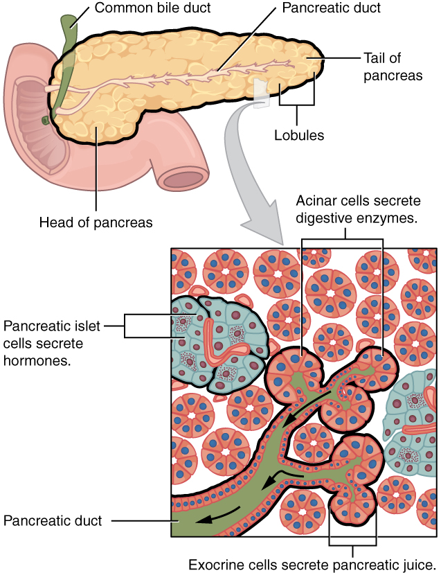
Why acute pancreatitis matters
Acute pancreatitis is one of the most common reasons for gastrointestinal hospitalization worldwide. The great majority of cases are mild and self-limited, but a substantial minority — roughly 20-30% — develop moderately severe or severe disease with local complications or organ dysfunction, and severe disease carries real mortality, concentrated in patients who develop infected pancreatic necrosis.[2] Gallstones and alcohol together account for the large majority of cases in most populations; hypertriglyceridemia, ERCP, drugs, and a genuinely long list of rarer causes make up the rest, and roughly 10-15% remain idiopathic even after a reasonable workup (see Risk Factors).[3]
Why chronic pancreatitis matters
Chronic pancreatitis's damage is cumulative and permanent — nothing in this chapter reverses established fibrosis or restores destroyed acinar or islet tissue. The clinical goal is instead to control pain, replace what the gland can no longer make (enzymes, and eventually insulin), prevent malnutrition and its downstream complications (fat-soluble vitamin deficiency, metabolic bone disease), and remove the ongoing insult — alcohol and tobacco chief among them — that keeps driving further damage.[4] This chapter is authored primarily from the International Association of Pancreatology's 2025 revised guidelines on acute pancreatitis — the newest and most comprehensive international synthesis available, spanning 18 domains and 96 clinical questions — cross-checked against the foundational revised Atlanta classification, the WSES 2019 severe-pancreatitis guidelines, the HaPanEU chronic pancreatitis guideline, and a genuinely Canadian consensus guideline on pancreatic exocrine insufficiency in primary care.[3][5]
Recognize the Condition
Acute pancreatitis
The classic presentation is severe, constant epigastric pain, often radiating straight through to the back, that characteristically improves when the patient leans forward or curls into a fetal position and worsens lying flat — though this "pancreatic posture" is a supportive clue, not a requirement. Nausea and vomiting are common, and low-grade fever, tachycardia, and epigastric or diffuse abdominal tenderness are typical on exam. Bowel sounds may be reduced from an associated ileus.
Two rare but classic physical findings signal a hemorrhagic, necrotizing course rather than routine interstitial pancreatitis: Cullen's sign (periumbilical ecchymosis, from retroperitoneal blood tracking along the falciform ligament) and Grey Turner's sign (flank ecchymosis, from retroperitoneal hemorrhage tracking laterally). Either finding should prompt reassessment for severe, necrotizing, or hemorrhagic disease rather than being treated as an incidental bruise.
Chronic pancreatitis
Recurrent or persistent epigastric pain, often radiating to the back and sometimes provoked by eating, is the most common presenting symptom, though a meaningful minority of patients — particularly later in the disease course — have little or no pain despite advanced structural damage ("burnout"), which does not mean the exocrine and endocrine failure have also stabilized. Steatorrhea (bulky, greasy, malodorous, difficult-to-flush stools) and unintentional weight loss are classic but genuinely late findings — clinically overt steatorrhea does not appear until pancreatic lipase output has fallen below roughly 10% of normal, meaning substantial, often clinically silent exocrine damage has already occurred by the time steatorrhea is the presenting complaint (see Assessment & Diagnosis for earlier-detection testing).[5] New-onset or difficult-to-control diabetes in a patient with a pancreatitis history — pancreatogenic (type 3c) diabetes — is itself a recognition clue, not only a late complication (see Special Populations).
The relationship between the two
Recurrent acute pancreatitis is itself one of the recognized pathways into chronic pancreatitis (see the TIGAR-O classification in Risk Factors) — a patient's second or third "acute" episode, particularly with a persistent etiology such as ongoing heavy alcohol use, deserves a structural and functional workup for evolving chronic disease rather than being treated as an isolated, unrelated event each time.
Risk Factors
Acute pancreatitis
- Gallstones — the single most common cause in most populations, including small stones and microlithiasis/biliary sludge too small to reliably see on standard ultrasound; obstruction at the ampulla is the presumed mechanism.
- Alcohol — typically heavy, sustained use rather than a single binge in a previously abstinent person; the second leading cause in most Western series.
- Hypertriglyceridemia — triglycerides above roughly 1000 mg/dL (11.3 mmol/L) carry a meaningfully increased risk, rising further above 2000 mg/dL (22.6 mmol/L); an under-recognized and often reversible cause (see Choose Initial Treatment and Special Populations).[6]
- Post-ERCP pancreatitis — the most common serious complication of ERCP itself; risk factors include difficult cannulation, pancreatic duct injection/instrumentation, and suspected sphincter of Oddi dysfunction (see Choose Initial Treatment for prophylaxis).[7]
- Drug-induced — a genuinely long list, including azathioprine/mercaptopurine, valproic acid, thiazide diuretics, mesalamine/sulfasalazine, didanosine, pentamidine, estrogens, and sulfonamide antibiotics; GLP-1 receptor agonists (semaglutide, liraglutide, dulaglutide, tirzepatide) carry a reported but not definitively causal association and should remain on the differential in a patient taking one who develops otherwise-unexplained pancreatitis.
- Trauma (including post-surgical), scorpion sting (rare outside endemic regions), and viral infection (mumps, coxsackievirus).
- Autoimmune pancreatitis — an IgG4-related systemic disease (type 1) or an idiopathic duct-centric process (type 2); consider specifically when imaging shows a diffusely enlarged, "sausage-shaped" gland or when pancreatitis coexists with other IgG4-related organ involvement.
- Genetic — hereditary pancreatitis (PRSS1 gain-of-function mutations), SPINK1, and CFTR mutations (see the Cystic Fibrosis chapter for CFTR-related exocrine disease) predispose to recurrent acute and eventual chronic pancreatitis, particularly with recurrent episodes at a young age or a strong family history.
- Idiopathic — roughly 10-15% even after a reasonable initial workup; pursue endoscopic ultrasound for occult microlithiasis or a structural anomaly (see Treatment Failure/Next Step) before permanently labelling a case idiopathic.
Chronic pancreatitis — the TIGAR-O framework
The TIGAR-O classification organizes chronic pancreatitis etiology into six categories and is a genuinely useful structure for a systematic workup rather than anchoring on "probably alcohol" alone.[4]
| Category | Examples |
|---|---|
| Toxic-metabolic | Alcohol (the leading cause worldwide, dose- and duration-dependent), tobacco smoking (an independent risk factor that also accelerates progression and acts synergistically with alcohol), hypercalcemia, hyperlipidemia, chronic kidney disease |
| Idiopathic | Early- and late-onset idiopathic chronic pancreatitis, tropical pancreatitis |
| Genetic | PRSS1 (hereditary pancreatitis), CFTR, SPINK1, chymotrypsin C |
| Autoimmune | Type 1 (IgG4-related) and type 2 (idiopathic duct-centric) autoimmune pancreatitis |
| Recurrent and severe acute pancreatitis | Post-necrotic (severe acute pancreatitis leaving structural damage), recurrent acute pancreatitis, vascular disease/ischemia, post-radiation |
| Obstructive | Pancreas divisum, sphincter of Oddi dysfunction, duct obstruction from a tumour or a post-traumatic/post-surgical stricture |
Alcohol and tobacco are by far the most common drivers in most populations, and — critically for counselling — their effects are synergistic, not simply additive: a patient who both smokes and drinks heavily faces a materially higher risk than either exposure alone.[4]
Red Flags & Refer
Escalate beyond routine pancreatitis management for any of the following:
- Persistent organ failure beyond 48 hours — cardiovascular (shock), respiratory (PaO2/FiO2 under 300), or renal (creatinine over roughly 1.9 mg/dL [170 μmol/L] after fluid resuscitation) — this defines severe acute pancreatitis under the revised Atlanta classification and needs critical-care-level support.[1]
- Cullen's or Grey Turner's sign — reassess urgently for hemorrhagic or necrotizing pancreatitis rather than treating either as an incidental bruise.
- Clinical deterioration or new fever after initial improvement, classically in the second week or later — the hallmark pattern of infected pancreatic necrosis, which changes the plan from supportive care to source control (see Choose Initial Treatment and Treatment Failure/Next Step).
- Concurrent cholangitis (Charcot's triad: right-upper-quadrant/epigastric pain, fever, jaundice) in gallstone pancreatitis — needs urgent ERCP, not routine pancreatitis management alone; see the Cholecystitis chapter for the full cholangitis differential and Choose Initial Treatment here for ERCP timing.
- Signs of abdominal compartment syndrome — a tensely distended abdomen, rising peak airway pressures, and oliguria despite adequate resuscitation — over-aggressive fluid resuscitation is itself a contributing cause (see Choose Initial Treatment).[8]
- Unexplained GI bleeding or a new abdominal bruit late in the course — evaluate for a splenic or gastroduodenal artery pseudoaneurysm eroding into a necrotic collection, a rare but life-threatening late complication.
- New jaundice, a sudden change in a stable chronic pancreatitis pain pattern, or new unexplained weight loss in chronic pancreatitis — chronic pancreatitis is itself a recognized risk factor for pancreatic adenocarcinoma; investigate for malignancy rather than assuming a routine disease flare.
- New variceal bleeding or unexplained splenomegaly in chronic pancreatitis — raises splenic vein thrombosis with left-sided ("sinistral") portal hypertension, a recognized chronic pancreatitis complication distinct from cirrhotic portal hypertension.
Assessment & Diagnosis
Diagnosing acute pancreatitis — the revised Atlanta "2 of 3" rule
The internationally adopted diagnostic standard requires 2 of the following 3 criteria: (1) abdominal pain consistent with acute pancreatitis, (2) serum lipase or amylase activity greater than 3 times the upper limit of normal, and (3) characteristic findings on cross-sectional imaging.[1][9] Lipase is preferred over amylase where both are available — it rises and stays elevated longer, and it is more specific, since amylase is also produced by the salivary glands and elevated in a wider range of non-pancreatic conditions. A patient with classic pain and a lipase over 3x the upper limit of normal does not routinely need a CT scan to confirm the diagnosis — 2 of 3 criteria are already met, and CT is reserved for diagnostic uncertainty, an atypical presentation, or assessing complications in a patient who is not improving as expected (see Imaging below).

Severity classification — the revised Atlanta 3-tier grade
| Grade | Definition |
|---|---|
| Mild | No organ failure, and no local or systemic complications. Typically resolves within a week with supportive care alone. |
| Moderately severe | Transient organ failure (resolving within 48 hours), local complications, and/or exacerbation of a coexisting comorbidity. |
| Severe | Persistent organ failure — cardiovascular, respiratory, or renal — lasting 48 hours or longer, in one or more organ systems. |
[1] Local complications, also defined by the revised Atlanta classification, are distinguished by content (fluid alone versus necrotic debris) and by timing (before versus after 4 weeks, by which point a collection typically develops a defined wall):
| Collection | Timing | Contents |
|---|---|---|
| Acute peripancreatic fluid collection (APFC) | Under 4 weeks | Fluid only, no defined wall |
| Pancreatic pseudocyst | 4 weeks or more | Fluid only, now with a defined, encapsulated wall |
| Acute necrotic collection (ANC) | Under 4 weeks | Fluid plus necrotic debris, no defined wall |
| Walled-off necrosis (WON) | 4 weeks or more | Fluid plus necrotic debris, now with a defined, encapsulated wall |
[1] This vocabulary matters clinically, not just academically — timing (before or after the collection is "walled off") is exactly what governs whether and when to intervene on infected necrosis (see Choose Initial Treatment).
Severity prediction — BISAP
The Bedside Index for Severity in Acute Pancreatitis (BISAP) is a simple, 5-variable score calculable from data available in the first 24 hours, without needing 48 hours of data collection the way older scores (Ranson's criteria, APACHE II) require — a genuine practical advantage that has made it the platform's preferred bedside tool, while Ranson's and APACHE II remain in wider legacy use and are referenced in some regional protocols.
| BISAP criterion (1 point each) | Threshold |
|---|---|
| Blood urea nitrogen | Over 25 mg/dL (8.9 mmol/L) |
| Impaired mental status | Any new disorientation, lethargy, or reduced Glasgow Coma Scale |
| SIRS | 2 or more systemic inflammatory response syndrome criteria present |
| Age | Over 60 years |
| Pleural effusion | Present on imaging |
A score of 3 or higher is associated with a substantially increased risk of in-hospital mortality (sensitivity approximately 56%, specificity approximately 91% in the original validation) and should prompt closer monitoring, a lower threshold for escalating the level of care, and reassessment of the fluid and nutrition plan below.[10] Trending hematocrit (hemoconcentration suggests inadequate resuscitation; a falling hematocrit with clinical worsening suggests hemorrhage) and C-reactive protein at 48 hours (a value over 150 mg/L is a well-validated marker of severe disease) are useful complementary bedside markers.
Imaging
| Modality | Role |
|---|---|
| Transabdominal ultrasound | Order in essentially every patient with a first episode of acute pancreatitis — not to diagnose the pancreatitis itself, but specifically to look for gallstones as the etiology, since finding them changes management (see Choose Initial Treatment on cholecystectomy timing).[3] |
| Contrast-enhanced CT (CECT) | Not required when clinical and laboratory criteria already confirm the diagnosis. Reserve for diagnostic uncertainty, or — ideally no earlier than 72 hours after symptom onset, when necrosis is more reliably demarcated — for a patient failing to improve as expected or clinically deteriorating, to assess for necrosis and local complications. |
| MRCP | Preferred over CT for characterizing the pancreatic and bile ducts without ionizing radiation or IV contrast; useful when choledocholithiasis is suspected but not confirmed, and safe in pregnancy. |
| Endoscopic ultrasound (EUS) | The most sensitive test for occult microlithiasis, small common bile duct stones, and early/minimal-change chronic pancreatitis; a reasonable next step in recurrent "idiopathic" acute pancreatitis before accepting that label (see Treatment Failure/Next Step). |
Diagnosing chronic pancreatitis
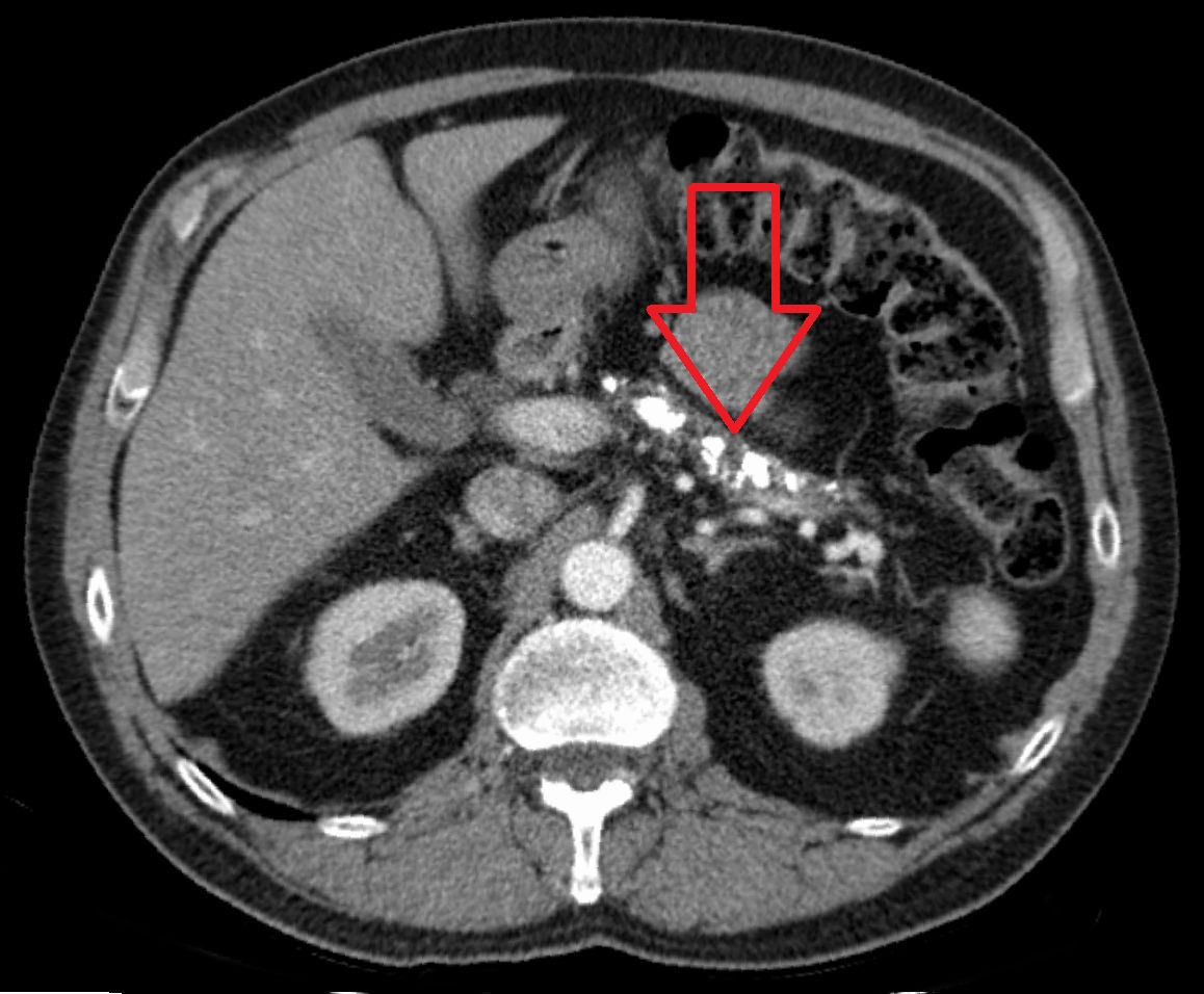
Cross-sectional imaging (CT or MRI/MRCP) findings — parenchymal calcifications (near-pathognomonic in advanced disease), pancreatic duct dilation or an irregular "chain of lakes" appearance, ductal strictures, and parenchymal atrophy — support the diagnosis, with EUS the most sensitive modality for earlier, minimal-change disease before calcifications or overt ductal changes have developed. Diagnostic ERCP has been largely superseded by MRCP and EUS given its own procedural risk, including a real risk of provoking acute pancreatitis.
Assessing exocrine function — do not wait for steatorrhea
Fecal elastase-1 is the standard, non-invasive first-line test for pancreatic exocrine insufficiency (PEI) — a low result (commonly under 200 mcg/g, with under 100 mcg/g suggesting more severe insufficiency) supports the diagnosis, but a single abnormal fecal elastase, on its own, should never be treated as diagnostic of PEI in isolation, and a predisposing condition such as chronic pancreatitis should never be treated as proof of PEI without actually testing — both are genuine, well-documented pitfalls.[5] The secretin-stimulated pancreatic function test is more sensitive and considered a reference standard where available, but it is invasive and not widely accessible. A 72-hour quantitative fecal fat collection is rarely used today given the burden on the patient and the lab. Blood levels of the fat-soluble vitamins (A, D, E, K) are a genuinely useful complementary marker — a deficiency associated with an underlying condition such as chronic pancreatitis is highly suggestive of clinically significant malabsorption even before overt steatorrhea appears.[5]
Treatment Targets
- Confirm the diagnosis efficiently using the revised Atlanta 2-of-3 rule, and grade severity (mild, moderately severe, severe) — grade drives fluid rate, level of care, and how closely to monitor for complications.
- Resuscitate with a moderate, goal-directed fluid strategy — not the older default of maximal early hydration, which current trial evidence shows causes harm without benefit.
- Control pain effectively with scheduled multimodal analgesia, including opioids where needed, without withholding effective analgesia over outdated sphincter-of-Oddi concerns.
- Feed early, by mouth as tolerated, rather than defaulting to prolonged bowel rest or reflexively placing a feeding tube.
- Identify the etiology specifically (biliary ultrasound in every patient, triglycerides, calcium, medication review) since etiology changes management — same-admission cholecystectomy for gallstones, insulin/fibrate for hypertriglyceridemia, drug discontinuation for a drug cause.
- Reserve antibiotics for confirmed or strongly suspected infected necrosis — not as prophylaxis against it.
- For necrotizing pancreatitis needing intervention, use a minimally invasive step-up approach and delay intervention until the collection is walled off wherever the patient's clinical trajectory allows.
- For chronic pancreatitis: control pain via a genuine stepwise ladder; correct exocrine insufficiency with correctly dosed and correctly counselled PERT; identify and treat pancreatogenic diabetes; prevent and correct fat-soluble vitamin deficiency and malnutrition; and remove the ongoing insult (alcohol, tobacco) driving further damage.
Choose Initial Treatment
Use lactated Ringer's solution in preference to normal saline where available — it is associated with less hyperchloremic metabolic acidosis and a signal toward reduced systemic inflammation. The randomized WATERFALL trial directly compared an aggressive strategy (a 20 mL/kg bolus over 2 hours, then 3 mL/kg/hour) with a moderate strategy (a 10 mL/kg bolus only if hypovolemic, then 1.5 mL/kg/hour): the aggressive arm caused significantly more fluid overload, with a trend toward more necrotizing pancreatitis and a longer hospital stay, and no outcome benefit — the trial was stopped early for safety.[8] Moderate, goal-directed resuscitation is now the evidence-based default — target urine output, resolving tachycardia, and a normalizing hematocrit/BUN trend, and reassess frequently rather than running a fixed aggressive rate by protocol.
Use scheduled multimodal analgesia (acetaminophen plus an opioid for moderate-severe pain) rather than PRN-only dosing. All opioids, including morphine, are acceptable — the older teaching that morphine should specifically be avoided in pancreatitis or biliary pain over sphincter of Oddi spasm concerns is outdated and not supported by outcome evidence, the same correction already documented in this platform's Cholecystitis chapter. Hydromorphone is a reasonable default; fentanyl is a reasonable alternative in significant renal impairment or hemodynamic instability.
Start oral feeding as soon as the patient's pain and nausea are improving and they can tolerate it — typically within 24-72 hours — rather than waiting for pain to fully resolve or for lipase to normalize, neither of which is required before refeeding. The randomized PYTHON trial compared very-early nasoenteric tube feeding (within 24 hours) with an oral diet started at 72 hours (with tube feeding available if the oral diet was not tolerated) in patients at high risk for complications: there was no difference in infection rate (25% vs 26%) or mortality, and the on-demand oral-diet group tolerated a full diet sooner — early tube feeding by default is not supported, and an oral diet, advanced as tolerated, is a genuinely appropriate first approach even in higher-risk patients.[11] When enteral access is needed, enteral nutrition (oral or tube) is preferred over parenteral nutrition even in severe or necrotizing pancreatitis — enteral feeding preserves gut mucosal integrity and is associated with fewer infectious complications; reserve parenteral nutrition for genuine enteral intolerance after an adequate trial, not as an early default for severe disease.[3]
Routine prophylactic antibiotics in predicted severe or necrotizing acute pancreatitis are not supported by current evidence and should not be given by default — this is one of the clearest reversals of older practice in this entire chapter.[9][3] Reserve antibiotics for a patient with clinical, laboratory, or imaging evidence of infected necrosis — classically, new fever or clinical deterioration after an initial period of improvement, typically in the second week or later, sometimes with gas within a necrotic collection on CT, ideally confirmed by CT-guided fine-needle aspiration culture where the diagnosis is genuinely uncertain. Choose an agent with good pancreatic tissue penetration — a carbapenem (imipenem-cilastatin or meropenem) is the preferred first choice; piperacillin-tazobactam is a reasonable alternative.[2]
When intervention on necrosis is needed, use a minimally invasive step-up approach — percutaneous catheter or endoscopic transluminal drainage first, escalating to minimally invasive necrosectomy only if drainage alone is insufficient — rather than primary open surgical necrosectomy. The randomized PANTER trial found the step-up approach reduced the composite of major complications or death from 69% to 40% compared with open necrosectomy, driven mainly by a large reduction in new-onset organ failure.[12] Delay intervention until the collection is walled off (typically 4 weeks or more) whenever the patient's clinical trajectory allows — "when in doubt, wait it out" — reserving earlier intervention for genuine, uncontrolled clinical deterioration or overwhelming sepsis rather than intervening on necrosis simply because it has been identified.[3]
ERCP is not routine for gallstone pancreatitis in the absence of concurrent cholangitis or persistent biliary obstruction. The randomized APEC trial tested urgent ERCP with sphincterotomy (within 24 hours) against conservative treatment specifically in predicted severe gallstone pancreatitis without cholangitis, and found no reduction in major complications or mortality (38% vs 44%) — ERCP should be reserved for a patient who actually has cholangitis or ongoing obstruction, not given routinely to every severe or predicted-severe case.[13] Mild gallstone pancreatitis perform cholecystectomy during the same hospital admission, once the patient is clinically improving, rather than discharging with a plan for interval surgery weeks later. The randomized PONCHO trial found same-admission cholecystectomy reduced recurrent gallstone-related complications or death from 17% (interval surgery, at 25-30 days) to 5% — a clear, guideline-changing result.[14] For more severe or necrotizing gallstone pancreatitis where surgery is not yet safe, cholecystectomy is reasonably deferred until inflammation has resolved, with ERCP and sphincterotomy considered as a bridge in a genuinely poor surgical candidate.
For any patient undergoing ERCP without a contraindication to NSAIDs, give rectal indomethacin or diclofenac 100 mg immediately before or after the procedure — a strong, guideline-level recommendation that meaningfully reduces post-ERCP pancreatitis incidence, and one of the most direct pharmacist-relevant prevention measures in this entire chapter.[7] Prophylactic pancreatic duct stenting is added in selected high-risk patients (e.g., inadvertent pancreatic guidewire passage, difficult cannulation).
IV insulin infusion (typically 0.1-0.3 units/kg/hour, with concurrent dextrose to prevent hypoglycemia once glucose normalizes) is standard first-line therapy — insulin activates lipoprotein lipase and accelerates triglyceride clearance. Apheresis (plasmapheresis) lowers triglycerides faster but has not been shown to improve mortality or morbidity over insulin alone, and current apheresis society guidance places it in a category where the optimal role is not firmly established — reserve it for severe disease with organ dysfunction rather than using it routinely. Once the acute episode resolves, start a fibrate and dietary fat restriction for secondary prevention — the Endocrine Society's guideline identifies fibrate therapy as first-line for severe/very severe hypertriglyceridemia, with a treatment goal of keeping triglycerides under 1000 mg/dL (11.3 mmol/L) to reduce recurrence risk.[6]
Start PERT for every patient with confirmed pancreatic exocrine insufficiency. Give it with the first bite of every meal and every fat-containing snack — not on a fixed clock independent of eating, and not only with "main meals." Start at at least 40,000 USP units of lipase per meal and at least 20,000 units per snack, and titrate upward to control steatorrhea, support nutritional markers, and relieve GI symptoms.[15] For a large meal, splitting the dose — some at the start and the remainder partway through — improves mixing with chyme.
Do not crush or chew enteric-coated microsphere/microtablet formulations — the coating protects the enzymes from gastric acid inactivation and is designed to dissolve in the alkaline duodenal environment; crushing destroys this protection and can also irritate the oral mucosa. A patient unable to swallow capsules whole may have them opened and the intact enteric-coated contents sprinkled onto a small amount of acidic soft food (pH under 4.5, such as applesauce) and swallowed immediately without chewing, followed by a drink of water or juice.
Persistent symptoms despite an adequate PERT dose and adherence a trial of concomitant acid suppression (a PPI, or an H2-receptor antagonist) is a reasonable next step — excess gastric acid can prematurely inactivate unprotected lipase and impair the enteric coating's intended duodenal-release timing, and correcting this can meaningfully improve response without simply escalating the enzyme dose further.[5]
Step 1 — alcohol and tobacco cessation (the single highest-yield intervention for both pain and disease progression), scheduled acetaminophen and/or an NSAID, and PERT itself (evidence for a direct analgesic effect via reduced cholecystokinin-driven pancreatic stimulation is mixed, but it is already indicated for exocrine insufficiency regardless). Step 2 — add an adjuvant neuromodulator for a centralized/neuropathic pain component: gabapentin or pregabalin, or duloxetine or amitriptyline. Step 3 — a weak opioid such as tramadol, preferred over jumping straight to a full-agonist opioid. Step 4 — for pain driven by a dilated or obstructed pancreatic duct, endoscopic therapy (stricture dilation/stenting, or extracorporeal shock-wave lithotripsy for large obstructing stones) or surgical drainage/resection; consider surgery genuinely early — after a maximum of about 5 endoscopic interventions rather than an indefinite endoscopic trial — since earlier surgical decompression may better preserve residual function than prolonged repeated endoscopy.[4] Step 5 — celiac plexus block/neurolysis for refractory pain in a patient who is not a surgical candidate, with the caveat that benefit is often modest and temporary. Avoid escalating to long-term full-agonist opioids as a default management strategy — evidence for durable benefit is limited and the opioid use disorder risk in this population is real — but do not withhold effective analgesia, including opioids, from a patient with genuinely refractory pain who has exhausted the steps above.[4]
Non-Pharmacologic Treatment
Acute pancreatitis
- Early oral feeding as tolerated, generally within 24-72 hours — not prolonged NPO/bowel rest (see Choose Initial Treatment).[11]
- Enteral (oral or tube) nutrition in preference to parenteral nutrition when tube feeding is genuinely needed.
- Same-admission cholecystectomy for mild gallstone pancreatitis once the patient is recovering.[14]
- ERCP with sphincterotomy, reserved for concurrent cholangitis or persistent biliary obstruction.[13]
- Step-up drainage and, if needed, minimally invasive necrosectomy for infected pancreatic necrosis, delayed until walled off where the clinical course allows.[12]
- Alcohol cessation counselling for alcohol-related pancreatitis, and smoking cessation counselling regardless of etiology, given tobacco's independent contribution to recurrence and progression.
Chronic pancreatitis
- Alcohol and tobacco cessation — the single most disease-modifying non-pharmacologic intervention available, altering the trajectory of both pain and further gland destruction, not just a general health recommendation.[4] Consider pharmacotherapy for alcohol use disorder (e.g., naltrexone) alongside counselling and, where available, addiction medicine referral.
- Do not routinely recommend a low-fat diet as the primary fix for malabsorption — patients with chronic pancreatitis are already at risk of malnutrition, and the correct first response to persistent steatorrhea is adequate PERT dosing (see Choose Initial Treatment), not fat restriction; an important exception is a patient with coexisting obesity, where some fat moderation may be reasonable alongside optimized PERT.[5]
- Small, more frequent meals may be better tolerated than three large meals.
- Registered dietitian referral for individualized nutritional counselling and monitoring.[5]
- Bone density (DEXA) screening given cumulative risk factors — malabsorption, chronic vitamin D/K deficiency, alcohol use, and reduced activity in more advanced disease.
- Psychosocial support — chronic pain and, often, coexisting substance use create a genuinely high treatment burden; involve psychology/psychiatry and social work proactively rather than only after adherence visibly breaks down.
Special Populations
Pregnancy
Gallstone disease is the most common cause of acute pancreatitis in pregnancy, typically presenting in the second or third trimester. General management principles are unchanged — moderate fluid resuscitation, adequate analgesia, early oral feeding as tolerated — with acetaminophen and opioids preferred for analgesia and NSAIDs avoided, particularly in the third trimester. MRCP (no ionizing radiation) is preferred over CT when duct imaging is needed. ERCP with appropriate fetal shielding is safe when genuinely indicated (concurrent cholangitis or persistent obstruction) and should not be withheld on the basis of pregnancy alone; laparoscopic cholecystectomy is likewise considered safe in any trimester where expertise is available, with elective/interval timing preferentially in the second trimester when surgery is not urgent.
Breastfeeding
Short-acting opioids are preferred where analgesia is needed, with monitoring for infant sedation; acetaminophen and pancrelipase (not systemically absorbed) are compatible. Confirm specific antibiotic and PPI compatibility against each drug's own Drug Database entry.
Pediatrics
Causes differ meaningfully from adults — trauma, biliary anomalies, systemic illness, drugs, and idiopathic disease are proportionally more common than gallstones or alcohol. The same revised Atlanta diagnostic and severity framework applies, adapted to paediatric norms. Consider hereditary pancreatitis (PRSS1) specifically in a child with recurrent acute pancreatitis and a strong family history; cystic fibrosis-related exocrine pancreatic insufficiency is covered in detail in this platform's Cystic Fibrosis chapter.
Older adults
Gallstone etiology and biliary complications are proportionally more common, presentation is more often blunted, and mortality from severe disease is higher — maintain a lower threshold for imaging and escalation, and review the full medication list specifically for a drug-induced contributor given the cumulative polypharmacy burden common in this population.
Pancreatogenic (type 3c) diabetes in chronic pancreatitis
This is mechanistically distinct from type 1 or type 2 diabetes — concurrent glucagon deficiency from islet destruction produces genuinely brittle glucose control with a materially higher hypoglycemia risk at any given insulin dose than typical type 2 diabetes. A trial of metformin is reasonable in earlier, milder pancreatogenic diabetes with preserved insulin secretion, but insulin (e.g., insulin glargine basal, insulin aspart prandial) becomes necessary once beta-cell reserve is inadequate, and should not be delayed simply because the patient does not fit a typical type 2 diabetes profile. As with cystic fibrosis-related diabetes, do not impose calorie or fat restriction to manage pancreatogenic diabetes — the malnutrition risk from underlying exocrine insufficiency takes priority, and insulin dosing should support adequate intake rather than restrict it.
Alcohol use disorder
Alcohol cessation is the single highest-yield intervention for both acute recurrence and chronic disease progression. Consider pharmacotherapy (e.g., naltrexone) alongside counselling and addiction medicine referral, and provide genuine psychosocial support for the behaviour change being asked of the patient rather than treating a single cessation discussion as sufficient.
Renal impairment
Dose-adjust meropenem, imipenem-cilastatin, piperacillin-tazobactam, gabapentin, and pregabalin for reduced renal function; use ketorolac (see the Cholecystitis chapter's drug table) cautiously if at all given nephrotoxicity risk during a period of ongoing volume shifts.
Hepatic impairment
Relevant particularly in alcohol-related chronic pancreatitis with coexisting liver disease — reduce the acetaminophen maximum daily dose, use duloxetine cautiously (avoid in significant hepatic impairment), and note that mild transaminase elevation can occur in acute pancreatitis itself and does not automatically indicate drug-related hepatotoxicity.
Monitoring & Follow-Up
Acute pancreatitis
- Serial vital signs, urine output, and abdominal exam, particularly through the first 48-72 hours while severity is still evolving.
- Trend BUN and hematocrit as resuscitation-adequacy markers, and CRP at 48 hours as a severity marker.[10]
- Reassess the BISAP score and overall clinical trajectory rather than relying on a single admission snapshot.
- Reimage only for a genuine clinical indication — failure to improve by about 72 hours, or new deterioration — not on a routine fixed schedule.
- Confirm the etiology workup was actually completed before discharge (biliary ultrasound, triglycerides, calcium, medication review) since it directly determines follow-up (same-admission cholecystectomy, fibrate initiation, drug discontinuation).
Chronic pancreatitis
- Reassess PERT dosing, timing, and adherence at every visit — persistent symptoms are more often a dosing/adherence/acid-suppression problem than a true treatment failure (see Treatment Failure/Next Step).[5]
- Annual (or more frequent, per symptoms) fat-soluble vitamin levels (A, 25-hydroxyvitamin D, E) and coagulation status if vitamin K status or liver disease is a concern.[5]
- Weight, BMI, and nutritional status trend at every visit.
- Bone density (DEXA) screening starting once cumulative risk factors are present.
- Glucose/HbA1c monitoring for pancreatogenic diabetes, recognizing the higher hypoglycemia risk at any given insulin dose.
- Reinforce alcohol and tobacco cessation at every visit — a single early discussion is not sufficient given how disease-modifying ongoing abstinence is.
- Maintain awareness of the elevated pancreatic cancer risk in chronic pancreatitis (particularly hereditary pancreatitis) and evaluate promptly, rather than reflexively, for any new red-flag symptom (see Red Flags & Refer).
Common Errors to Avoid
- Defaulting to aggressive, high-volume fluid resuscitation instead of moderate, goal-directed resuscitation — current trial evidence shows this causes more fluid overload without benefit.
- Prolonged NPO/"bowel rest" instead of early oral refeeding once the patient is tolerating it.
- Giving prophylactic antibiotics for predicted severe or necrotizing pancreatitis without confirmed or strongly suspected infection.
- Ordering routine urgent ERCP for gallstone pancreatitis without confirmed cholangitis or ongoing biliary obstruction.
- Delaying cholecystectomy to "let inflammation settle" for mild gallstone pancreatitis, rather than proceeding during the same admission.
- Reflexively avoiding morphine for pancreatitis or biliary pain over outdated sphincter-of-Oddi-spasm concerns.
- Reaching for octreotide as a routine adjunct — evidence does not support it in acute pancreatitis.
- Choosing primary open necrosectomy over a minimally invasive step-up approach for infected necrosis, or intervening before a collection is walled off without a genuine indication to act sooner.
- Dosing pancreatic enzymes on a fixed clock schedule instead of with the first bite of every fat-containing meal and snack.
- Crushing or chewing enteric-coated PERT formulations.
- Recommending a routine low-fat diet as the primary fix for chronic pancreatitis malabsorption instead of adequately dosing PERT.
- Waiting for overt steatorrhea to suspect pancreatic exocrine insufficiency — it appears only after enzyme output has already fallen below roughly 10% of normal, so earlier detection (fecal elastase, fat-soluble vitamin levels) matters.
- Anchoring on gallstones or alcohol as the only possible causes and missing hypertriglyceridemia or a drug-induced etiology.
- Escalating to long-term full-agonist opioids for chronic pancreatitis pain by default, rather than working through the adjuvant and endoscopic/surgical steps of the pain ladder first.
- Treating a single abnormal fecal elastase, or the mere presence of a predisposing condition like chronic pancreatitis, as diagnostic of exocrine insufficiency without the accompanying clinical picture.
Clinical Decision Algorithm
Drug Tables
Acute Pancreatitis — Fluid Resuscitation, Analgesia, and Antiemesis
Moderate, goal-directed fluid resuscitation and scheduled multimodal analgesia are the evidence-based defaults — see Choose Initial Treatment for the WATERFALL trial rationale.
| Drug | Dosage | Adverse effects | Drug interactions | Comments |
|---|---|---|---|---|
| Lactated Ringer's Injection | Adults: 10 mL/kg bolus if hypovolemic, then 1.5 mL/kg/hour | Fluid overload with over-resuscitation; hyperkalemia in significant renal impairment | None clinically significant | Preferred over normal saline; bolus only if hypovolemic, then ~1.5 mL/kg/hour, reassessed frequently. |
| Hydromorphone | Adults: 0.2-1 mg IV every 3-4 hours, titrated | Respiratory depression, sedation, constipation, nausea | Additive CNS/respiratory depression with other sedatives | Reasonable default scheduled opioid for moderate-severe pain. |
| Morphine | Adults: 2-4 mg IV every 3-4 hours, titrated | Respiratory depression, sedation, constipation, histamine-related flushing | Additive CNS/respiratory depression with other sedatives | Fully acceptable — the sphincter-of-Oddi avoidance dogma is outdated. |
| Fentanyl | Adults: Individualized IV dosing/infusion, titrated | Respiratory depression, sedation, chest wall rigidity at high rapid doses | Additive CNS/respiratory depression; CYP3A4 substrate | Preferred in significant renal impairment or hemodynamic instability. |
| Acetaminophen | Adults: 975-1000 mg PO/IV every 6 hours, maximum 4 g/day | Hepatotoxicity with overdose or significant hepatic impairment | None significant at standard doses | Scheduled, opioid-sparing; reduce maximum daily dose in alcohol-related liver disease. |
| Ondansetron | Adults: 4-8 mg IV/PO, may repeat per the product monograph | Headache, QTc prolongation at higher/repeated doses | Additive QTc prolongation with other QTc-prolonging drugs | First-line antiemetic. |
| Metoclopramide | Adults: 10 mg IV/PO every 6-8 hours | Extrapyramidal symptoms, QTc prolongation, restlessness | Additive extrapyramidal effects with other dopamine antagonists | Alternative antiemetic; limit duration given tardive dyskinesia risk with prolonged use. |
Confirmed or Strongly Suspected Infected Pancreatic Necrosis — Antibiotic Therapy
Not for prophylaxis in predicted severe or necrotizing disease — reserve strictly for confirmed or strongly suspected infection (see Choose Initial Treatment).
| Drug | Dosage | Adverse effects | Drug interactions | Comments |
|---|---|---|---|---|
| Meropenem | Adults: 1 g IV every 8 hours, renally adjusted | GI upset, hypersensitivity, rare seizures at high doses/renal impairment | May decrease valproic acid levels significantly | Good pancreatic tissue penetration; preferred first-choice carbapenem. |
| Imipenem-cilastatin | Adults: 500 mg IV every 6 hours, renally adjusted | GI upset, hypersensitivity, seizure risk (higher than meropenem at high doses/renal impairment) | May decrease valproic acid levels | Reasonable alternative carbapenem with equivalent strict reservation for confirmed infection. |
| Piperacillin-tazobactam | Adults: 3.375-4.5 g IV every 6 hours, renally adjusted | GI upset, rash, interstitial nephritis | May increase methotrexate levels | Non-carbapenem alternative with adequate pancreatic penetration and gram-negative/anaerobic coverage. |
Hypertriglyceridemia-Induced Acute Pancreatitis
Insulin infusion is first-line during the acute episode; fibrate therapy is for secondary prevention once the episode resolves — see Choose Initial Treatment.
| Drug | Dosage | Adverse effects | Drug interactions | Comments |
|---|---|---|---|---|
| Regular insulin (IV infusion) | Adults: 0.1-0.3 units/kg/hour IV infusion, titrated to glucose and triglyceride response | Hypoglycemia | Additive hypoglycemia risk with other glucose-lowering therapy | Accelerates triglyceride clearance via lipoprotein lipase activation; pair with concurrent dextrose once glucose normalizes. |
| Fenofibrate | Adults: 145 mg PO once daily, per renal function | Myalgia, elevated transaminases, gallstones with long-term use | Potentiates warfarin; caution combining with a statin | First-line per the Endocrine Society guideline; goal triglycerides under 1000 mg/dL (11.3 mmol/L). |
| Gemfibrozil | Adults: 600 mg PO twice daily | Myalgia, GI upset, gallstones | Materially higher myopathy/rhabdomyolysis risk than fenofibrate when combined with a statin — avoid where possible | Alternative fibrate for secondary prevention. |
Chronic Pancreatitis — Pancreatic Enzyme Replacement Therapy and Fat-Soluble Vitamins
For every patient with confirmed pancreatic exocrine insufficiency. Dose PERT with the first bite of every fat-containing meal or snack, never crushed or chewed — see Choose Initial Treatment.
| Drug | Dosage | Adverse effects | Drug interactions | Comments |
|---|---|---|---|---|
| Pancrelipase | Adults, per meal: At least 40,000 lipase units, titrated upward Adults, per snack: At least 20,000 lipase units | Perioral irritation, GI upset (nausea, abdominal pain, constipation or diarrhea); fibrosing colonopathy reported at sustained supratherapeutic doses | None clinically significant at recommended doses | At least 40,000 lipase units/meal and 20,000 units/snack to start; titrate to steatorrhea control and nutritional response. |
| Pantoprazole | Adults: 40 mg PO once daily | Long-term use: hypomagnesemia, vitamin B12 deficiency, fracture risk | Reduces absorption of drugs requiring an acidic environment (e.g., some azole antifungals) | Adjunct trial for persistent symptoms despite adequate PERT dosing and adherence — reduces premature enzyme inactivation. |
| Vitamin D3 | Typical: Individualized, guided by serum level monitoring | Hypercalcemia with excessive dosing (rare at typical replacement doses) | None clinically significant | Dose to serum 25-hydroxyvitamin D level; malabsorption often requires above-average doses. |
| Vitamin K1 | Typical: Individualized, per coagulation status | Generally well tolerated | Reduces the anticoagulant effect of warfarin | Guided by coagulation status, particularly with coexisting liver disease. |
Chronic Pancreatitis Pain — Stepwise Adjuvant and Opioid Therapy
Follow the stepwise ladder in Choose Initial Treatment — adjuvant neuromodulators and a weak opioid (tramadol) before a full-agonist opioid.
| Drug | Dosage | Adverse effects | Drug interactions | Comments |
|---|---|---|---|---|
| Gabapentin | Adults: Start 100-300 mg PO at bedtime, titrate as tolerated | Sedation, dizziness, peripheral edema | Additive CNS depression with other sedatives; reduced absorption with antacids | Step 2 adjuvant; renally adjust. |
| Pregabalin | Adults: Start 75 mg PO twice daily, titrate as tolerated | Sedation, dizziness, peripheral edema, weight gain | Additive CNS depression with other sedatives | Step 2 adjuvant, alternative to gabapentin; renally adjust. |
| Duloxetine | Adults: 30-60 mg PO once daily | Nausea, dry mouth, elevated blood pressure, hepatotoxicity | Avoid with serotonergic drugs (SSRIs/SNRIs/MAOIs, tramadol) given serotonin syndrome risk | Step 2 adjuvant; avoid in significant hepatic impairment. |
| Amitriptyline | Adults: Start 10-25 mg PO at bedtime, titrate as tolerated | Anticholinergic effects, sedation, QTc prolongation | Avoid with serotonergic drugs given serotonin syndrome risk; additive anticholinergic burden | Step 2 adjuvant; use cautiously in older adults given anticholinergic burden. |
| Tramadol | Adults: 50 mg PO every 6 hours as needed, titrate | Seizure risk at high doses, dependence | Serotonin syndrome risk with serotonergic drugs (including duloxetine/amitriptyline above) | Step 3 — preferred over a full-agonist opioid as the first opioid trialled. |
| Oxycodone | Adults: 5-10 mg PO every 4-6 hours as needed, titrate | Respiratory depression, sedation, constipation, dependence | Additive CNS/respiratory depression with other sedatives | Reserve for genuinely refractory pain after steps 1-4; avoid as a default long-term strategy. |
Not Recommended in Routine Acute Pancreatitis Management
Included for teaching contrast — evidence-based guidelines do not support routine use.
| Drug | Dosage | Adverse effects | Drug interactions | Comments |
|---|---|---|---|---|
| Octreotide | Not applicable: Not recommended for routine use | GI upset, biliary sludge/gallstones with sustained use, hyperglycemia | None clinically significant at typical short-course doses | Does not improve outcomes in acute pancreatitis; not a routine adjunct despite historical use to reduce pancreatic secretion. |
References
- Classification of acute pancreatitis — 2012: revision of the Atlanta classification and definitions by international consensus — Gut, Acute Pancreatitis Classification Working Group (Banks PA, Bollen TL, Dervenis C, et al.). Gut 2013;62(1):102-111
- 2019 WSES guidelines for the management of severe acute pancreatitis — World Journal of Emergency Surgery, World Society of Emergency Surgery (Leppäniemi A, Tolonen M, Tarasconi A, et al., incl. Kirkpatrick AW). World J Emerg Surg 2019;14:27
- International Association of Pancreatology Revised Guidelines on Acute Pancreatitis 2025 — Pancreatology, International Association of Pancreatology (supported and endorsed by the American Pancreatic Association, European Pancreatic Club, Indian Pancreas Club, and Japan Pancreas Society). Pancreatology 2025;25(6):770-814
- United European Gastroenterology evidence-based guidelines for the diagnosis and therapy of chronic pancreatitis (HaPanEU) — United European Gastroenterology Journal (Löhr JM, Dominguez-Munoz E, Rosendahl J, et al.). United European Gastroenterol J 2017;5(2):153-199
- Diagnosis and management of pancreatic exocrine insufficiency (PEI) in primary care: consensus guidance of a Canadian expert panel — Current Medical Research and Opinion (Durie P, Baillargeon JD, Bouchard S, Donnellan F, Zepeda-Gomez S, Teshima C). Curr Med Res Opin 2018;34(1):25-33
- Evaluation and Treatment of Hypertriglyceridemia: An Endocrine Society Clinical Practice Guideline — Journal of Clinical Endocrinology & Metabolism, The Endocrine Society (Berglund L, Brunzell JD, Goldberg AC, et al.). J Clin Endocrinol Metab 2012;97(9):2969-2989
- ERCP-related adverse events: European Society of Gastrointestinal Endoscopy (ESGE) Guideline (prophylaxis of post-ERCP pancreatitis) — Endoscopy, European Society of Gastrointestinal Endoscopy (Dumonceau JM, Kapral C, Aabakken L, et al.). Endoscopy 2020;52(2):127-149
- Aggressive or Moderate Fluid Resuscitation in Acute Pancreatitis — New England Journal of Medicine (de-Madaria E, Buxbaum JL, Maisonneuve P, et al.). N Engl J Med 2022;387(11):989-1000
- IAP/APA evidence-based guidelines for the management of acute pancreatitis — Pancreatology, International Association of Pancreatology / American Pancreatic Association Acute Pancreatitis Guidelines Working Group. Pancreatology 2013;13(4 Suppl 2):e1-e15
- The early prediction of mortality in acute pancreatitis: a large population-based study — Gut (Wu BU, Johannes RS, Sun X, Tabak Y, Conwell DL, Banks PA). Gut 2008;57(12):1698-1703
- Early versus On-Demand Nasoenteric Tube Feeding in Acute Pancreatitis — New England Journal of Medicine (Bakker OJ, van Brunschot S, van Santvoort HC, et al.). N Engl J Med 2014;371(21):1983-1993
- A step-up approach or open necrosectomy for necrotizing pancreatitis — New England Journal of Medicine (van Santvoort HC, Besselink M, Bakker OJ, et al.). N Engl J Med 2010;362(16):1491-1502
- Urgent endoscopic retrograde cholangiopancreatography with sphincterotomy versus conservative treatment in predicted severe acute gallstone pancreatitis (APEC): a multicentre randomised controlled trial — The Lancet (Schepers NJ, Hallensleben NDL, Besselink MG, et al.). Lancet 2020;396(10245):167-176
- Same-admission versus interval cholecystectomy for mild gallstone pancreatitis (PONCHO): a multicentre randomised controlled trial — The Lancet (da Costa DW, Bouwense SA, Schepers NJ, et al.). Lancet 2015;386(10000):1261-1268
- AGA Clinical Practice Update on the Epidemiology, Evaluation, and Management of Exocrine Pancreatic Insufficiency: Expert Review — Gastroenterology, American Gastroenterological Association (Whitcomb DC, Buchner AM, Forsmark CE). Gastroenterology 2023;165(5):1292-1301
Professional reference only. Apply clinical judgment and verify patient-specific decisions against current authoritative guidance and product information.