Plantar Warts (Verruca Plantaris)
Clinical Snapshot
Plantar warts (verruca plantaris) are HPV infections of the keratinocytes of the sole, most often caused by HPV type 1, followed by types 2, 3, 4, 27, and 57.[1] What sets them apart clinically is not the virus but the location: the constant axial load of body weight on the sole pushes the wart inward rather than letting it project outward, producing a flatter, thickened, callus-like plaque that hides most of its true depth beneath a compacted keratotic cap — the opposite growth pattern from the papilliform, outward-projecting texture of a common wart on the hand, finger, or knee (covered in its own chapter). This endophytic growth is also why plantar warts are so often mistaken for an ordinary corn or callus at a glance, and why they tend to be disproportionately painful for their visible size — the compressed lesion sits directly under a weight-bearing point and is compounded by friction with every step.[1]
Natural history also differs from common warts in a way that matters for counselling: in the primary-care trial underlying the Canadian Family Physician evidence review this chapter draws on, no patient older than 12 years experienced spontaneous resolution of a plantar wart during the study period, and cryotherapy and salicylic acid performed comparably for plantar warts specifically (cryotherapy cured 30%, salicylic acid 33%, no treatment 23%, at 13 weeks) rather than cryotherapy showing the clearer advantage it does for warts elsewhere on the body.[2] In practice, this means watchful waiting is a less reliably rewarding strategy here than for a common wart, and active treatment for a plantar wart generally needs to be more intensive — higher-concentration or occluded salicylic acid, more aggressive and more frequently repeated cryotherapy — simply to get through the thicker skin of the sole. The single highest-value screening question before recommending anything, though, is not about the wart at all: does this patient have diabetes, peripheral neuropathy, or peripheral arterial disease, since self-treatment of any foot lesion in that population carries a real risk of an unnoticed injury progressing to ulceration or infection (see Red Flags/Refer).[3]
Recognize the Condition
Typical appearance
A plantar wart presents as a sharply demarcated, thickened, roughened plaque on the sole, often appearing deceptively flat and callus-like at first glance because the pressure of standing and walking pushes it into the skin rather than allowing it to project outward. Two findings on gentle paring confirm the diagnosis and separate it from a callus: the wart interrupts and deviates the normal dermatoglyphic skin lines running across the sole, and paring reveals small red-black puncta — thrombosed, dilated dermal capillaries, the same "seeds" seen in warts elsewhere — which characteristically produce pinpoint bleeding when pared, a finding that on its own reliably distinguishes a plantar wart from a corn or callus, which pares down to a smooth, bloodless keratin plug instead.[4] Plantar warts are frequently described by patients as feeling like there is a small stone lodged in the shoe at the affected spot, and direct palpation — pressing straight down onto the lesion — reproduces the pain, since compression and the friction of ambulation are what drive the discomfort.[1]
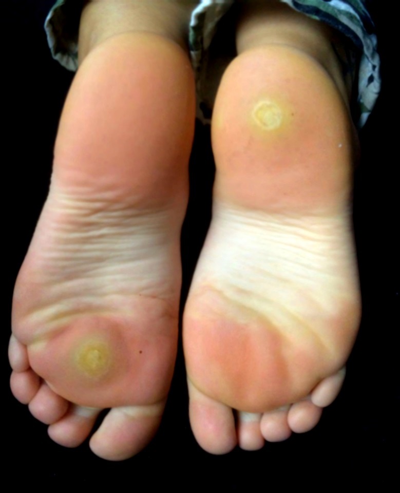
Mosaic warts
When several plantar warts, typically caused by HPV type 2, coalesce into a single closely grouped plaque, the result is a mosaic wart — a larger, confluent area rather than one discrete lesion, generally more resistant to treatment and more likely to cause significant pain across a broader area of the sole than a single plantar wart would.[4] Mosaic warts are also more likely to recur after destructive treatment, including after surgical removal, since residual virus at the margin of a large treated area can seed new adjacent lesions.

Where they occur
Plantar warts occur at weight-bearing points — the metatarsal heads, the heel, and the plantar surface of the toes — as well as at non-weight-bearing sites on the sole where they are simply less likely to cause pain despite being present. A wart at a weight-bearing point is typically more symptomatic and more likely to prompt a visit than one tucked into the arch.
Risk Factors
- Barefoot exposure to damp communal surfaces — locker rooms, public pool decks, and communal showers — where HPV survives on wet flooring and gains entry through skin that is macerated or has minor breaks from that same moisture.
- Participation in activities where bare feet are routine: swimming, gym and fitness-centre use, and contact sports such as wrestling.
- Adolescents and young adults have the highest incidence, but unlike common warts, spontaneous resolution of a plantar wart becomes markedly less likely with age — essentially absent past age 12 in the trial data this chapter draws on — so age alone should not be read as reassurance that a plantar wart will simply go away.[2]
- An existing callus at a pressure point can mask an early, small plantar wart underneath it, delaying recognition until the lesion is more established and more symptomatic.
- Immunosuppression (solid-organ transplant, HIV, other cellular immunodeficiency) — as with warts elsewhere, associated with more numerous, more extensive, and more treatment-refractory disease; see the Common Warts chapter for the shared underlying mechanism.
- Occupations or activities involving prolonged standing, walking, or running, which increase both plantar pressure and the friction that makes an established wart symptomatic.
- A personal history of plantar warts, or ongoing autoinoculation from an untreated lesion elsewhere on the same foot.
Red Flags & Refer
The single most important screening question in this chapter comes before any product recommendation: does this patient have diabetes, peripheral neuropathy of any cause, or peripheral arterial disease? If so, do not recommend self-treatment or self-debridement of a plantar wart — Diabetes Canada's foot care guideline is explicit that corns, calluses, ingrown toenails, warts, splinters, and other foot wounds in a patient with diabetes should not be self-treated, and should instead be assessed by a doctor or foot care specialist (a foot care nurse, podiatrist, or chiropodist).[3] The reasoning applies with equal force to neuropathy and peripheral arterial disease from any cause: reduced sensation means a patient may not feel the tissue damage from a keratolytic acid or an inadvertent nick from paring, and reduced circulation means that even a small resulting injury heals poorly and can progress to infection or, in severe cases, a limb-threatening complication before it is even noticed.[3] Footwear counselling remains appropriate in this group; active treatment and self-debridement do not.
- A single, longstanding lesion that is enlarging, ulcerating, bleeding spontaneously (not just with paring), or has failed repeated adequate courses of destructive treatment — a chronic, treatment-resistant "plantar wart" is one of the recognized ways both verrucous carcinoma (epithelioma cuniculatum) and acral lentiginous or amelanotic melanoma present, and both are well documented to be initially misdiagnosed and treated as an ordinary wart, sometimes for a prolonged period, before the correct diagnosis is made. Biopsy a lesion with this pattern rather than continuing empiric destructive therapy.
- New pigmentation, an irregular or expanding pigmented streak, or any lesion that simply does not look like the others being treated — refer for dermoscopic and, if indicated, histologic assessment rather than assuming it is just an unusually stubborn wart.
- Significant pain limiting weight-bearing or gait, which may need more active offloading and a lower threshold for podiatry involvement than a typical lesion.
- Signs of infection — spreading redness, warmth, swelling, purulent drainage, or fever — need prompt assessment rather than continued self-care.
- Extensive mosaic disease, or disease in an immunocompromised patient, not responding to an adequate first-line trial — refer earlier to dermatology or podiatry rather than working through a prolonged standard OTC course.
Assessment & Diagnosis
History
- Barefoot exposure — locker rooms, pool decks, communal showers, contact sports — and duration and growth pattern of the lesion(s).
- Screen explicitly for diabetes, peripheral neuropathy, and peripheral arterial disease before recommending any self-care or self-debridement — this determines the entire management pathway (see Red Flags/Refer).
- Prior treatment: which product or procedure, how it was applied (was the surface pared first, and how aggressively?), and for how long, since an incomplete or too-gentle trial is a common reason a plantar lesion looks like a "treatment failure" when it has simply never received a regimen intense enough for the sole's thicker skin.
- Pain characteristics, using the same bedside maneuver detailed fully in the Corns and Calluses chapter's comparison table: pain reproduced by direct, straight-down pressure onto the lesion points toward a plantar wart, while pain reproduced by squeezing the lesion from side to side (lateral compression) points toward a corn instead — the opposite pattern from what many clinicians instinctively expect, and worth restating here from the wart side of that same distinction.
- Functional impact — is the lesion changing how the patient walks or bears weight, and for how long has that been the case.
Examination
Gently pare the surface and confirm the 2 findings described in Recognize the Condition: interrupted skin lines and pinpoint bleeding from thrombosed capillary puncta, distinguishing a plantar wart from the continuous skin lines and bloodless keratin core of a corn or callus.[4] Note whether the presentation is a single discrete lesion or a coalescing mosaic plaque, and note its position relative to the weight-bearing points of the sole (metatarsal heads, heel, plantar toe surfaces), which affects both symptom severity and offloading strategy.
Differential diagnosis
| Condition | Features that point away from a plantar wart |
|---|---|
| Corn or callus | Continuous, uninterrupted skin lines and a smooth, bloodless keratin core on paring, with pain reproduced by lateral compression rather than direct pressure — see the full comparison table in the Corns and Calluses chapter for the complete bedside differential. |
| Plantar fibroma | A firm, slow-growing nodule fixed to the deeper plantar fascia (often in the arch, a non-weight-bearing area), without the surface hyperkeratosis, interrupted skin lines, or thrombosed puncta of a wart. |
| Foreign-body granuloma / retained splinter | A history of a penetrating injury at that site; imaging may show a retained foreign body, and the lesion does not show the wart's characteristic surface changes on paring. |
| Porokeratosis plantaris discreta | A discrete, often very tender, translucent keratotic plug at a pressure point, without disrupted skin lines or thrombosed capillaries on paring. |
| Verrucous carcinoma (epithelioma cuniculatum) | A slow-growing, long-standing, warty plaque on the sole that has typically already failed one or more prior courses of destructive wart treatment; deep biopsy is required for diagnosis, since its bland, verrucous histology can itself be mistaken for an ordinary wart. Suspect this in any lesion that has been present a long time, keeps growing, and has not responded to adequate treatment. |
| Acral lentiginous / amelanotic melanoma | A pigmented, irregularly pigmented, or non-pigmented lesion on the sole that is changing, enlarging, or simply does not look like a typical wart; this location-specific melanoma subtype is well documented to be frequently misdiagnosed initially as a wart, callus, or non-healing ulcer, with diagnostic delay directly worsening outcomes — biopsy rather than treat empirically when a lesion is atypical. |
Most classic plantar warts are diagnosed clinically on the pare-test findings above without further testing. Reserve biopsy for a lesion that is atypical, pigmented, rapidly changing, or that has genuinely failed an adequate course of destructive therapy — persistence through adequate treatment is itself a diagnostic red flag, not just a treatment-intensity problem.
Treatment Targets
- Set expectations correctly from the first visit: plantar warts are less likely to resolve spontaneously than warts elsewhere, especially past childhood, so active treatment is a reasonable default recommendation rather than a fallback.[2]
- Match treatment intensity to the sole's thicker skin — a regimen borrowed unmodified from a hand or knee wart is a common, avoidable reason for apparent treatment failure here.
- Relieve pain and pressure and preserve normal weight-bearing and gait during treatment.
- Screen every patient for diabetes, peripheral neuropathy, and peripheral arterial disease before recommending any self-care or self-debridement, and route that population to professional foot care instead.
- Limit spread within the same foot (autoinoculation, mosaic formation) and to other people through communal barefoot exposure.
- Minimize scarring, particularly with refractory destructive options — a surgical scar at a weight-bearing point on the sole can itself become a new, permanent source of pain, unlike a scar on the hand or knee.
- Recognize early when a lesion needs to move beyond self-care: diagnostic uncertainty, an atypical or non-responding "wart," or a patient whose foot status makes self-treatment unsafe regardless of the lesion itself.
Choose Initial Treatment
Step 1 — screen before recommending anything
Diabetes, peripheral neuropathy, or peripheral arterial disease do not recommend salicylic acid or self-debridement — refer to a foot care specialist instead.[3] No red flags present proceed with active treatment below; given how unreliable spontaneous resolution is for plantar warts past childhood, discuss watchful waiting honestly as a less rewarding option here than for a common wart, rather than presenting it as an equally attractive default.[2]
Step 2 — topical salicylic acid, matched to plantar skin thickness
1st line topical salicylic acid, generally at the higher end of available OTC concentrations (up to 40%) given how much thicker the plantar stratum corneum is than skin elsewhere. Soak the foot in warm water for 5-10 minutes, then pare the softened surface down aggressively — to the point of just reaching pinpoint bleeding — before each application, since inadequate paring is one of the most common reasons a plantar salicylic acid trial appears to fail.[4] Apply to the wart only, protecting surrounding normal skin, and occlude with a bandage, tape, or a cut-to-size 40% salicylic acid plaster; some regimens extend occlusion for several days at a time between debridement and reapplication rather than removing and reapplying daily, reflecting how much longer it takes an active agent to penetrate the sole's thicker skin. In the primary-care trial underlying this chapter's core evidence, salicylic acid performed at least as well as cryotherapy for plantar warts specifically (33% cured vs 30% at 13 weeks), unlike the clearer cryotherapy advantage seen for warts elsewhere on the body — worth knowing so a plantar-wart patient is not steered toward cryotherapy on the (here, incorrect) assumption that it is clearly more effective.[2] Set the expectation explicitly: a full trial takes 6-12 weeks of consistent, adherately aggressive paring and application, and often longer than the same regimen would take for a hand or knee wart.
Step 3 — cryotherapy: an equally reasonable alternative, with plantar-specific technique
1st line alternative clinic-administered liquid nitrogen cryotherapy. Two technique points are specific to the plantar location: pare the wart before freezing, which increases clearance, and use the freeze-thaw-freeze technique — after the freeze halo fully disappears, repeat the freeze once more — a more aggressive protocol than is typically needed for a common wart elsewhere.[4] In a second trial of 229 patients over age 12 with mostly recalcitrant plantar warts, cryotherapy and 50% salicylic acid performed identically (both 14% cured at 12 weeks), underscoring that recalcitrant plantar disease is genuinely harder to clear with either first-line modality alone and often needs combination or escalation.[2] Repeat every 1-3 weeks, and expect more sessions than for a comparable common wart given plantar skin thickness. OTC home cryotherapy is available but, as with common warts, its coolant is substantially less cold than clinic-applied liquid nitrogen and is not well established to match professional treatment, an even more relevant limitation given how much colder and more aggressive plantar treatment typically needs to be.
Step 4 — combination therapy for a slow responder
Combination salicylic acid plus cryotherapy is a reasonable next step for a plantar wart responding slowly to either modality alone, rather than switching agents entirely or simply repeating the same regimen at the same intensity.
Step 5 — refractory plantar warts: options that need a referral
Refractory to an adequate, appropriately intense trial of salicylic acid and/or cryotherapy refer for cantharidin (in-office application under an occlusive dressing, producing a delayed blister at 24-48 hours; application itself is painless, which makes it a genuinely useful option for children who cannot tolerate the pain of aggressive plantar cryotherapy, and case series specifically in recalcitrant plantar warts report good effectiveness with this approach), contact immunotherapy (SADBE or diphenylcyclopropenone, specialist-administered sensitization followed by topical application to provoke a local immune reaction), or laser therapy (pulsed dye or Nd:YAG laser, targeting the wart's feeding capillaries; generally combined with paring of the hyperkeratotic cap, with a comparatively low risk of scarring relative to surgical options). Minor surgery (curettage and cautery, or excision) is a genuine option but deserves a specific plantar caution that does not apply the same way elsewhere on the body: scarring is particularly problematic on the sole of the foot, and recurrence after surgical removal can run as high as 30%[4] — and unlike a hand or knee scar, a surgical scar sitting at a weight-bearing point on the sole can itself become a new, permanent source of pain with every step. Reserve surgical options for lesions that have genuinely failed the less-scarring refractory options above, and counsel patients on this trade-off explicitly before they consent to it.
Non-Pharmacologic Treatment
- Wear sandals or water shoes in locker rooms, on pool decks, and in communal showers while a plantar wart is active, both to avoid spreading the virus to others on shared wet surfaces and to reduce the chance of seeding a new lesion elsewhere on the same foot.
- Do not pick or scratch at a plantar wart, and do not share pumice stones, emery boards, nail files, or towels — the same autoinoculation and transmission principles that apply to common warts apply here, with the added risk that a plantar lesion is already at a friction-prone, weight-bearing site.
- Offload the lesion with a donut-shaped foam or felt pad (a hole cut to surround the wart) or a cushioned insole during active treatment, which reduces pain from day-to-day pressure and friction while the wart is being treated — the same principle used for corns and calluses, applied here to reduce discomfort during an active infection rather than to correct a mechanical cause.
- Pare down the thickened surface regularly with a pumice stone or emery board (kept separate from any file used on normal nails or skin) between active-treatment applications — this alone improves comfort and, unlike with a corn or callus, is also part of what makes concurrent salicylic acid or cryotherapy actually penetrate the sole's thicker skin.
- Duct tape occlusion has the same genuinely mixed evidence base described in the Common Warts chapter, but for plantar warts specifically it is more often used as an occlusive adjunct layered on top of salicylic acid — to help keep a higher-concentration product in prolonged contact with thick plantar skin — than as a stand-alone therapy; be honest with patients that the evidence for benefit beyond that adjunct role is inconsistent.
- Set the treatment timeline explicitly and generously: plantar warts often take longer to clear than a comparable common wart given the intensity of treatment required, and premature discontinuation before a full 6-12 week course (or longer) is a common, avoidable reason for apparent failure.
Special Populations
Diabetes, peripheral neuropathy, and peripheral arterial disease
This is the central safety point of this chapter, covered fully in Red Flags/Refer: do not recommend salicylic acid or self-debridement for a plantar wart in a patient with diabetes, peripheral neuropathy of any cause, or peripheral arterial disease. Diabetes Canada's foot care guideline is explicit that warts, along with corns, calluses, ingrown toenails, and other foot wounds, should be treated by a doctor or foot care specialist rather than self-treated in this population, given the combined risk of an unnoticed injury (from reduced sensation) progressing to a poorly healing, potentially serious complication (from reduced circulation).[3] Loss of protective sensation is itself a significant, independent predictor of future foot ulcer and amputation, which is why routine foot examinations in this population specifically screen for it — a useful context point for explaining to a patient why a plantar wart is being handled differently than it would be on someone without diabetes.[3] Footwear counselling remains appropriate; active treatment and self-debridement do not.
Pediatrics
Children and adolescents have the highest incidence of plantar warts, and salicylic acid is generally preferred for young children because it can be applied at home by a caregiver without the pain of cryotherapy. Because plantar treatment typically needs to be more aggressive than for a common wart — deeper paring, more intense or repeated freezing — cantharidin's painless in-office application is a particularly useful refractory option specifically for young children who cannot tolerate an intensified cryotherapy protocol on a weight-bearing area.
Pregnancy and breastfeeding
Topical salicylic acid applied to a small, localized plantar lesion has minimal systemic absorption and is generally considered acceptable; the larger treatment area and longer occlusion sometimes used for a stubborn plantar wart argue for staying with the smallest effective area and duration rather than defaulting to the most aggressive regimen. Cryotherapy is a local, non-systemic procedure and is a reasonable option in both pregnancy and breastfeeding. Cantharidin, imiquimod, and contact immunotherapy have limited pregnancy-specific safety data — favour salicylic acid or cryotherapy first, and involve the prescriber directly if one of these is genuinely needed.
Athletes and on-foot occupations
Plantar warts disproportionately affect people who are barefoot in communal wet spaces (swimmers, wrestlers, gym users) and people on their feet for long stretches at a time. Blistering from cryotherapy or the delayed blister from cantharidin can genuinely interfere with training, competition, or a physically demanding job for several days — worth discussing explicitly when timing treatment, rather than leaving a patient surprised by a painful blister before an event.
Immunocompromised patients
As with warts elsewhere, transplant recipients, patients with HIV, and others with cellular immunodeficiency develop more numerous, more extensive, and more treatment-refractory plantar and mosaic warts, and conventional first-line therapy is less reliably effective. Treat actively and use a lower threshold to refer early for dermatology- or podiatry-directed combination therapy rather than working through a prolonged standard OTC trial first.
Older adults
Age-related thinning of the protective plantar fat pad increases pressure-related pain from any plantar lesion, including a wart, and a painful plantar lesion that changes gait carries a real fall risk in this group — the same consideration the Corns and Calluses chapter raises for that condition applies equally here. Screen carefully for diabetes, neuropathy, and peripheral arterial disease before recommending self-care, since these are also more prevalent with advancing age.
Monitoring & Follow-Up
Reassess a salicylic acid course at the end of a full 6-12 week trial, and expect that a plantar course may reasonably run longer than that for a common wart given the intensity of paring and occlusion required. At follow-up, confirm the patient has actually been paring aggressively to pinpoint bleeding before each application and maintaining occlusion, since a technique borrowed from a common-wart regimen (light paring, no occlusion) is a common reason for an apparent plateau here.
For a patient undergoing cryotherapy, review response and tolerability every 1-3 weeks, and confirm the freeze-thaw-freeze technique and pre-treatment paring are actually being used — a single light freeze without paring first under-treats a plantar wart relative to warts elsewhere.[4] Watch specifically for excessive blistering that is genuinely impairing weight-bearing or gait, which may call for a less aggressive interval or additional offloading between sessions.
For patients with diabetes, peripheral neuropathy, or peripheral arterial disease, there is no self-monitoring plan — confirm at follow-up contact that the patient has actually been seen by a foot care specialist rather than continuing to watch a lesion at home.[3]
At every visit, reinforce footwear-in-communal-areas counselling (sandals in locker rooms, pool decks, communal showers) to prevent both spread to others and reinfection or new lesion formation on the same foot.
Common Errors to Avoid
- Treating a plantar wart with a common-wart-intensity regimen — light daily salicylic acid with no occlusion, or a single gentle cryotherapy freeze with no pre-treatment paring — and concluding the drug failed when the real problem was under-treatment for the sole's thicker skin.
- Recommending salicylic acid or self-debridement to a patient with diabetes, peripheral neuropathy, or peripheral arterial disease without screening for these conditions first.[3]
- Assuming cryotherapy is clearly the more effective option here the way it can be for common warts elsewhere — the plantar-specific trial data show salicylic acid performing at least as well.[2]
- Confusing a plantar wart for a corn or callus (or vice versa) without checking skin-line continuity, pinpoint bleeding on paring, and the direct-pressure-versus-lateral-pinch pain distinction detailed in the Corns and Calluses chapter.
- Treating a longstanding, single, enlarging, or treatment-resistant "plantar wart" indefinitely with repeated destructive therapy rather than considering verrucous carcinoma or acral melanoma and pursuing biopsy.
- Reaching for surgical excision or curettage as an early refractory option without discussing the plantar-specific scarring and recurrence risk, and the possibility that the resulting scar itself becomes a new source of weight-bearing pain.
- Endorsing unsupervised home "bathroom surgery" with a blade — genuinely more dangerous on a weight-bearing, friction-prone area than elsewhere on the body.
- Not counselling on footwear in communal wet areas alongside whichever active treatment is chosen, leaving an ongoing source of reinfection or transmission unaddressed.
Clinical Decision Algorithm
Drug Tables
How to use this table: Doses are adult doses and require patient-specific confirmation (renal/hepatic function, weight, allergy history). Pediatric dosing is weight-based per the current product monograph unless stated. Drug pricing is intentionally omitted. Select any drug name to open its complete Pharmasist Drug Database page.
Keratolytics (Topical, Self-Applied)
First-line active treatment for most plantar warts. Requires more aggressive paring (to pinpoint bleeding) and typically longer occlusion than the same product used for a common wart elsewhere on the body, given the thickness of plantar skin.
| Drug | Dosage | Adverse effects | Drug interactions | Comments |
|---|---|---|---|---|
| Salicylic acid | OTC concentration: Up to 40%, applied and occluded, 6-12+ weeks | Local irritation, burning; chemical burn to surrounding normal skin if not protected | None clinically significant given minimal systemic absorption at typical localized use | Soak 5-10 minutes, pare aggressively to pinpoint bleeding, apply to the wart only (protect surrounding skin), and occlude — often for several days at a time between debridement and reapplication. A genuine trial needs 6-12 weeks or longer. |
Cryotherapy
For plantar warts, cure rates with salicylic acid at least match cryotherapy — unlike warts elsewhere on the body, where cryotherapy tends to have the edge. A more aggressive freeze protocol than for a common wart is typically needed.
| Drug | Dosage | Adverse effects | Drug interactions | Comments |
|---|---|---|---|---|
| Liquid nitrogen (clinic-administered, freeze-thaw-freeze technique) | Interval: Every 1-3 weeks until resolved | Pain, blistering, occasional permanent hypopigmentation or scarring | None | Pare the wart before freezing to increase clearance; after the freeze halo fully disappears, repeat the freeze once more (freeze-thaw-freeze). Expect more sessions than for a comparable common wart. |
| OTC home cryotherapy (dimethyl ether/propane spray) | Use: Per product directions, typically every 1-2 weeks | Local pain, blistering, skin irritation | None | Reaches only about -20°C versus about -100°C for clinic-applied liquid nitrogen — an even more relevant limitation given how aggressive plantar cryotherapy typically needs to be. |
Office-Administered / Specialist Refractory-Wart Therapies
Reserve for plantar warts that have not responded to an adequately intense, adherent trial of salicylic acid and/or cryotherapy. Favour the lower-scarring options before considering surgery, given the plantar-specific scarring and recurrence risk described in Choose Initial Treatment.
| Drug | Dosage | Adverse effects | Drug interactions | Comments |
|---|---|---|---|---|
| Cantharidin | Use: In-office application only | Delayed blistering at the application site 24-48 hours later, painful once formed, especially on a weight-bearing area | None | A vesicant applied in-office under occlusion, not for home use. Application itself is painless, making it useful in young children who cannot tolerate an intensified cryotherapy protocol. |
| Contact immunotherapy (SADBE or diphenylcyclopropenone) | Use: Specialist-administered sensitization, then topical application | Local dermatitis at the treated site (the intended mechanism), pruritus | None | The patient is first sensitized, then the agent is applied topically to provoke a local delayed-type hypersensitivity reaction. Specialist-administered; useful for extensive/mosaic or immunocompromised-patient disease. |
| Laser therapy (pulsed dye or Nd:YAG) | Use: Specialist-administered, typically more than 1 session | Local pain, purpura, occasional transient hypopigmentation | None | Targets the dilated capillaries supplying the wart, generally combined with paring of the hyperkeratotic cap. Comparatively low scarring risk relative to surgical excision. |
| Minor surgery (curettage and cautery, or excision) | Use: Specialist-administered, after less-scarring refractory options have failed | Scarring is particularly problematic on the sole of the foot; recurrence can run as high as 30% | None | A last-resort refractory option. A surgical scar at a weight-bearing point can itself become a new, permanent source of pain — discuss this trade-off explicitly before the patient consents. |
| Imiquimod | Use: Per prescriber direction | Local irritation, erythema; flu-like symptoms with extensive application | None clinically significant at localized use | Off-label; evidence for nongenital warts generally is limited (27-30% clearance in small uncontrolled studies) and even less specific to the plantar location — a specialist-directed option, not routine first-line. |
References
- Wart — StatPearls Publishing (Al Aboud AM, Nigam PK). StatPearls, last updated August 14, 2023
- What works best for nongenital warts? — Canadian Family Physician, Tools for Practice (adapted from the Alberta College of Family Physicians' PEER evidence program) (Finley C, Korownyk C, Kolber MR). Can Fam Physician 2016;62(12):997
- Foot Care — Diabetes Canada Clinical Practice Guidelines, Chapter 32 (Embil JM, Albalawi Z, Bowering K, Trepman E). Diabetes Canada CPG, Chapter 32, 2018
- Treatment of Nongenital Cutaneous Warts — American Family Physician (Mulhem E, Pinelis S). Am Fam Physician 2011;84(3):288-293
Professional reference only. Apply clinical judgment and verify patient-specific decisions against current authoritative guidance and product information.