Pseudotumor Cerebri (Idiopathic Intracranial Hypertension)
Clinical Snapshot
Pseudotumor cerebri — now more precisely termed idiopathic intracranial hypertension (IIH) when no secondary cause is identified, and grouped under the broader umbrella term pseudotumor cerebri syndrome (PTCS) that also includes cases with a genuinely identifiable secondary cause — is a disorder of elevated intracranial pressure (ICP) occurring without a mass lesion, hydrocephalus, or venous sinus thrombosis to explain it.[1] It is, overwhelmingly, a disease of obese women of reproductive age: more than 93% of affected patients are obese, the median age at onset is 34 years, and the annual incidence among obese women of childbearing age specifically reaches roughly 20 per 100,000 — a striking, genuinely useful epidemiologic pattern that should raise suspicion the moment a patient fitting that profile presents with a new headache.[2] This chapter is built around a single organizing idea that should reframe how every encounter is approached: headache is what brings the patient to the pharmacy or the clinic, but vision is what the treatment plan actually has to protect. Papilledema — swelling of the optic disc from the transmitted pressure — occurs in up to 95% of cases, and it is asymptomatic vision loss, not the headache, that causes permanent harm when the diagnosis or the treatment escalation is delayed.[2] A second organizing idea, and this chapter's other centerpiece, is that a meaningful share of intracranial hypertension is not idiopathic at all: a specific, genuinely memorable list of medications — tetracycline-class antibiotics (especially minocycline), vitamin A and its retinoid relatives, recombinant growth hormone, and several others — can cause the identical clinical syndrome, and recognizing that list is one of the most concrete, high-value contributions a pharmacist can make to this diagnosis (see Risk Factors).[3]
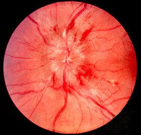
The classic presentation is a headache in an obese woman of reproductive age, but the diagnosis is made — and vision is protected or lost — on the basis of the eye exam and the lumbar puncture, not the headache history alone. See Assessment & Diagnosis for the full revised diagnostic criteria.
Recognize the Condition
Headache
Headache is the presenting complaint in the substantial majority of patients, and it is frequently the feature that dominates the clinical picture — daily or near-daily, often severe enough to be genuinely disabling, and lacking a single specific pattern that distinguishes it from migraine or tension-type headache on history alone.[2] It can be worsened by the maneuvers that raise intracranial pressure further — lying flat, Valsalva, coughing, bending forward — and can wake a patient from sleep. This nonspecificity is itself a genuine diagnostic trap: a patient can be treated for presumed migraine for months while papilledema-driven optic nerve damage accumulates silently in the background, which is exactly why the eye exam belongs in the workup of any new, atypical, or progressively worsening headache in the classic epidemiologic group (obese women of reproductive age) rather than being reserved for patients who already report visual symptoms.
Papilledema
Papilledema — optic disc swelling specifically caused by elevated intracranial pressure — is the physical finding around which the entire diagnostic framework is built (see Assessment & Diagnosis), and is present in up to 95% of cases.[2] It ranges from subtle disc-margin blurring that is easy to miss without dilated fundoscopy or fundus photography, to severe swelling with peripapillary hemorrhages, cotton-wool spots, and exudates — a spectrum commonly graded clinically using the Frisén scale (grade 0, normal, through grade 5, severe swelling with obscuration of major vessels). Higher-grade papilledema, and papilledema that fails to improve with treatment, both correlate with a greater risk of permanent visual loss, which is why serial, standardized documentation (ideally with fundus photography and optical coherence tomography, not just a written description) matters far more here than it does in most other headache-adjacent complaints (see Monitoring & Follow-Up).
Transient visual obscurations
Transient visual obscurations (TVOs) — brief, seconds-long episodes of graying, dimming, or complete blackout of vision in one or both eyes — are reported by a large share of patients and are a direct consequence of transient disc ischemia from the swollen nerve head. They are frequently positional, provoked by bending over, standing up quickly, or Valsalva, and while they are genuinely common and do not, on their own, indicate irreversible damage, their frequency is a useful marker of disease activity worth asking about explicitly and tracking over time.
Pulsatile tinnitus
Pulsatile (or "synchronous") tinnitus — a whooshing, rushing sound the patient hears in time with their own heartbeat — is reported by roughly 60% of patients and is considered the single most specific symptom of the condition, reflecting turbulent blood flow across a narrowed or externally compressed venous sinus.[2] A simple, genuinely useful bedside maneuver is gentle jugular venous compression on the symptomatic side, which transiently abolishes the sound in true pulsatile tinnitus of this type — a distinguishing feature from the far more common, non-pulsatile, non-specific tinnitus that has nothing to do with intracranial pressure.
Sixth cranial nerve (abducens) palsy and other neurologic findings
Horizontal diplopia from a unilateral or bilateral sixth cranial nerve (abducens) palsy occurs in a meaningful minority of patients. This is a genuinely important "false localizing sign" — it results from the long, vulnerable intracranial course of the abducens nerve being stretched or compressed by elevated pressure generally, not from a lesion actually located along the nerve's path, and its presence does not change the diagnostic approach (see Assessment & Diagnosis). Beyond this, the neurologic examination should otherwise be normal; any other focal deficit, seizure, or altered level of consciousness is a red flag that points away from idiopathic disease and toward a structural lesion or another process entirely (see Red Flags for Referral). Neck, shoulder, and interscapular pain — sometimes radicular in character — is also reported by a minority of patients and is thought to reflect meningeal or nerve-root traction from elevated CSF pressure.
Visual field loss — the finding that matters most, and the one patients notice least
This is the single most important teaching point in this entire chapter: visual field loss in this condition is typically insidious, painless, and can progress to a genuinely severe degree before a patient spontaneously reports any visual complaint at all. The earliest, most common pattern is an enlarged physiologic blind spot, followed by an inferonasal field defect, and — without adequate treatment — progression toward generalized constriction of the visual field and, eventually, severe and sometimes irreversible vision loss. Relying on "the patient says their vision is fine" as reassurance is a genuine, well-documented error; the visual field must be formally, objectively tested (see Assessment & Diagnosis and Monitoring & Follow-Up) rather than inferred from symptoms.
Fulminant idiopathic intracranial hypertension
A distinct, higher-risk subgroup — fulminant IIH — presents with severe visual loss developing rapidly, within about four weeks of symptom onset, sometimes accompanied by markedly elevated opening pressures and severe papilledema at first presentation. This pattern behaves differently from typical, gradually progressive disease: the usual stepwise medical titration described in Choose Initial Treatment is too slow to reliably prevent severe, permanent vision loss, and recognizing this pattern early enough to fast-track surgical evaluation — rather than waiting to see whether acetazolamide "catches up" — is a genuinely consequential clinical decision (see Special Populations and Treatment Failure/Next Step).
Risk Factors
The two demographic risk factors that define the classic phenotype
- Obesity — present in more than 93% of patients — and, independently, a recent increase in body weight (even a relatively modest gain, well short of frank obesity, is associated with increased risk), suggesting a genuine dose-response relationship between adiposity and intracranial pressure rather than a simple binary obese/non-obese distinction.[2]
- Female sex and reproductive age (roughly the 20s to 40s) — the single most consistent demographic pattern described across the literature, with a median age at onset of 34 years.[2]
Drug-induced and secondary intracranial hypertension — a genuine pharmacist-centered risk factor
Tetracycline-class antibiotics — doxycycline, minocycline, and tetracycline
This is the single most clinically actionable secondary cause on this list, precisely because these are commonly prescribed, often for weeks-to-months-long courses of acne, rosacea, or other indications. Minocycline in particular carries the strongest reported association among the tetracyclines, attributed in part to its higher lipophilicity and greater blood-brain-barrier penetration relative to doxycycline and tetracycline.[3] Onset is genuinely variable — reported latency ranges from about two weeks to more than six months after starting the drug — so a temporal relationship is not always obvious, and a new headache with visual symptoms in a patient on a tetracycline for acne deserves the same diagnostic workup as any other case, not a reflexive dismissal because the drug has "already been tolerated for a while."[3]
Vitamin A and retinoids — isotretinoin, all-trans retinoic acid (tretinoin), and hypervitaminosis A
Vitamin A and its synthetic retinoid relatives are among the most strongly and mechanistically plausible drug-induced causes, given vitamin A's own established role in CSF physiology; isotretinoin (used for severe cystic acne) and all-trans retinoic acid/tretinoin (used in the treatment of acute promyelocytic leukemia, among other indications) are the two most clinically relevant agents, alongside frank hypervitaminosis A from excessive supplementation.[3] As with the tetracyclines, this is a real-world reconciliation point: a young patient started on isotretinoin for severe acne who develops a new headache should not be told to "give it more time" without at least a directed history and fundoscopic screening.
Growth hormone
Recombinant growth hormone (somatropin) is a well-recognized cause, disproportionately reported in children and adolescents being treated for growth hormone deficiency, Turner syndrome, or other approved pediatric indications — a genuinely important reason to keep a low threshold for fundoscopic screening in any child on growth hormone therapy who develops a new headache (see Special Populations).[3]
Other implicated agents
A further, genuinely worth-knowing list includes lithium, danazol, tamoxifen, cyclosporine, and levothyroxine over-replacement (particularly relevant in children who are over-treated for congenital hypothyroidism), along with rapid corticosteroid dose reduction or withdrawal after a prolonged course — it is the abrupt drop in corticosteroid exposure, not ongoing corticosteroid use itself, that has been linked to rebound intracranial hypertension.[3] Older literature also implicates nalidixic acid, a quinolone rarely used today. None of these associations are as strong or as reproducible as the tetracycline-class or retinoid associations above, but each is worth keeping on a working differential during medication reconciliation.
Other secondary causes to exclude before accepting an idiopathic diagnosis
- Cerebral venous sinus thrombosis — excluded with MR venography as part of the standard neuroimaging workup (see Assessment & Diagnosis); this is the single most important non-drug secondary cause not to miss, since its own treatment (anticoagulation) is entirely different.
- Obstructive sleep apnea — a recognized aggravating and, in some patients, contributory factor, and worth screening for independently of weight (see Non-Pharmacologic Treatment).
- Chronic kidney disease, systemic lupus erythematosus, and other systemic inflammatory or endocrine conditions (Addison disease, hypoparathyroidism, polycystic ovary syndrome) have each been associated with secondary intracranial hypertension in case reports and smaller series, and are reasonable to consider when the classic demographic or drug-exposure pattern is absent.
Red Flags & Refer
Any patient with suspected papilledema, or with headache plus visual symptoms in the classic demographic (an obese woman of reproductive age), needs urgent same-day or next-available ophthalmology or neuro-ophthalmology assessment. This is not a "start a drug and reassess in a month" presentation — vision loss in this condition is frequently silent until it is severe, and the diagnostic workup itself (neuroimaging before lumbar puncture) is genuinely time-sensitive.
- Rapidly progressive, severe vision loss developing over days to a few weeks (the fulminant pattern): a same-day emergency — do not wait for a standard outpatient ophthalmology appointment or for a trial of acetazolamide to take effect; this pattern needs expedited neuro-ophthalmology and neurosurgical involvement in parallel (see Special Populations).
- Any focal neurologic deficit, seizure, or altered level of consciousness: not typical for idiopathic disease — reconsider a mass lesion, cerebral venous sinus thrombosis, or another intracranial process, and pursue emergent neuroimaging rather than a routine outpatient workup.
- New headache and visual symptoms in a pregnant patient: needs the same urgent ophthalmologic and diagnostic workup as a non-pregnant patient — pregnancy does not lower the priority of ruling out papilledema and elevated ICP, and additionally raises pre-eclampsia/eclampsia on the differential (see Special Populations).
- Any patient-reported change in visual acuity, color vision, or peripheral vision, however mild: a genuine escalation trigger given how insidious visual field loss typically is — do not defer formal ophthalmologic assessment on the assumption that a mild complaint means mild disease.
- A patient recently started on a known causative medication (a tetracycline, a retinoid, growth hormone, or another agent from Risk Factors) who develops a new headache: warrants the same urgent fundoscopic and diagnostic evaluation as any other suspected case — the presence of a plausible drug cause does not make the underlying elevated-pressure process any less dangerous to the optic nerve.
Assessment & Diagnosis
The revised diagnostic criteria for pseudotumor cerebri syndrome
The modern, modified Dandy criteria — formally the revised diagnostic criteria for pseudotumor cerebri syndrome (PTCS) — require, for a definite diagnosis in a patient with papilledema: papilledema on examination; a normal neurologic examination other than cranial nerve abnormalities (a sixth-nerve palsy is allowed and does not exclude the diagnosis — see Recognize the Condition); neuroimaging (MRI, with MR venography in most patients) showing a normal brain without hydrocephalus, a mass, or a structural lesion, and without abnormal meningeal enhancement — the same imaging that also directly excludes cerebral venous sinus thrombosis; normal cerebrospinal fluid composition; and an elevated lumbar puncture opening pressure, measured with the patient relaxed in the lateral decubitus position, of more than 250 mm CSF in adults (or more than 280 mm CSF in children, with 250 mm CSF used in a non-sedated, non-obese child).[1][2] Supportive but not required neuroimaging findings — an empty or partially empty sella, flattening of the posterior sclera, distension of the perioptic subarachnoid space, and transverse venous sinus stenosis — strengthen the diagnosis when present but are not themselves diagnostic.[1] When papilledema is absent, the same diagnosis can still be made if a sixth-nerve palsy is present alongside the other criteria; without either papilledema or a sixth-nerve palsy, the diagnosis can only be suggested, not confirmed, and requires at least three of the supportive neuroimaging findings above.[1]
Neuroimaging comes before the lumbar puncture, not after
MRI of the brain (with MR venography) should be obtained and reviewed before lumbar puncture is performed, both to exclude a mass lesion that would make lumbar puncture dangerous and to directly exclude cerebral venous sinus thrombosis — the single most important secondary cause not to miss, and one whose own treatment (anticoagulation) is entirely different from the management described in Choose Initial Treatment.
The lumbar puncture — technique matters
Opening pressure must be measured with the patient in the lateral decubitus position, legs extended and genuinely relaxed (not actively flexed against resistance, and not during a Valsalva maneuver or breath-holding), since a poorly performed or artifactually elevated measurement can produce a falsely elevated — or falsely reassuring — reading. CSF should also be sent for cell count, protein, and glucose; genuinely normal composition is part of the diagnostic criteria, and any abnormality should prompt reconsideration of an infectious, inflammatory, or malignant process rather than idiopathic disease.
Medication reconciliation belongs inside the diagnostic workup, not after it
Before a diagnosis of idiopathic (primary) disease is accepted, the patient's full medication list should be reviewed specifically against the agents in Risk Factors — this is not a peripheral detail but a core, integral step of arriving at an accurate diagnosis, since a case caused by a recently started tetracycline or retinoid is managed differently (see Choose Initial Treatment and Special Populations) from genuinely idiopathic disease, even though both meet the same PTCS diagnostic criteria on lumbar puncture and imaging.[3]
The ophthalmologic workup that drives ongoing management
Beyond confirming papilledema on fundoscopy, formal quantitative assessment should include automated (Humphrey) visual field perimetry, optical coherence tomography of the retinal nerve fibre layer to objectively quantify disc edema, and baseline fundus photography for serial comparison — all three are what actually drive the treatment and escalation decisions in the sections that follow, not the headache itself (see Monitoring & Follow-Up).
| Feature | Idiopathic intracranial hypertension | Drug-induced/secondary PTCS | Structural lesion or venous sinus thrombosis |
|---|---|---|---|
| Typical patient | Obese woman, 20s-40s | Any age/sex; recent exposure to a causative agent (see Risk Factors) | Any age/sex |
| Neurologic exam | Normal aside from possible sixth-nerve palsy | Normal aside from possible sixth-nerve palsy | May show focal deficits |
| Neuroimaging | Normal parenchyma; no mass, hydrocephalus, or venous thrombosis | Normal parenchyma; no mass, hydrocephalus, or venous thrombosis | Mass, hydrocephalus, or filling defect on MR venography |
| CSF composition | Normal | Normal | Often abnormal (infection, malignancy) |
| Management | Weight loss + acetazolamide (see Choose Initial Treatment) | Discontinue/substitute the causative agent, plus standard IIH-directed therapy as needed | Entirely different — treat the underlying lesion or anticoagulate |
Treatment Targets
- Preserve vision — the primary, non-negotiable treatment target, tracked objectively with visual fields and optical coherence tomography rather than inferred from symptoms (see Recognize the Condition and Monitoring & Follow-Up).
- Reduce intracranial pressure toward a level that halts papilledema progression and reverses active visual field loss.
- Identify and remove or substitute any causative medication, in coordination with the prescriber, rather than treating drug-induced disease purely with add-on pharmacotherapy.
- Achieve meaningful, sustained weight loss (a genuinely disease-modifying goal, not an adjunct to drug therapy) in patients with obesity or recent weight gain.
- Control headache as its own, separate axis of morbidity, recognizing that headache can persist even after papilledema and visual field loss have resolved.
- Recognize the fulminant, rapidly progressive phenotype early enough to fast-track surgical evaluation rather than relying on medical therapy alone to act quickly enough.
Choose Initial Treatment
Vision is the outcome that determines treatment intensity — not headache severity
Treatment decisions in this condition should be driven primarily by the objective visual findings (papilledema grade, visual field mean deviation, visual acuity) rather than by how severe the patient's headache is. A patient with a debilitating headache but a stable, mild visual field defect and a patient with a mild headache but progressive visual field loss are not on the same treatment track — the second patient needs faster escalation, even though the first patient may be, understandably, the more vocal one in clinic.
Weight loss — a genuinely disease-modifying intervention, not just supportive advice
Weight loss has long been described as the cornerstone of management for a reason: it is not merely adjunctive to drug therapy, it independently lowers intracranial pressure, and the evidence for this has grown considerably more rigorous over time.[2] In the landmark placebo-controlled acetazolamide trial itself, both study arms — including the diet-alone, placebo arm — were enrolled in a supervised low-sodium weight-reduction program, and a subsequent randomized trial specifically comparing bariatric surgery against a structured community weight-management program in patients with active idiopathic intracranial hypertension found that bariatric surgery produced substantially greater and more durable weight loss and a correspondingly greater, sustained reduction in intracranial pressure at 12 months.[4][5] In practice, this means a genuine, early, and structured weight-management referral — with bariatric surgery evaluation for eligible patients whose disease remains active despite standard measures — belongs in the initial treatment plan alongside pharmacotherapy, not as an afterthought raised only once drug therapy has already failed.
Acetazolamide — first-line pharmacotherapy
Acetazolamide, a carbonic anhydrase inhibitor that reduces cerebrospinal fluid production, is first-line pharmacotherapy on the strength of the Idiopathic Intracranial Hypertension Treatment Trial (IIHTT): in patients with mild visual loss at baseline, acetazolamide added to a supervised low-sodium weight-reduction diet produced a modest but statistically significant improvement in visual field function (perimetric mean deviation) at 6 months compared with diet alone, alongside greater reductions in papilledema grade and in CSF opening pressure.[4] A practical starting approach is 250 mg orally twice daily (or an extended-release formulation dosed once or twice daily), titrated upward over days to a couple of weeks as tolerated toward the higher doses used in the trial protocol — up to a maximum of 4 g/day if genuinely tolerated, though many patients achieve good control, and most patients in the trial were maintained, at a considerably lower dose.[4] Counsel proactively, before the patient calls back alarmed. Paresthesias of the fingers, toes, and around the mouth are extremely common, dose-related, and not dangerous — they reflect the drug's carbonic-anhydrase-inhibiting mechanism, not an allergic reaction, and should not by themselves prompt discontinuation. A metallic or otherwise altered taste, particularly with carbonated beverages, is similarly common and benign. Fatigue and mild gastrointestinal upset are also expected. Genuinely worth monitoring for, rather than just mentioning, are metabolic acidosis and hypokalemia (periodic basic metabolic panel — see Monitoring & Follow-Up) and an increased risk of renal calculi.
The sulfonamide "allergy" question
Acetazolamide is a non-antibiotic sulfonamide, and the historical concern about cross-reactivity with antimicrobial sulfonamides (e.g., sulfamethoxazole) in a patient with a labelled "sulfa allergy" is now understood to be considerably weaker than once assumed — the antigenic determinants differ meaningfully between the antibiotic and non-antibiotic sulfonamide classes. This should not be used to reflexively withhold a genuinely first-line, vision-protecting therapy from a patient with a mild prior reaction (e.g., a rash) to a sulfonamide antibiotic; a case-by-case risk discussion, reserving real caution for patients with a history of a severe reaction such as Stevens-Johnson syndrome or toxic epidermal necrolysis, is the more accurate and more clinically useful approach.
Second-line and adjunct pharmacotherapy
Furosemide is a reasonable second-line or add-on diuretic option when acetazolamide alone is insufficient or not tolerated, though its supporting evidence base is considerably weaker than acetazolamide's. Topiramate is a genuinely useful alternative, particularly when headache has a migrainous quality or when the patient would benefit from its own independent weight-loss effect: it carries mild carbonic-anhydrase-inhibiting activity of its own in addition to its established migraine-preventive and appetite-suppressant properties, giving it a plausible dual mechanism in this specific condition.[6] Topiramate carries its own, separate teratogenicity concern (oral clefts) and needs the same reproductive-health counselling given for any teratogenic agent in a woman of reproductive age (see Special Populations).
Corticosteroids — a short-term surgical bridge only, never maintenance therapy
Corticosteroids (e.g., a short oral dexamethasone course, or IV methylprednisolone in a hospitalized patient) have a narrow, specific role as a temporary bridge to lower intracranial pressure while awaiting definitive surgical intervention in fulminant or severely progressive disease — they are not appropriate as ongoing maintenance therapy. Chronic corticosteroid use is genuinely counterproductive here: it promotes further weight gain (working directly against the disease-modifying goal above) and can produce rebound intracranial hypertension when the dose is tapered or stopped (see Risk Factors), making an already difficult disease harder to control if used beyond a short, specialist-directed bridge.
Non-Pharmacologic Treatment
- Refer promptly to a structured weight-management program (dietitian involvement, a supervised low-sodium, calorie-reduced diet) as active, disease-modifying treatment — not a lifestyle suggestion appended at the end of the visit.
- Discuss bariatric surgery evaluation for medically eligible patients whose disease remains active despite standard weight-management measures, given the substantially greater and more durable weight loss and intracranial-pressure reduction demonstrated against community weight management in a dedicated randomized trial.[5]
- Identify and discontinue or substitute any causative medication (see Risk Factors) in direct coordination with the prescribing clinician — do not stop an essential therapy such as isotretinoin for severe cystic acne or lithium for bipolar disorder unilaterally; work out an alternative or a bridging plan together.
- Screen for and treat obstructive sleep apnea when present, given its recognized role as an aggravating factor independent of weight.
- Arrange serial, standardized visual field testing and optical coherence tomography rather than relying on symptom review alone to judge disease activity (see Monitoring & Follow-Up).
- Manage headache as its own problem once the underlying pressure is being addressed, and actively avoid medication-overuse headache from frequent as-needed analgesic use.
Surgical options for medically refractory disease or fulminant vision loss
Optic nerve sheath fenestration (ONSF)
A surgical incision (fenestration) is made in the dural sheath surrounding the optic nerve, allowing CSF to decompress locally around the nerve itself. ONSF is directed specifically at protecting vision rather than at lowering intracranial pressure globally or treating headache, and in comparative outcome data it has shown a generally favourable, low-complication profile with meaningful rates of visual field and visual acuity improvement or stabilization.[7] It is a reasonable first surgical choice when vision, rather than headache, is the dominant problem.
Cerebrospinal fluid shunting (ventriculoperitoneal or lumboperitoneal)
A shunt diverts CSF from the ventricular system or lumbar subarachnoid space to the peritoneal cavity, directly and globally lowering intracranial pressure — addressing headache as well as papilledema, unlike ONSF. The trade-off, seen consistently across comparative series, is a substantially higher rate of mechanical complications, shunt failure, and the need for surgical revision compared with ONSF.[7] Shunting is a reasonable choice when headache itself is severe and disabling, or when rapid, global pressure reduction is needed, but the higher revision burden is worth setting as an explicit expectation with the patient before surgery.
Venous sinus stenting
In patients found on catheter venography to have a focal transverse venous sinus stenosis with a significant pressure gradient across it, endovascular stent placement across the stenosis is an increasingly used option, with published outcome series showing meaningful improvement in headache, papilledema, and visual symptoms, alongside a genuine, specific risk of in-stent or stent-adjacent restenosis that can require re-intervention.[7] This option specifically depends on demonstrating both an anatomic stenosis and a physiologically significant pressure gradient — it is not interchangeable with ONSF or shunting in a patient without that specific finding.
Special Populations
Pregnancy
Pregnancy does not fundamentally change the diagnostic urgency or approach described above — a pregnant patient with new headache and visual symptoms in the classic pattern still needs the same fundoscopic and, when indicated, lumbar-puncture-based workup, alongside consideration of pre-eclampsia/eclampsia on the differential (see Red Flags for Referral). Acetazolamide has historically been used cautiously in pregnancy, reflecting an older signal of limb-reduction defects in high-dose animal studies; accumulating human observational data have been reassuring, and many centres now use it when the benefit of protecting vision clearly outweighs the residual theoretical risk, particularly beyond the first trimester — this is a genuine shared, specialist-directed decision rather than a default avoidance. Topiramate, by contrast, carries a better-established human teratogenicity signal (oral clefts) and is generally avoided in pregnancy where an alternative exists. Structured weight-loss intervention is deferred until after delivery. Surgical options are reserved for vision-threatening or fulminant presentations, as they would be outside pregnancy, with obstetric input on timing and anesthesia considerations.
Pediatrics
The disease looks meaningfully different in children, especially prepubertal children: the strong female predominance and obesity association seen in adults are considerably less consistent before puberty, and a secondary cause — most notably growth hormone therapy or, less commonly, another agent from Risk Factors — should be actively sought rather than assumed absent.[3] The lumbar puncture opening pressure threshold used for diagnosis also differs from the adult cutoff (see Assessment & Diagnosis). Children on growth hormone therapy who develop a new headache warrant a low threshold for fundoscopic screening given the recognized association. The same vision-protective priorities and monitoring intensity described throughout this chapter apply regardless of age.
Fulminant disease
Fulminant idiopathic intracranial hypertension — severe visual loss developing within about four weeks of symptom onset — should not be managed on the standard stepwise medical-therapy timeline described in Choose Initial Treatment, because that timeline is built around gradual dose titration that is too slow to reliably prevent severe, permanent visual loss in this specific presentation. Expedited neuro-ophthalmology and neurosurgical evaluation, a short corticosteroid bridge where directed by the treating specialist, and early surgical intervention (see Non-Pharmacologic Treatment) should proceed in parallel with — not sequentially after — a trial of maximal medical therapy.
Drug-induced and secondary disease
Once a plausible causative agent from Risk Factors is identified, the central management step is discontinuing or substituting that agent in direct coordination with the prescribing clinician, rather than simply adding acetazolamide on top of a drug that continues to drive the underlying process.[3] Resolution can lag meaningfully behind discontinuation — do not assume the process is finished the moment the drug is stopped, and continue the same standard visual monitoring described in Monitoring & Follow-Up until papilledema and visual field findings have genuinely normalized, not just until symptoms have improved. If findings fail to improve after a reasonable period following discontinuation, reconsider whether the patient in fact has coincidental idiopathic disease that happens to have appeared around the same time as the medication exposure, and manage accordingly (see Treatment Failure/Next Step).
Monitoring & Follow-Up
- Obtain formal, automated visual field perimetry at baseline and at every follow-up visit while disease is active; space visits further apart only once the visual fields, papilledema grade, and opening-pressure-driving symptoms have been stable for a sustained period.
- Track optical coherence tomography of the retinal nerve fibre layer and serial fundus photography alongside perimetry — together, these three objective measures, not the headache history, are what should drive escalation or de-escalation decisions (see Treatment Failure/Next Step).
- Check a basic metabolic panel periodically in any patient on acetazolamide (or acetazolamide plus topiramate or furosemide) to monitor for metabolic acidosis and hypokalemia, and ask directly about flank pain or other symptoms of renal calculi.
- Track weight trend and engagement with the weight-management program as an active treatment metric, not an incidental data point.
- Keep a headache diary or equivalent tracking, and specifically screen for medication-overuse headache developing from frequent as-needed analgesic use.
- Reconcile medications at every visit specifically for newly added agents from Risk Factors — a genuinely easy, high-value check that is simple to miss once a patient is established in a monitoring routine.
- In pregnancy, coordinate visual and pressure-related monitoring jointly with obstetrics rather than as an entirely separate track (see Special Populations).
Common Errors to Avoid
- Treating headache severity as the primary marker of disease activity and treatment adequacy, rather than the objective visual findings that actually determine long-term outcome (see Treatment Targets).
- Reassuring a patient because they report no visual symptoms, without formal visual field testing — visual field loss in this condition is characteristically insidious and can be substantial before it is subjectively noticed (see Recognize the Condition).
- Performing or ordering a lumbar puncture before neuroimaging has excluded a mass lesion and, specifically, cerebral venous sinus thrombosis.
- Skipping medication reconciliation against the drug-induced causes in Risk Factors at the time of diagnosis, and only considering it later if standard therapy fails to work as expected.
- Reflexively withholding acetazolamide from a patient with a mild, non-severe "sulfa allergy" label without an individualized risk discussion (see Choose Initial Treatment).
- Mistaking acetazolamide's expected paresthesias or taste disturbance for an allergic reaction and stopping a genuinely effective, first-line, vision-protecting therapy as a result.
- Using chronic oral corticosteroids as maintenance therapy — this promotes the weight gain the rest of the treatment plan is working against and risks rebound intracranial hypertension on withdrawal (see Choose Initial Treatment).
- Treating the fulminant, rapidly progressive phenotype on the same gradual medical-titration timeline as typical disease, rather than fast-tracking surgical evaluation (see Special Populations).
- Prescribing topiramate to a woman of reproductive potential without an explicit teratogenicity and contraception discussion.
- Discontinuing a causative medication unilaterally, without coordinating an alternative or a bridging plan with the prescribing clinician, in a patient on an essential therapy (isotretinoin for severe acne, lithium for bipolar disorder).
- Treating weight loss as a vague lifestyle suggestion appended at the end of the visit rather than as an active, evidence-based, disease-modifying component of the treatment plan in its own right (see Choose Initial Treatment).
- Assuming a drug-induced case is fully resolved the moment the causative medication is stopped, rather than continuing standard visual monitoring until objective findings normalize (see Special Populations).
Clinical Decision Algorithm
Drug Tables
How to use this table: Doses are adult doses and require patient-specific confirmation (renal/hepatic function, weight, allergy history). Pediatric dosing is weight-based per the current product monograph unless stated. Drug pricing is intentionally omitted. Select any drug name to open its complete Pharmasist Drug Database page.
First-Line Pharmacotherapy
Acetazolamide, added to a supervised weight-reduction program, is first-line pharmacotherapy on the strength of the placebo-controlled Idiopathic Intracranial Hypertension Treatment Trial (IIHTT).
| Drug | Dosage | Adverse effects | Drug interactions | Comments |
|---|---|---|---|---|
| Acetazolamide | Starting dose: 250 mg PO twice daily Titration: Increase over days to a couple of weeks as tolerated toward the IIHTT protocol range Maximum studied: Up to 4 g/day if genuinely tolerated; many patients are well controlled at a lower maintenance dose | Paresthesias (fingers, toes, perioral), dysgeusia (especially with carbonated beverages), fatigue, GI upset, metabolic acidosis, hypokalemia, increased risk of renal calculi | Additive metabolic acidosis with topiramate; monitor electrolytes closely if combined with furosemide or other diuretics | Paresthesias and taste disturbance are expected, dose-related, non-allergic effects — counsel proactively rather than waiting for the patient to call back alarmed; periodic renal function/electrolyte monitoring recommended |
Second-Line and Adjunct Pharmacotherapy
Used when acetazolamide alone is insufficient or not tolerated, or when a dual-purpose agent is desirable.
| Drug | Dosage | Adverse effects | Drug interactions | Comments |
|---|---|---|---|---|
| Furosemide | Adults: 20-40 mg PO once or twice daily, per specialist direction | Hypokalemia, hyponatremia, dehydration, dizziness | Additive electrolyte depletion when combined with acetazolamide | Weaker evidence base than acetazolamide for this specific indication; a reasonable add-on or alternative |
| Topiramate | Starting dose: 25 mg PO nightly Titration: Increase by 25 mg increments every 1-2 weeks toward 100 mg/day or higher as tolerated and directed | Paresthesias, cognitive slowing/word-finding difficulty, weight loss, nephrolithiasis, metabolic acidosis (additive with acetazolamide) | Additive metabolic acidosis with acetazolamide; may reduce the efficacy of combined oral contraceptives at higher doses | A genuinely useful dual-purpose option when headache has a migrainous quality, given independent migraine-preventive and weight-loss effects on top of mild carbonic-anhydrase-inhibiting activity; confirm effective contraception given its own teratogenicity risk (oral clefts) |
Short-Term Surgical Bridge (Specialist-Directed)
Reserved for fulminant or rapidly progressive disease while definitive surgical treatment is arranged — never used as chronic maintenance therapy.
| Drug | Dosage | Adverse effects | Drug interactions | Comments |
|---|---|---|---|---|
| Dexamethasone (oral) | Bridge course: Short, specialist-directed course; tapered promptly once definitive treatment is completed | Hyperglycemia, insomnia, mood change, weight gain with prolonged use, rebound intracranial hypertension if stopped abruptly after a prolonged course | Additive hyperglycemia with other agents affecting glucose control | Reserved for a short, specialist-directed bridge to definitive surgical treatment — ongoing use is directly counterproductive to the weight-loss-based treatment plan |
Medications and Agents Associated With Secondary Intracranial Hypertension
Not a treatment table — a genuinely pharmacist-relevant medication-reconciliation reference. Confirming or excluding one of these exposures is an integral part of the diagnostic workup (see Assessment & Diagnosis), and management centers on discontinuing or substituting the agent in coordination with the prescriber, not simply adding standard pharmacotherapy on top of it (see Special Populations).
| Drug | Dosage | Adverse effects | Drug interactions | Comments |
|---|---|---|---|---|
| Doxycycline | Reported latency: 2 weeks to more than 6 months after starting therapy | Reported cause of drug-induced intracranial hypertension, typically with headache, transient visual obscurations, or pulsatile tinnitus | Mechanism not fully established; proposed interference with CSF reabsorption at the arachnoid granulations | Common, often long-course exposure (acne, rosacea) makes this a genuinely high-yield medication-reconciliation target; do not dismiss a temporal relationship just because the drug has already been tolerated for weeks |
| Minocycline | Reported latency: 2 weeks to more than 6 months after starting therapy | Strongest reported association among the tetracyclines | Same proposed CSF-reabsorption mechanism as other tetracyclines, more pronounced given greater lipophilicity and blood-brain-barrier penetration | Same acne/rosacea-driven exposure pattern as doxycycline; a genuinely important reconciliation target |
| Tetracycline | Reported latency: Variable; same general pattern as the rest of the class | Weaker but real reported association, same class effect as doxycycline and minocycline | Same proposed mechanism as other tetracyclines | Less commonly prescribed today than doxycycline or minocycline, but still a recognized cause |
| Isotretinoin | Reported exposure: At standard therapeutic doses used for severe cystic acne | A well-recognized cause; often used for severe cystic acne in the same young, at-risk demographic as idiopathic disease itself, which can obscure the causal relationship | Vitamin A/retinoid-class mechanism, related to vitamin A's own established role in CSF physiology | Do not stop isotretinoin unilaterally for severe acne — coordinate with the prescribing dermatologist on an alternative or bridging plan |
| Tretinoin (all-trans retinoic acid) | Reported exposure: Most robustly described with systemic, higher-dose use | Reported particularly with high-dose systemic use (e.g., acute promyelocytic leukemia induction) as well as topical/dermatologic use | Vitamin A/retinoid-class mechanism | In the oncology setting this occurs against a background of a seriously ill patient already being closely monitored; maintain a low threshold for fundoscopic screening regardless |
| Somatropin (recombinant growth hormone) | Reported latency: Most often within the first months of therapy | Disproportionately reported in children and adolescents treated for growth hormone deficiency, Turner syndrome, or other pediatric indications | Proposed growth-hormone-mediated effect on CSF production/absorption | Keep a low threshold for fundoscopic screening in any child on growth hormone therapy who develops a new headache |
| Lithium carbonate | Reported exposure: Variable; reported at standard therapeutic lithium levels | A less strongly established but recognized association | Mechanism not well characterized | Do not stop lithium unilaterally for bipolar disorder — coordinate with the prescribing clinician on an alternative or bridging plan |
| Danazol | Reported exposure: Variable | Reported association, considerably less common exposure today given the drug's declining use | Mechanism not well characterized | Consider alongside other hormonal agents when reconciling medications in an atypical presentation |
| Tamoxifen | Reported exposure: Standard adjuvant dosing | Reported association, generally in the context of standard adjuvant breast cancer dosing | Mechanism not well characterized; proposed hormonal/antiestrogenic pathway | Coordinate any suspected causal relationship with the prescribing oncologist rather than stopping unilaterally |
| Cyclosporine | Reported exposure: Standard immunosuppressive dosing | Reported association, typically in the transplant or autoimmune-disease dosing context | Mechanism not well characterized | Coordinate any suspected causal relationship with the prescribing specialist given the typically indispensable nature of this therapy |
| Levothyroxine (over-replacement) | Reported exposure: Supratherapeutic dosing/over-replacement, not standard replacement | Reported specifically with over-replacement rather than standard replacement dosing, particularly described in children treated for congenital hypothyroidism | Thyroid-hormone-mediated mechanism, not well characterized | A dose/level review, not necessarily discontinuation, is usually the correct first step given that thyroid hormone replacement itself remains indicated |
References
- Revised diagnostic criteria for the pseudotumor cerebri syndrome in adults and children — Neurology, American Academy of Neurology (Friedman DI, Liu GT, Digre KB). Neurology 2013;81(13):1159-1165
- Idiopathic intracranial hypertension — CMAJ, Canadian Medical Association Journal (Belliveau MJ, ten Hove MW). CMAJ 2011;183(16):1881
- Drug-Induced Intracranial Hypertension: A Systematic Review and Critical Assessment of Drug-Induced Causes — American Journal of Clinical Dermatology (Tan MG, Worley B, Kim WB, Ten Hove M, Beecker J). Am J Clin Dermatol 2020;21(2):163-172
- Effect of acetazolamide on visual function in patients with idiopathic intracranial hypertension and mild visual loss: the idiopathic intracranial hypertension treatment trial — JAMA (Wall M, McDermott MP, Kieburtz KD, et al; NORDIC Idiopathic Intracranial Hypertension Study Group Writing Committee). JAMA 2014;311(16):1641-1651
- Effectiveness of Bariatric Surgery vs Community Weight Management Intervention for the Treatment of Idiopathic Intracranial Hypertension: A Randomized Clinical Trial — JAMA Neurology (Mollan SP, Mitchell JL, Ottridge RS, et al; Sinclair AJ). JAMA Neurol 2021;78(6):678-686
- European Headache Federation guideline on idiopathic intracranial hypertension — The Journal of Headache and Pain (Hoffmann J, Mollan SP, Paemeleire K, Lampl C, Jensen RH, Sinclair AJ). J Headache Pain 2018;19(1):93
- Meta-Analysis of CSF Diversion Procedures and Dural Venous Sinus Stenting in the Setting of Medically Refractory Idiopathic Intracranial Hypertension — American Journal of Neuroradiology (Satti SR, Leishangthem L, Chaudry MI). AJNR Am J Neuroradiol 2015;36(10):1899-1904
Professional reference only. Apply clinical judgment and verify patient-specific decisions against current authoritative guidance and product information.