Ruptured Eardrum (Tympanic Membrane Perforation)
Clinical Snapshot
The tympanic membrane (eardrum) is a thin, cone-shaped membrane that separates the external ear canal from the middle ear and functions as the first stage of sound conduction — it vibrates in response to incoming sound waves and transmits that vibration through the ossicular chain to the inner ear.[1] A tympanic membrane perforation is a tear or hole in this membrane that creates a direct, abnormal communication between the ear canal and the middle ear space, disrupting normal sound conduction and — critically for how this membrane must be treated afterward — removing the barrier that normally protects the middle and inner ear from whatever is placed in the ear canal.[1][2]
Three broad causes account for essentially all cases: infection (acute or chronic otitis media, the leading cause overall), trauma (ear canal instrumentation — cotton-tipped applicators specifically are responsible for roughly 45-60% of traumatic perforations — foreign bodies, slap or blast injury, and barotrauma from diving or air travel), and iatrogenic causes (myringotomy with or without pressure-equalization tube placement, tympanoplasty, stapedectomy, or cochlear implantation).[1][2][3] See Risk Factors for the detailed breakdown.
The great majority of these perforations are genuinely low-risk and self-limited: roughly 80% heal spontaneously, and in one prospective study of traumatic perforations specifically, 72% of small perforations (less than a quarter of the eardrum's surface) closed within a month of conservative care alone, versus about 25% of larger perforations in the same timeframe.[1][2] What actually determines the plan is not whether a perforation is present, but two other things: whether any red flag is present that changes this from routine expectant management to an urgent referral (see Red Flags for Referral), and — the centerpiece of this chapter — making sure that if any ear drop is used while the drum is open, it is a round-window-safe formulation rather than an ototoxic one (see Choose Initial Treatment).
Recognize the Condition
Classic presentation
The textbook history is sudden, sharp ear pain that is often followed by a distinct sense of relief — the pressure behind an infected or barotrauma-distended eardrum resolves the instant the membrane tears, and patients frequently describe the pain easing right at the moment they also notice drainage or a "popping" sensation. This pain-then-relief pattern is a genuinely useful, specific clue on history, particularly when a patient describes an antecedent ear infection or a discrete traumatic event (a slap to the ear, a cotton swab pushed too far, a dive, a loud blast) immediately beforehand.
Alongside pain, expect some combination of:
- Otorrhea — drainage that may be bloody (fresh traumatic tear), clear/serous (barotrauma), or purulent (an infectious cause, or a traumatic perforation that has become secondarily infected).[1][2]
- Conductive hearing loss — muffled hearing or a sense of fullness, proportional in general to perforation size; larger perforations cause more noticeable hearing loss.[1]
- Tinnitus — a ringing or buzzing sensation, common but nonspecific.[1]
- Vertigo or dizziness — can occur transiently with the initial injury, but persistent or significant vertigo points toward inner-ear (labyrinthine) involvement rather than a routine, uncomplicated perforation — see Red Flags for Referral.[1][2]
Chronic or asymptomatic presentations
Not every perforation announces itself acutely. A long-standing perforation — one that never fully closed, or one from years earlier — can present with only mild, insidious hearing impairment, or be found incidentally on an unrelated ear exam.[1] These patients still need the same water-precaution counseling and ototoxic-drop-avoidance rule as an acute perforation; a long-standing perforation is not a "healed" one just because it is no longer painful.
Two common patterns
Traumatic vs. infectious presentations
Traumatic pattern
An acute, discrete event — cotton-swab or other instrumentation injury, a slap or blow to the ear, a diving or flying-related pressure event, or a blast — immediately followed by sharp pain, often bloody otorrhea, and a subjective "pop" or sudden hearing change. Water sports and diving are recognized specific risk factors in their own right, independent of instrumentation injury.[2][3]
Infectious (otitis-media) pattern
A preceding or concurrent picture of acute otitis media — fever, preceding ear pain, systemic illness in a child — followed by sudden pain relief and the appearance of purulent (and possibly blood-tinged) drainage as the built-up middle-ear pressure and infected fluid vent out through the newly perforated membrane. This pattern still needs systemic antibiotic treatment for the underlying otitis media, not just topical care of the perforation itself — see Pharmacologic Treatment Options and Special Populations.[4]
What this is not
Persistent or worsening vertigo, facial weakness or asymmetry, or hearing loss that is out of proportion to what otoscopy shows should not be filed away as "just a ruptured eardrum" — these point toward inner-ear or facial-nerve involvement, or an alternative or additional diagnosis, and need prompt referral rather than routine expectant management. See Red Flags for Referral.
Risk Factors
- Ear canal instrumentation with cotton-tipped applicators (Q-tips) — the single leading traumatic cause. A large United States emergency-department study attributed 61.2% of traumatic perforations to foreign-body instrumentation overall, with cotton-tipped applicators responsible for 44.9% of those instrumentation injuries specifically (hairpins and toys were the next most common objects); a Canadian clinical practice note similarly attributes roughly 60% of ear injuries to cotton-tipped applicators.[3][2] Children age 0-5 were especially likely to be injured this way (85.8% of their perforations were instrumentation-related in the same study).[3]
- Acute or chronic otitis media — the leading cause overall across all age groups when infectious and traumatic causes are considered together; built-up purulent middle-ear pressure ruptures the membrane, which is often followed by pain relief as pressure vents.[1]
- Water trauma and water sports — direct water pressure injury (diving, forceful water entering the ear) accounted for 18.1% of traumatic perforations in the same United States ED series, and was the leading mechanism specifically in teenagers 13-18 years old (38.1% of their injuries), a notable exception to the younger-child instrumentation-dominant pattern.[3][2]
- Blunt trauma to the head or ear (a slap, a blow, a fall onto a water surface) — 12.1% of traumatic perforations in the same series; a slap or "kiss injury" compresses air in the canal against the drum with enough force to tear it.[3][1]
- Barotrauma — rapid pressure changes from air travel (particularly during descent) or scuba diving, especially when the eustachian tube cannot equalize pressure quickly enough.[1][2]
- Blast or acoustic trauma — explosions, gunfire, or other extreme acoustic pressure events; a genuine occupational and military risk factor, and a less common but real civilian one (fireworks, industrial accidents).[3][1]
- Falls — a smaller but recognized traumatic mechanism (3.9% in the United States ED series).[3]
- Iatrogenic causes — myringotomy with or without pressure-equalization (tympanostomy) tube placement (up to 3% of children undergoing tube placement develop a persistent perforation as a complication), tympanoplasty, stapedectomy, ossicular chain reconstruction, or cochlear implantation.[1]
- Caustic or thermal injury — chemical or slag burns to the canal and drum are an uncommon but recognized cause.[1]
- Male sex and younger-to-middle adult age — epidemiologic studies report a male-to-female ratio of roughly 1.5:1 to 2:1 and a mean patient age in the mid-to-late 20s, though children make up a substantial minority (roughly 25%) of cases, largely reflecting the instrumentation-injury pattern above.[1]
Red Flags & Refer
Most tympanic membrane perforations genuinely are routine, low-risk, and self-limited — but any of the following changes the plan from expectant management and dry-ear precautions to a prompt or urgent referral:
- Perforation still present at 3 months despite appropriate dry-ear precautions and treatment of any infection — this is the specific, evidence-based trigger for ENT referral in the Canadian practice guidance used as this chapter's primary source, since spontaneous closure becomes progressively less likely the longer a perforation persists.[2]
- Significant subjective hearing loss — out of proportion to a small perforation, or simply concerning to the patient — warrants audiometric evaluation and referral rather than reassurance alone.[2][1]
- Vertigo or dizziness that is persistent or significant, rather than a brief sensation immediately at the moment of injury — suggests possible inner-ear (labyrinthine) involvement and needs urgent evaluation.[2][1]
- Facial nerve weakness or asymmetry — a genuine otologic emergency; the facial nerve runs through the middle ear and temporal bone in close proximity to structures that can be affected by significant trauma or infection here.[2]
- A tuning-fork test that lateralizes to the unaffected ear (Weber test) — this pattern suggests a sensorineural component rather than the expected conductive pattern of simple perforation, and needs audiometric confirmation and referral rather than assumption that hearing loss is "just" the perforation.[2][1]
- Retained foreign body in the ear canal or visible on otoscopy — needs professional removal, not attempted home removal.[2]
- Purulent otorrhea that does not respond to 2 weeks of appropriate topical antibiotic therapy — treat as a signal to reassess and refer rather than simply extending or repeating the same topical course.[2]
- Signs suggesting cholesteatoma — a deep retraction pocket, squamous (skin) debris in the middle ear, or foul-smelling chronic discharge — a destructive process that needs specialist assessment rather than routine perforation follow-up.[1]
- Large, subtotal, or marginal perforations, or perforations occurring in the setting of chronic suppurative otitis media — inherently less likely to close spontaneously and reasonably referred earlier rather than observed for the full 3 months by default.[1]
- Recurrent perforation (reperforation) — carries an increased risk of further complications including infection and cholesteatoma, and warrants specialist involvement rather than repeated simple observation.[1]
- A perforation caused by a blast, a significant blow, or any mechanism with a plausible risk of ossicular chain disruption or inner-ear injury — evaluate promptly rather than assuming this will behave like a routine, isolated membrane tear.[1]
Assessment & Diagnosis
Diagnosis is clinical and otoscopic
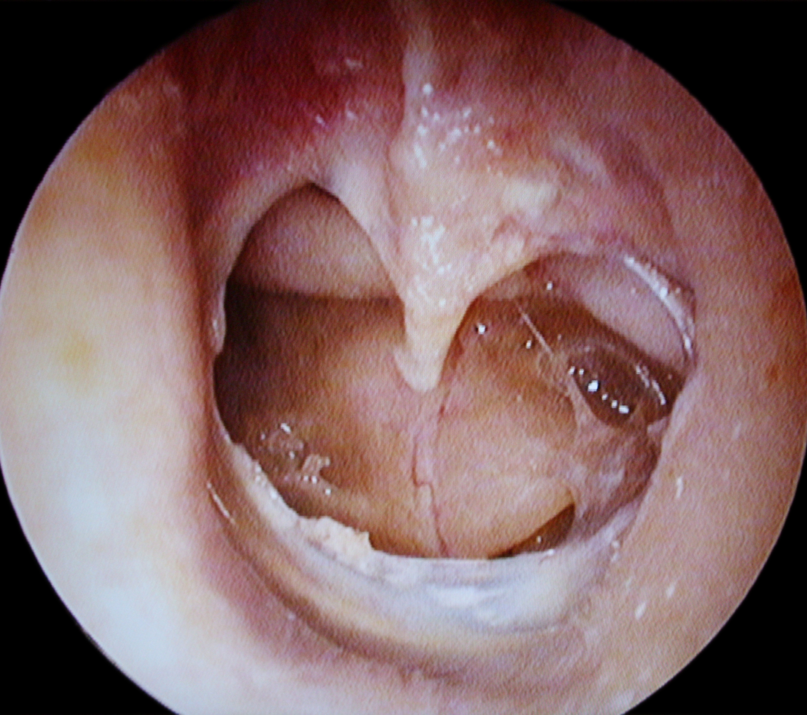
Otoscopy alone is usually sufficient to establish the diagnosis — a visible tear or hole in the tympanic membrane, with documentation of its location, approximate size (small/moderate/large/subtotal), and any visible middle-ear findings (discharge character, squamous debris, granulation tissue, or a retraction pocket).[1][2] Where the perforation itself is difficult to visualize directly (very small, or obscured by debris or discharge), absent tympanic membrane movement on pneumatic otoscopy is a useful confirmatory sign, since an intact drum should move visibly with gentle pressure changes and a perforated one will not.[2]
Tuning fork tests
Weber and Rinne tests support and characterize the clinical picture.[1] A perforation classically produces a conductive pattern — Weber lateralizes toward the affected ear, and Rinne is negative (bone conduction greater than air conduction) on that side. A Weber test lateralizing away from the affected ear (toward the good ear) is the pattern that does not fit simple perforation and points toward a sensorineural component instead — this is one of the specific red flags in this chapter that changes the plan to prompt referral.[2][1]
Audiometry and tympanometry
Formal pure-tone audiometry and tympanometry provide definitive, quantitative evaluation and are recommended both at initial assessment (when hearing loss is a concern) and after the healing period to confirm resolution — Canadian practice guidance specifically recommends audiometric testing at 3 months post-injury to verify healing, coinciding with the same 3-month mark that triggers ENT referral if the perforation has not closed.[2][1]
Imaging is not routinely needed
Computed tomography of the temporal bone is reserved for cases where an additional complication is suspected — possible cholesteatoma, suspected ossicular chain disruption, or another structural concern — rather than for routine, uncomplicated perforations.[1]
Differential diagnosis
| Conditions that can mimic, coexist with, or be mistaken for tympanic membrane perforation | |
|---|---|
| Acute otitis media (without perforation) | Chronic suppurative otitis media |
| Otitis media with effusion | Otitis externa |
| Cholesteatoma | Bullous myringitis |
| Tympanosclerosis | Otosclerosis |
| Middle ear osteoma | A patent tympanostomy tube (mimics a small central perforation on cursory exam) |
Not every patient presenting with otorrhea has a perforation — otitis externa alone, or a discharging tympanostomy tube, can look similar without a true membrane tear; otoscopy is what actually distinguishes them.[1]
Treatment Targets
- Prevent contamination of the exposed middle ear with water or pathogens during the healing window, since the perforation removes the eardrum's normal protective barrier.
- Control any active otorrhea/infection with an appropriately selected topical (and, where indicated, systemic) antibiotic.
- Never expose the middle ear and round window to an ototoxic agent — this is the single highest-stakes error to avoid in managing this condition, and applies to every otic drop recommendation or dispensing decision made while a perforation is present or even simply unconfirmed.
- Support spontaneous healing with dry-ear precautions rather than defaulting to unnecessary intervention — most perforations close on their own within weeks.
- Set a clear, evidence-based timeline for reassessment (audiometry and otoscopy around 3 months) rather than open-ended, unstructured follow-up.
- Recognize the red flags that mean this specific case is not routine — persistent perforation, significant hearing loss, vertigo, facial weakness, suspected cholesteatoma — and refer promptly rather than continuing to observe.
- Educate patients clearly on what not to do: no ear syringing/irrigation, no cotton swabs, no swimming or diving until healed and cleared.
- Refer appropriately for surgical repair (tympanoplasty/myringoplasty) when a perforation does not close on its own, rather than leaving a non-healing perforation unaddressed indefinitely.
Choose Initial Treatment
The round window is directly exposed through a perforated eardrum — never use, dispense, or recommend an ototoxic otic preparation
For a drop placed in the ear canal to reach and damage the inner ear, it has to cross the eardrum — normally via a perforation, a tympanostomy tube, or a graft that has not fully sealed — and then diffuse across the round window membrane into the inner ear fluids.[5] An intact eardrum is normally an excellent barrier against this; a perforated one is not a barrier at all.[2][6] Never use with a known, suspected, or unconfirmed perforation any otic preparation containing an aminoglycoside (neomycin, gentamicin, tobramycin, framycetin) — a documented case series of topical gentamicin used against a non-intact tympanic membrane produced permanent vestibulotoxicity, and Canadian clinical guidance states plainly that "aminoglycosides should be avoided as they are ototoxic" in this setting.[5][2] This rule applies even when the product is a familiar, widely available combination antibiotic-steroid otic drop — familiarity is not the same as safety here, and the ingredient list, not the brand recognition, is what has to be checked every time. See Pharmacologic Treatment Options and Drug Tables for the specific safe-versus-avoid comparison.
If the ear canal is dry and there is no active infection
Most uncomplicated perforations, no active otorrhea no topical or systemic drug therapy is needed at all — dry-ear precautions alone (see Non-Pharmacologic Management) support spontaneous closure, which occurs in roughly 80% of perforations overall, and in about 72% of small traumatic perforations within a month.[1][2] Do not reach for a prophylactic ear drop "just in case" — it adds ototoxicity risk for no established benefit in an uninfected, dry ear.
If there is active purulent otorrhea
First-line for purulent drainage through a perforation a fluoroquinolone-based topical otic preparation — ciprofloxacin-containing drops (with or without a combined corticosteroid, e.g., ciprofloxacin/dexamethasone or ciprofloxacin/fluocinolone) are the round-window-safe, evidence-supported choice, and this is true whether the perforation is spontaneous/traumatic or the result of a tympanostomy tube.[2][6] Randomized trials comparing topical ofloxacin, ciprofloxacin, or ciprofloxacin-dexamethasone against oral antibiotics for otorrhea through a non-intact tympanic membrane found topical therapy achieved superior clinical cure rates (77-96% versus 30-67% with systemic therapy alone in one guideline's cited trials) with fewer systemic adverse effects.[6] A typical course is a fixed, short duration (commonly around 7 days per standard product labelling for the combination otic suspensions) rather than open-ended use — prolonged or repeated courses of any topical quinolone drop have themselves been associated with otomycosis (fungal overgrowth) in the ear canal, so this is not a "the more the better" therapy either.[6] See Pharmacologic Treatment Options for the specific drug table.
If the perforation is due to acute otitis media in a child
Otitis-media-associated perforation with purulent otorrhea, especially in children treat the underlying acute otitis media with prompt systemic antimicrobial therapy per standard pediatric AOM guidance — Canadian Paediatric Society guidance is explicit that all children with a perforated eardrum presenting with AOM symptoms should be treated promptly with systemic antimicrobials (amoxicillin first-line) and examined for associated complications, rather than being managed with topical therapy alone.[4] A topical fluoroquinolone drop can still reasonably be added for local symptom control and to directly treat the otorrhea, but it does not substitute for the systemic antibiotic decision in this specific etiology.[4][6] See Special Populations and Pharmacologic Treatment Options.
What never has a place in this pathway
Do not perform or recommend ear syringing or irrigation when a perforation is known or suspected — this is explicitly contraindicated, since forcing fluid through the defect risks pain, infection, and further injury to already-vulnerable middle- and inner-ear structures.[2] This is the same contraindication that governs cerumen management around a perforation, applied here to a confirmed rather than merely suspected defect.
Non-Pharmacologic Treatment
Dry-ear (water) precautions — the other genuinely practical counseling point
- Keep the affected ear dry until it has fully healed and been confirmed closed on follow-up exam. Water entering an open middle ear through a perforation carries pathogens directly into a space that is normally sterile and protected.[2][1]
- For showering or bathing, a practical, low-cost method is a small ball of cotton lightly coated in petroleum jelly, or a properly fitted silicone earplug, placed at the entrance of the ear canal to block water — not pushed in deeply.[2]
- Avoid swimming, diving, and any deliberate submersion of the head until the perforation is confirmed healed on follow-up otoscopy — this includes recreational swimming, not only diving specifically.[1][2]
- Avoid activities involving rapid pressure changes — air travel (particularly descent) and scuba diving — during the acute healing period, since these are recognized causes of perforation in their own right and can also aggravate or reopen a healing one.[1]
- Do not attempt ear syringing, irrigation, or cerumen removal by any method that introduces fluid into the canal while a perforation is present — refer wax removal to a clinician using non-irrigation methods (curette or suction) instead.[2]
- Do not insert cotton swabs or any other object into the ear canal — this is both a leading original cause of traumatic perforation and a genuine risk of reinjuring a perforation that is in the process of healing.[3][2]
A genuine, important nuance: this is different from the "no water precautions needed" advice sometimes heard for tympanostomy tubes
Patients (or their caregivers) may have heard that children with surgically placed tympanostomy tubes generally do not need routine water precautions — this is accurate for that specific, distinct clinical context: a well-established guideline recommends against routine, prophylactic water precautions for children with tympanostomy tubes specifically, based on trial evidence showing minimal benefit for uncomplicated, non-draining tubes.[6] That recommendation does not transfer to an acute, unhealed traumatic or infectious perforation, and the same guideline explicitly carves out exceptions where water precautions remain appropriate even for tubes — active otorrhea, recurrent or prolonged otorrhea, and a history of problems with prior water exposure.[6] A freshly perforated, actively healing eardrum should be treated as one of those higher-caution situations, consistent with the dry-ear precautions above, not as a routine tube in a quiet, non-draining ear.
Analgesia
Simple analgesia (acetaminophen or, if no contraindication, ibuprofen) is appropriate for pain associated with the initial injury or infection, at standard adult or weight-based pediatric dosing. Persistent or worsening pain despite adequate analgesia and appropriate topical/systemic treatment of any infection should prompt reassessment rather than escalating analgesia alone.
Special Populations
Pediatrics
Children make up roughly a quarter of all tympanic membrane perforation cases, and the two dominant mechanisms differ sharply by age: young children (0-5 years) are most often injured by ear canal instrumentation (cotton-tipped applicators specifically), while teenagers more often perforate through water trauma.[1][3] Otitis-media-associated perforation is also common in this age group; the Canadian Paediatric Society is explicit that children presenting with AOM symptoms and a perforated eardrum need prompt systemic antimicrobial therapy (amoxicillin first-line), examined for associated complications, rather than topical therapy alone.[4] Children with pressure-equalization (tympanostomy) tubes are a related but distinct population — up to 3% develop a persistent perforation as a tube complication, and the specific, evidence-based recommendation against routine water precautions applies to quiet, non-draining tubes in this population specifically, not to an acute, actively healing perforation of any cause.[1][6] See Non-Pharmacologic Management for that distinction spelled out in full.
Divers and frequent flyers
Barotrauma from diving or air travel is a recognized, preventable cause of perforation.[1][2] Patients with a history of eustachian tube dysfunction or difficulty equalizing pressure benefit from proactive counseling (gentle equalization techniques, avoiding diving or flying with an active upper respiratory infection or congestion) as primary prevention, and should be counseled to avoid diving and flying entirely during the active healing period after a perforation, given both the direct barotrauma risk to the healing membrane and the general water-exposure risk of diving specifically.[1]
Occupational and blast exposure
Military personnel, industrial workers, and others with occupational exposure to blast or extreme acoustic pressure are a distinct risk group; a blast-related perforation should prompt a broader evaluation for associated injury (tympanic membrane perforation from a blast is often accompanied by inner-ear injury, and hearing protection counseling is a genuine, practical prevention opportunity for this population going forward).[3][1]
Immunocompromise
An open middle ear in an immunocompromised patient carries a materially higher infection risk from any water or pathogen exposure; apply dry-ear precautions and the round-window-safe drop selection rule with a lower threshold for professional (rather than purely home) management and a lower threshold for referral if infection develops or persists.
Pregnancy and breastfeeding
Topical otic fluoroquinolone drops (ciprofloxacin/dexamethasone, ciprofloxacin/fluocinolone) have minimal systemic absorption from otic administration and are reasonable options when clinically indicated; confirm current product-specific guidance for the individual patient. Standard AOM-associated systemic antibiotic choices (amoxicillin, amoxicillin-clavulanate) are well-established as compatible with pregnancy and breastfeeding at usual doses. Acetaminophen remains the preferred analgesic in pregnancy; ibuprofen requires the same standard trimester-specific caution applied to any NSAID use in pregnancy, unrelated to the ear condition itself.
Monitoring & Follow-Up
Set the follow-up expectation clearly and early: most perforations are expected to close spontaneously within weeks, and Canadian practice guidance specifically recommends audiometric testing at 3 months post-injury to confirm healing — the same 3-month mark at which a perforation that has not closed becomes the trigger for ENT referral.[2] Otoscopy alongside that audiometric check confirms both anatomic closure and functional hearing recovery, since hearing outcomes can be somewhat variable and are not perfectly predictable from perforation size alone.[1]
For a perforation being managed with topical fluoroquinolone therapy for active otorrhea, reassess after the treatment course (commonly around 7 days): resolved drainage with an otherwise uncomplicated course can proceed to routine expectant follow-up, while otorrhea persisting beyond 2 weeks of appropriate topical antibiotic therapy is a specific, defined trigger to reassess and refer rather than simply repeating or extending the same course.[2]
Counsel every patient to return promptly, rather than waiting for the scheduled follow-up, if they develop new or worsening pain, new drainage after a quiet interval, dizziness, facial weakness, or a hearing change — these can indicate secondary infection, a complication, or an alternative diagnosis rather than the expected course of a healing perforation.
Dry-ear precautions and the ototoxic-drop-avoidance rule both remain in force for the entire interval until healing is otoscopically confirmed — not just for the first few days or until pain resolves, since a perforation can still be open well after symptoms have settled.
Common Errors to Avoid
- Recommending or dispensing an aminoglycoside-containing (or other ototoxic-ingredient) otic drop without first asking about or examining for a perforation — the single highest-stakes error covered in this chapter; the round window has no protection once the eardrum is open.[5][2]
- Assuming a familiar, commonly stocked combination antibiotic-steroid otic drop is automatically safe simply because it is a normal, everyday otic product — familiarity is not the same as an ototoxicity check; the ingredient list has to be read every time.
- Performing or recommending ear syringing/irrigation, or cerumen removal by irrigation, when a perforation is known or suspected.[2]
- Recommending a prophylactic "just in case" ear drop for a dry, uninfected, healing perforation — this adds ototoxicity risk without an established benefit.
- Treating cotton-swab use as harmless routine ear hygiene rather than actively counseling against it, both as a leading original cause of perforation and as a genuine risk of reinjuring a healing one.[3][2]
- Applying the "routine water precautions are not needed" guidance for quiet, non-draining tympanostomy tubes to an acute, actively healing traumatic or infectious perforation — these are genuinely different clinical situations with different evidence behind them.[6]
- Assuming that pain resolution means healing is complete — a perforation can remain open well after acute pain has settled, and dry-ear precautions need to continue until otoscopically confirmed closure.
- Treating a child's AOM-associated perforation with topical therapy alone rather than the systemic antimicrobial therapy that guidance specifically calls for in this population.[4]
- Dismissing vertigo, facial weakness, or hearing loss out of proportion to exam findings as expected features of a routine perforation rather than recognizing them as red flags.
- Continuing the same topical antibiotic course indefinitely rather than reassessing and referring once otorrhea has persisted beyond 2 weeks of appropriate therapy.[2]
- Prescribing prolonged or repeated courses of topical quinolone drops without reason — associated with otomycosis, and not supported for open-ended use.[6]
Clinical Decision Algorithm
Drug Tables
Round-Window-Safe Otic Antibiotics (fluoroquinolone-based) — Preferred with a Perforated or Non-Intact Eardrum
These do not contain an aminoglycoside and are the correct first-line choice for purulent otorrhea through a known or suspected perforation, including tympanostomy tube otorrhea.
| Drug | Dosage | Adverse effects | Drug interactions | Comments |
|---|---|---|---|---|
| Ciprofloxacin / dexamethasone otic | Adults and children: 4 drops into the affected ear twice daily for 7 days | Local stinging; taste disturbance if drops reach the pharynx via the eustachian tube | None significant with otic use | First-line; safe with a non-intact tympanic membrane. Confirm current product-specific labelling for the patient's age and indication. |
| Ciprofloxacin / fluocinolone acetonide otic | Adults and children: 4 drops into the affected ear twice daily for 7 days | Local stinging or irritation | None significant with otic use | Specifically labelled for tympanostomy tube otorrhea in children — a real-world example of a fluoroquinolone otic product designed for exactly this non-intact-eardrum scenario. |
| Ofloxacin otic 0.3% | Adults and children: Per product labelling | Local stinging | None significant with otic use | A genuinely round-window-safe fluoroquinolone otic option used elsewhere for this indication. This platform's Drug Database entry for ofloxacin documents the ophthalmic solution only — confirm the actual product and its labelled route before dispensing. |
Ototoxic or Irritant Otic Preparations — AVOID with a Perforated or Non-Intact Eardrum
The round window is directly exposed once the eardrum is open. Confirm the eardrum is intact — otoscopically, or by clear history — before ever recommending or dispensing any of the following.
| Drug | Dosage | Adverse effects | Drug interactions | Comments |
|---|---|---|---|---|
| Framycetin / dexamethasone / gramicidin otic-ophthalmic | Do not use with a perforation: Not applicable | Ototoxicity (vestibular and/or cochlear) if it reaches the round window through a perforation | None significant otherwise | Contains framycetin, an aminoglycoside. A familiar, genuinely otic-labelled combination product for otitis externa with an intact drum — but the wrong choice once the eardrum is not intact. |
| Aminoglycoside-containing otic drops generally (neomycin/polymyxin B-based combinations, gentamicin otic, tobramycin otic) | Do not use with a perforation: Not applicable | Ototoxicity (cochlear and/or vestibular); a documented case series exists for topical gentamicin used against a non-intact membrane | None significant otherwise | Commonly reached for empirically for "ear infection" or otitis externa. Default to a fluoroquinolone-based product instead whenever perforation status cannot be confirmed. |
| Acetic acid 2% otic solution | Do not use with a perforation: Not applicable | Direct irritation of exposed middle-ear mucosa; pain on instillation into a non-intact ear | None significant otherwise | Avoided for a different reason than the aminoglycosides above — an irritant/chemical concern for exposed middle-ear tissue, not classic round-window ototoxicity — but the dispensing advice is the same. |
Systemic Antibiotics for Otitis-Media-Associated Perforation
The presence of a perforation does not change which oral antibiotic is preferred for the underlying acute otitis media — it changes the urgency of treating it. Canadian Paediatric Society guidance: children with a perforated eardrum and AOM symptoms should be treated promptly with systemic antimicrobials, not topical therapy alone.
| Drug | Dosage | Adverse effects | Drug interactions | Comments |
|---|---|---|---|---|
| Amoxicillin | Adults: Standard adult dosing Children: Weight-based pediatric dosing per current guidance | GI upset, hypersensitivity reactions | Reduced efficacy of some hormonal contraceptives (theoretical); reduced efficacy with concurrent doxycycline | First-line for AOM-associated perforation, particularly in children. |
| Amoxicillin / clavulanic acid | Adults: Standard adult dosing Children: Weight-based pediatric dosing per current guidance | GI upset (higher rate than amoxicillin alone), hypersensitivity reactions | Same class-level interaction profile as amoxicillin | Second-line for treatment failure, recent amoxicillin exposure, or a concurrent purulent conjunctivitis picture. |
Analgesia for Associated Discomfort
Adjunctive only — not a treatment for the perforation or any underlying infection. Persistent or worsening pain despite adequate analgesia needs reassessment rather than escalating analgesia alone.
| Drug | Dosage | Adverse effects | Drug interactions | Comments |
|---|---|---|---|---|
| Acetaminophen | Adults: Standard adult dosing Children: Weight-based pediatric dosing | Hepatotoxicity with overdose or significant hepatic impairment | None significant at standard doses | First-line analgesic, including in pregnancy. |
| Ibuprofen | Adults: Standard adult dosing Children: Weight-based pediatric dosing | GI upset, renal impairment with prolonged use | Increased bleeding risk with anticoagulants; avoid with other NSAIDs | Reasonable alternative if acetaminophen is not appropriate or insufficient; standard NSAID-in-pregnancy cautions apply, unrelated to the ear condition itself. |
References
- Tympanic Membrane Perforation — StatPearls Publishing (Sutton AE, Weimer AD). StatPearls, updated December 1, 2025
- Traumatic tympanic membrane perforations — Canadian Medical Association Journal (Harvie M, Roy CF, Gurberg J). CMAJ 2024;196(3):E100
- Traumatic Tympanic Membrane Perforations Diagnosed in Emergency Departments — JAMA Otolaryngology-Head & Neck Surgery (Carniol ET, Bresler A, Shaigany K, Svider P, Baredes S, Eloy JA, Ying YM). JAMA Otolaryngol Head Neck Surg 2018;144(2):136-139
- Management of acute otitis media in children six months of age and older — Canadian Paediatric Society, Infectious Diseases and Immunization Committee (Le Saux N, Robinson JL). Paediatr Child Health 2016;21(1):39-44, reaffirmed November 21, 2024
- Ototoxicity and topical eardrops — Clinical Otolaryngology and Allied Sciences (Marais J, Rutka JA). Clin Otolaryngol Allied Sci 1998;23(4):360-367
- Clinical Practice Guideline: Tympanostomy Tubes in Children (Update) — American Academy of Otolaryngology-Head and Neck Surgery Foundation (Rosenfeld RM, Tunkel DE, Schwartz SR, et al.). Otolaryngol Head Neck Surg 2022;166(1_suppl):S1-S55
Professional reference only. Apply clinical judgment and verify patient-specific decisions against current authoritative guidance and product information.