Systemic Vasculitis
Systemic Vasculitis
References
- 2012 Revised International Chapel Hill Consensus Conference Nomenclature of Vasculitides — Arthritis & Rheumatism (Jennette JC, Falk RJ, Bacon PA, et al.). Arthritis Rheum 2013;65(1):1-11
- 2021 American College of Rheumatology/Vasculitis Foundation Guideline for the Management of Giant Cell Arteritis and Takayasu Arteritis — Arthritis & Rheumatology (Maz M, Chung SA, Abril A, et al.). Arthritis Rheumatol 2021;73(8):1349-1365
- 2022 American College of Rheumatology/EULAR classification criteria for Takayasu arteritis — Annals of the Rheumatic Diseases (Grayson PC, Ponte C, Suppiah R, et al.). Ann Rheum Dis 2022;81(12):1654-1660
- CanVasc Consensus Recommendations for the Management of Antineutrophil Cytoplasm Antibody-associated Vasculitis: 2020 Update — Journal of Rheumatology (Mendel A, Ennis D, Go E, et al.; CanVasc). J Rheumatol 2021;48(4):555-566
- EULAR/PRINTO/PRES criteria for Henoch-Schönlein purpura, childhood polyarteritis nodosa, childhood Wegener granulomatosis and childhood Takayasu arteritis: Ankara 2008. Part II: Final classification criteria — Annals of the Rheumatic Diseases (Ozen S, Pistorio A, Iusan SM, et al.; Paediatric Rheumatology International Trials Organisation). Ann Rheum Dis 2010;69(5):798-806
- EULAR recommendations for the management of ANCA-associated vasculitis: 2022 update — Annals of the Rheumatic Diseases (Hellmich B, Sanchez-Alamo B, Schirmer JH, et al.). Ann Rheum Dis 2024;83(1):30-47
- 2022 American College of Rheumatology/EULAR classification criteria for giant cell arteritis — Annals of the Rheumatic Diseases (Ponte C, Grayson PC, Robson JC, et al.). Ann Rheum Dis 2022;81(12):1647-1653
- Trial of Tocilizumab in Giant-Cell Arteritis — New England Journal of Medicine (Stone JH, Tuckwell K, Dimonaco S, et al.; GiACTA). N Engl J Med 2017;377(4):317-328
- Rituximab versus Cyclophosphamide for ANCA-Associated Vasculitis — New England Journal of Medicine (Stone JH, Merkel PA, Spiera R, et al.; RAVE-ITN Research Group). N Engl J Med 2010;363(3):221-232
- Rituximab versus Cyclophosphamide in ANCA-Associated Renal Vasculitis — New England Journal of Medicine (Jones RB, Cohen Tervaert JW, Hauser T, et al.; RITUXVAS Investigators). N Engl J Med 2010;363(3):211-220
- Plasma Exchange and Glucocorticoids in Severe ANCA-Associated Vasculitis — New England Journal of Medicine (Walsh M, Merkel PA, Peh CA, et al.; PEXIVAS Investigators). N Engl J Med 2020;382(7):622-631
- Rituximab versus Azathioprine for Maintenance in ANCA-Associated Vasculitis — New England Journal of Medicine (Guillevin L, Pagnoux C, Karras A, et al.; MAINRITSAN Investigators). N Engl J Med 2014;371(19):1771-1780
- Avacopan for the Treatment of ANCA-Associated Vasculitis — RETRACTED June 29, 2026 — New England Journal of Medicine (Jayne DRW, Merkel PA, Schall TJ, Bekker P) — retraction notice: data integrity concerns identified by FDA investigation. N Engl J Med 2021;384:599-609 (retracted 2026)
- TAVNEOS (avacopan) and Health Canada's Review of Data Integrity Concerns — Health Canada. Health Canada Advisory, July 7, 2026
- Mepolizumab or Placebo for Eosinophilic Granulomatosis with Polyangiitis — New England Journal of Medicine (Wechsler ME, Akuthota P, Jayne D, et al.; MIRRA Trial Investigators). N Engl J Med 2017;376(20):1921-1932
Clinical Snapshot
Systemic vasculitis is inflammation and necrosis of blood vessel walls that impairs blood flow through the affected vessel, producing organ damage from ischemia rather than from the vessel wall itself. The diseases grouped under this heading share that mechanism but otherwise differ enormously in typical patient age, vessel calibre, organ targets, and treatment — which is exactly why the vessel-size framework below, from the 2012 Revised International Chapel Hill Consensus Conference (CHCC) Nomenclature of Vasculitides, is the organizing structure for this entire chapter rather than an optional preamble.[1]
| Vessel size | Diseases covered in this chapter | Typical vessels involved |
|---|---|---|
| Large vessel | Giant cell arteritis; Takayasu arteritis | Aorta and its major branches |
| Medium vessel | Polyarteritis nodosa; Kawasaki disease (see this platform's dedicated Kawasaki Disease chapter — not duplicated here) | Main visceral arteries and their initial branches |
| Small vessel — ANCA-associated | Granulomatosis with polyangiitis; microscopic polyangiitis; eosinophilic granulomatosis with polyangiitis | Intraparenchymal arteries, arterioles, capillaries, venules |
| Small vessel — immune-complex | IgA vasculitis (Henoch-Schönlein purpura) | Arterioles, capillaries, venules, via vessel-wall IgA immune-complex deposition rather than ANCA |
Two practical consequences follow directly from this framework. First, age is itself diagnostic: giant cell arteritis essentially never occurs under age 50, Takayasu arteritis classically presents under age 40, and IgA vasculitis is overwhelmingly a disease of childhood — a patient's age alone should narrow the differential before a single test is ordered. Second, vessel size predicts the dominant clinical picture: large-vessel disease produces claudication, bruits, and asymmetric pulses/pressures; medium-vessel disease produces mononeuritis multiplex, visceral microaneurysms, and skin nodules; small-vessel disease produces palpable purpura, glomerulonephritis, and pulmonary hemorrhage. Recognizing which pattern a patient fits is the single fastest route to the right diagnosis.
Why giant cell arteritis and ANCA-associated vasculitis anchor this chapter
Every vasculitis in this chapter matters, but two deserve to be treated as centrepieces because delay in recognizing and treating them causes irreversible harm on a timescale of hours to weeks rather than months: giant cell arteritis, where delayed glucocorticoid therapy causes permanent vision loss (see Red Flags & Refer and Choose Initial Treatment), and ANCA-associated vasculitis, where delayed or under-intensive immunosuppression allows progression to end-stage kidney disease or fatal pulmonary hemorrhage, while over-cautious avoidance of the necessary immunosuppression's real infection risk is itself a genuine, preventable cause of death (see Choose Initial Treatment and Special Populations).
Recognize the Condition
Large-vessel vasculitis
Giant cell arteritis (GCA)
The most common systemic vasculitis in adults over 50, with incidence rising steeply with age and a genuine female predominance (roughly 2-3:1); most common in populations of Northern European ancestry.[2] Classic features include a new, often temporal, headache; scalp tenderness (pain on combing hair or resting the head on a pillow); jaw or tongue claudication (pain with chewing that eases with rest — a genuinely specific finding, caused by ischemia of the muscles of mastication); and constitutional symptoms (fever, weight loss, malaise). Visual symptoms are the finding that changes everything — amaurosis fugax (transient monocular vision loss), diplopia, or any acute visual disturbance in this clinical context reflects arteritic anterior ischemic optic neuropathy or central retinal artery occlusion and is a direct precursor to permanent, irreversible blindness, sometimes within hours (see Red Flags & Refer). Roughly 40-60% of GCA patients have overlapping polymyalgia rheumatica (proximal shoulder and hip girdle pain and stiffness, worse in the morning) at diagnosis, and a patient presenting with apparently isolated polymyalgia rheumatica should always be screened for GCA symptoms specifically, since the two exist on a spectrum rather than as fully separate diseases.
Takayasu arteritis
A large-vessel vasculitis that is, in most other respects, GCA's younger mirror image: classically presenting under age 40, with a strong female predominance (roughly 8-9:1) and a materially higher incidence in patients of Asian ancestry, though it occurs worldwide.[3] Early disease produces nonspecific constitutional symptoms that are easy to miss; the diagnosis becomes apparent as large-vessel stenosis, occlusion, or aneurysm develops, producing limb claudication (classically upper-limb, from subclavian involvement), a measurable blood pressure discrepancy of more than 10 mmHg between arms, diminished or absent peripheral pulses (the historical name "pulseless disease"), carotidynia, vascular bruits (subclavian, carotid, abdominal aorta), and, in more advanced disease, hypertension from renal artery involvement or aortic regurgitation from aortic root dilation. Because early symptoms are nonspecific and pulse/pressure asymmetry can be subtle, diagnosis is frequently delayed by months to years from symptom onset — a genuine, well-documented pattern that argues for actively checking bilateral blood pressures and pulses in any young patient with unexplained constitutional symptoms and elevated inflammatory markers.
Medium-vessel vasculitis
Polyarteritis nodosa (PAN)
A necrotizing vasculitis of medium-sized muscular arteries that is, critically, not ANCA-associated and does not cause glomerulonephritis — both genuinely important distinguishing points from the small-vessel ANCA-associated vasculitides covered below, since PAN spares the glomerular capillaries even as it can produce severe hypertension from renal artery microaneurysms and infarction.[1] Classic features include mononeuritis multiplex (asymmetric, stepwise sensorimotor neuropathy — a foot drop or wrist drop appearing over days is a hallmark), livedo reticularis, tender subcutaneous nodules along artery courses, testicular pain (from testicular artery involvement — a finding that should prompt vasculitis workup rather than reflexive urologic referral alone), and gastrointestinal ischemia that can progress to bowel infarction or perforation. A genuinely important subset is hepatitis B-associated PAN, historically responsible for a substantial proportion of cases before universal HBV vaccination reduced its incidence; every new PAN diagnosis should include hepatitis B surface antigen testing, since HBV-associated disease is managed with a fundamentally different, antiviral-led strategy rather than the immunosuppression used for idiopathic disease (see Choose Initial Treatment).
Kawasaki disease — cross-reference
Kawasaki disease is the medium-vessel vasculitis of childhood under the Chapel Hill classification and this platform's existing Kawasaki Disease chapter covers its full diagnostic criteria, IVIG and aspirin dosing, and coronary-artery-aneurysm surveillance pathway in complete detail — that content is not repeated here.
Small-vessel ANCA-associated vasculitis (AAV)
The three AAV subtypes share pauci-immune small-vessel inflammation (minimal immune-complex deposition on biopsy, distinguishing them from IgA vasculitis) but differ meaningfully in organ pattern and typical ANCA serology (see Assessment & Diagnosis for the full serology table).
Granulomatosis with polyangiitis (GPA, formerly Wegener granulomatosis)
Defined by granulomatous inflammation of the respiratory tract in addition to small-vessel vasculitis. Upper airway disease is genuinely distinctive: chronic sinusitis unresponsive to usual therapy, saddle-nose deformity from destruction of nasal cartilage, subglottic stenosis producing stridor or voice change, and otitis media/mastoiditis. Lower respiratory tract disease produces pulmonary nodules (which can cavitate) and, in severe disease, diffuse alveolar hemorrhage. Renal disease is pauci-immune crescentic glomerulonephritis, often clinically silent until advanced. Roughly 90% of active generalized GPA is PR3-ANCA/c-ANCA positive.[4]
Microscopic polyangiitis (MPA)
Small-vessel vasculitis without the granulomatous upper-airway disease that defines GPA. The classic, genuinely dangerous presentation is a pulmonary-renal syndrome — simultaneous diffuse alveolar hemorrhage (hemoptysis, dyspnea, falling hemoglobin) and pauci-immune crescentic glomerulonephritis (rising creatinine, active urinary sediment) — which is a medical emergency requiring immediate, aggressive treatment (see Red Flags & Refer). Roughly 70% of MPA is MPO-ANCA/p-ANCA positive.[4]
Eosinophilic granulomatosis with polyangiitis (EGPA, formerly Churg-Strauss syndrome)
The AAV subtype most likely to be missed early, because it begins as apparent severe, late-onset asthma rather than as obvious vasculitis. EGPA classically evolves through overlapping phases: a prodromal allergic phase (adult-onset asthma, allergic rhinitis, nasal polyps, sometimes for years before diagnosis); an eosinophilic phase (peripheral eosinophilia and eosinophilic tissue infiltration — pulmonary infiltrates, eosinophilic gastroenteritis); and a vasculitic phase (mononeuritis multiplex, palpable purpura, and, most consequentially for mortality, eosinophilic myocarditis and cardiomyopathy). Only roughly 30-40% of EGPA is ANCA-positive (usually MPO/p-ANCA when positive), and this serologic split is not incidental: ANCA-positive EGPA behaves more like a genuine vasculitis, with more renal and neurologic involvement, while ANCA-negative EGPA behaves more like a severe eosinophilic organ-infiltrative disease, with more cardiac and pulmonary involvement — a distinction that increasingly shapes treatment choice (see Choose Initial Treatment).
Small-vessel immune-complex vasculitis
IgA vasculitis (Henoch-Schönlein purpura)
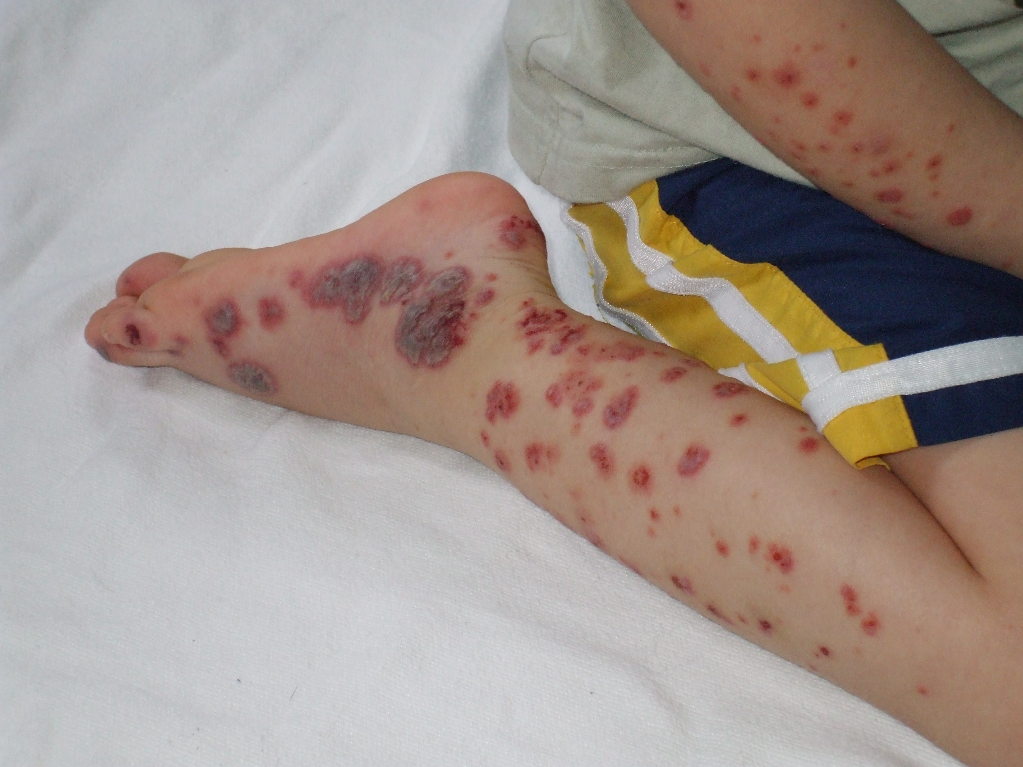
IgA vasculitis is the most common vasculitis of childhood, with a peak incidence between ages 3 and 15 and a generally self-limited course that stands in deliberate contrast to the chronic, relapsing, immunosuppression-dependent adult AAV subtypes above — this distinction should shape expectations and counselling from the first visit.[5] It follows an upper respiratory infection in a substantial proportion of cases and is mediated by IgA1 immune-complex deposition in small vessels, rather than ANCA. The classic tetrad is palpable purpura (non-thrombocytopenic, classically on the buttocks and lower extremities, sometimes with associated edema), arthralgia or arthritis (usually large joints, non-deforming), abdominal pain (from bowel wall edema and hemorrhage, which can be severe and is a recognized precipitant of intussusception in children — see Red Flags & Refer), and renal involvement (hematuria with or without proteinuria, histologically and pathophysiologically continuous with IgA nephropathy — see the platform's IgA Nephropathy and Glomerulonephritis chapters for the shared renal workup). Most children recover fully within 4-6 weeks without any disease-modifying therapy; the renal trajectory, not the skin or joint findings, is what determines long-term prognosis and the small minority of patients who progress to chronic kidney disease.
Risk Factors
- Age over 50 — an essentially required feature of giant cell arteritis; GCA in a patient under 50 should prompt diagnostic reconsideration rather than acceptance.[2]
- Age under 40, female sex, and Asian ancestry — the classic Takayasu arteritis demographic, though the disease occurs across ancestries.[3]
- A history of polymyalgia rheumatica — a genuine risk marker for concurrent or subsequent GCA, not a separate, unrelated diagnosis.
- Northern European ancestry, for GCA specifically.
- Hepatitis B infection — the defining risk factor for the HBV-associated PAN subset, now uncommon in populations with high HBV vaccination coverage but still worth actively excluding in every new PAN diagnosis.
- A personal history of adult-onset asthma, allergic rhinitis, or nasal polyps — the prodromal-phase risk pattern that precedes eosinophilic granulomatosis with polyangiitis, sometimes by years.
- Childhood age (peak 3-15 years) and a preceding upper respiratory infection — the typical IgA vasculitis risk pattern; a minority of cases follow a specific medication exposure or streptococcal infection.
- PR3-ANCA/c-ANCA positivity specifically — associated with a higher relapse rate than MPO-ANCA/p-ANCA-positive disease across the AAV spectrum, a genuine prognostic distinction that should inform the intensity and duration of maintenance therapy (see Choose Initial Treatment and Monitoring & Follow-Up).[6]
Red Flags & Refer
Any new visual symptom in a patient with suspected giant cell arteritis is an ophthalmologic and rheumatologic emergency. Amaurosis fugax, diplopia, or any acute visual disturbance occurring with new headache, jaw claudication, scalp tenderness, or unexplained constitutional symptoms and elevated inflammatory markers in a patient over 50 requires same-day high-dose glucocorticoid therapy and same-day ophthalmology assessment — not routine referral, and not deferral pending a scheduled biopsy (see Choose Initial Treatment and the Clinical Decision Algorithm). Vision loss that has already occurred is usually permanent; the entire purpose of urgent treatment is to protect the contralateral eye and any remaining vision in the affected eye, since bilateral, sequential involvement within days is a recognized and devastating pattern if treatment is delayed.
Beyond the GCA visual emergency, the following also demand urgent, same-day escalation rather than routine follow-up:
- Hemoptysis with a rising creatinine or active urinary sediment in a patient with suspected or known AAV — this pulmonary-renal syndrome pattern (classically MPA, but seen in GPA as well) reflects diffuse alveolar hemorrhage plus rapidly progressive glomerulonephritis and requires immediate hospital-based, specialist-directed treatment.
- A rapidly rising creatinine or new active urinary sediment (dysmorphic red cells, red cell casts) in any patient with known or suspected AAV — treat as rapidly progressive glomerulonephritis until proven otherwise; delay materially worsens the chance of renal recovery.
- A new, stepwise mononeuropathy (foot drop, wrist drop, or similar) in a patient with a systemic illness consistent with PAN or EGPA — this is mononeuritis multiplex until proven otherwise and warrants urgent specialist assessment.
- New or worsening limb ischemia, a new bruit with an ischemic limb, or evidence of aortic dissection or severe aortic regurgitation in a patient with known or suspected Takayasu arteritis.
- Acute abdominal pain with peritoneal signs in a patient with PAN or severe IgA vasculitis — evaluate urgently for bowel ischemia or perforation.
- Severe, colicky abdominal pain in a child with IgA vasculitis, particularly with vomiting or blood in the stool — evaluate for intussusception, a recognized and specifically actionable complication.
- New chest pain, dyspnea, or arrhythmia in a patient with EGPA, especially ANCA-negative disease — evaluate urgently for eosinophilic myocarditis, the leading driver of EGPA mortality.
- Fever in any patient on cyclophosphamide, rituximab, or high-dose glucocorticoids — treat as a potential serious infection in an immunosuppressed host until proven otherwise, given the genuine infection risk this therapy carries (see Choose Initial Treatment and Special Populations).
Assessment & Diagnosis
Giant cell arteritis
ESR and CRP are markedly elevated in the great majority of GCA cases and remain a reasonable first-line screen, but a normal ESR does not exclude the diagnosis — a small but genuine minority (on the order of a few percent) of biopsy-confirmed GCA presents with a normal ESR, so a compelling clinical picture should not be dismissed on inflammatory markers alone.[2] Temporal artery biopsy remains the diagnostic reference standard, but two practical points matter more clinically than the biopsy result itself: first, glucocorticoid therapy should never be delayed to obtain it (see Choose Initial Treatment), and second, biopsy sensitivity is preserved for roughly two weeks after starting glucocorticoids, so treating first and biopsying promptly after does not meaningfully compromise diagnostic yield. Skip lesions mean a negative unilateral biopsy does not fully exclude GCA; a longer biopsy segment (at least 1-2 cm), contralateral biopsy in a persistently high-suspicion patient, or vascular ultrasound demonstrating the "halo sign" (a hypoechoic ring around the temporal artery lumen from wall edema) as either an adjunct or, where locally validated, an alternative to biopsy are all reasonable per current guidance.[2] The 2022 ACR/EULAR classification criteria require age 50 or older at diagnosis plus a cumulative score of 6 or more points across weighted clinical, laboratory, and imaging/biopsy items (examples include morning stiffness in shoulders/neck, sudden visual loss, jaw/tongue claudication, new temporal artery abnormality on examination, ESR ≥50 or CRP ≥10 mg/L, and a positive biopsy or halo sign on ultrasound), though this tool is intended for research classification and structured clinical reasoning rather than as a bedside diagnostic checklist that overrides clinical judgment.[7]
Takayasu arteritis
Diagnosis rests on vascular imaging — CT angiography, MR angiography, or PET-CT — demonstrating wall thickening, stenosis, occlusion, or aneurysm of the aorta or its major branches, together with the clinical/examination findings in Recognize the Condition (pulse and blood pressure asymmetry, bruits, claudication).[3] Inflammatory markers are frequently, but not universally, elevated and correlate imperfectly with actual disease activity on imaging — a genuinely important limitation, since normal inflammatory markers do not reliably indicate quiescent vessel disease in Takayasu arteritis specifically. The 2022 ACR/EULAR classification criteria require age 60 or younger at diagnosis, evidence of vasculitis on imaging, plus a weighted point total from clinical and imaging criteria (examples include female sex, angina, limb claudication, bruits, pulse or blood pressure asymmetry, and the specific pattern of arterial involvement on imaging).[3]
ANCA serology — the central diagnostic tool for small-vessel ANCA-associated vasculitis
ANCA testing combines indirect immunofluorescence (which yields the classic c-ANCA/p-ANCA staining pattern) with antigen-specific ELISA testing for proteinase 3 (PR3) and myeloperoxidase (MPO) antibodies; current practice relies primarily on the ELISA result, with the immunofluorescence pattern as a supportive, historically named correlate.[4]
| ANCA pattern | Classic disease association | Practical notes |
|---|---|---|
| PR3-ANCA (c-ANCA pattern) | Granulomatosis with polyangiitis (~90% of active generalized disease) | Associated with a materially higher relapse rate than MPO-ANCA-positive disease — a genuine factor in choosing maintenance intensity and duration.[6] |
| MPO-ANCA (p-ANCA pattern) | Microscopic polyangiitis (~70%); a minority of granulomatosis with polyangiitis; eosinophilic granulomatosis with polyangiitis when ANCA-positive | Somewhat lower relapse rate than PR3-ANCA disease, though relapse still occurs and maintenance therapy remains necessary. |
| ANCA-negative | ~60-70% of eosinophilic granulomatosis with polyangiitis; a minority of granulomatosis with polyangiitis and microscopic polyangiitis | A negative ANCA does not exclude AAV — this is a genuine, easy-to-miss diagnostic pitfall (see Common Errors to Avoid); diagnosis in this setting rests on the clinical picture plus biopsy. |
Renal biopsy showing pauci-immune (minimal immune deposit) crescentic glomerulonephritis is the confirmatory test where renal involvement is present and is often pursued even in ANCA-positive disease to establish a baseline chronicity/activity index that guides both initial treatment intensity and prognosis.
Polyarteritis nodosa
ANCA serology is characteristically negative in PAN — a genuine, clinically useful distinguishing feature from the AAV subtypes above.[1] Diagnosis rests on conventional angiography demonstrating the classic microaneurysms and areas of stenosis in medium-sized visceral arteries (renal, mesenteric, hepatic), or biopsy of clinically involved tissue (sural nerve, skin, muscle) showing necrotizing arteritis of medium vessels without glomerulonephritis. Hepatitis B surface antigen testing is mandatory in every new diagnosis, since it fundamentally changes the treatment approach (see Choose Initial Treatment).
Eosinophilic granulomatosis with polyangiitis
Peripheral eosinophilia (typically >1.5 x 10⁹/L, or >10% of the differential) in a patient with the asthma/allergic-rhinitis prodrome described in Recognize the Condition is the key laboratory clue; biopsy of an accessible involved site (skin, nerve, lung) showing eosinophilic tissue infiltration with or without frank vasculitis supports the diagnosis. Cardiac assessment (ECG, troponin, and echocardiography or cardiac MRI where myocarditis is suspected) is warranted given its outsized contribution to mortality, particularly in ANCA-negative disease.
IgA vasculitis
Diagnosis in a child with the classic tetrad (palpable purpura, arthralgia, abdominal pain, renal involvement) is usually clinical, without requiring biopsy. The EULAR/PRINTO/PRES (Ankara 2008) criteria require palpable purpura (mandatory) plus at least one of: abdominal pain, histopathology showing typical leukocytoclastic vasculitis with predominant IgA deposition (or proliferative glomerulonephritis with IgA deposition), arthritis or arthralgia, or renal involvement (hematuria and/or proteinuria).[5] Skin biopsy with immunofluorescence for IgA deposition is reserved for atypical presentations (adult onset, atypical distribution, or diagnostic uncertainty) rather than routine practice. Urinalysis for hematuria/proteinuria at diagnosis and through follow-up is essential in every case, since renal involvement — not the skin or joint findings — determines long-term outcome (see Monitoring & Follow-Up, and the platform's IgA Nephropathy and Glomerulonephritis chapters for the shared renal workup).
Treatment Targets
- Prevent irreversible vision loss in giant cell arteritis by starting high-dose glucocorticoid therapy immediately upon clinical suspicion, without waiting for biopsy confirmation — the single most time-critical target in this chapter (see Choose Initial Treatment).
- Induce remission in ANCA-associated vasculitis promptly and completely, using an intensity matched to disease severity, to prevent progression to end-stage kidney disease or fatal pulmonary hemorrhage.
- Transition every AAV patient from induction to a distinct, lower-intensity maintenance regimen rather than continuing induction-intensity immunosuppression indefinitely, balancing relapse prevention against cumulative treatment toxicity.
- Minimize cumulative glucocorticoid exposure across every vasculitis in this chapter through the fastest safe taper and, where evidence supports it, an early steroid-sparing agent (tocilizumab in GCA; rituximab, methotrexate, or azathioprine in AAV) — cumulative steroid toxicity is itself a major, preventable source of long-term morbidity.
- Proactively prevent infection during induction-intensity immunosuppression, with Pneumocystis jirovecii pneumonia prophylaxis as the single most specific, actionable pharmacist-level intervention (see Choose Initial Treatment).
- Recognize hepatitis B-associated polyarteritis nodosa as a distinct disease requiring an antiviral-led, not immunosuppression-led, strategy.
- Support the generally self-limited course of pediatric IgA vasculitis without over-treating it as though it were adult AAV, while still monitoring the renal trajectory that determines its minority of genuinely chronic outcomes.
Choose Initial Treatment
This is the single most important directive in this chapter. Once GCA is clinically suspected — especially with any visual symptom — start high-dose glucocorticoids the same day, in the clinic or emergency department, before temporal artery biopsy has been performed or even scheduled.[2] Biopsy sensitivity is preserved for roughly two weeks after glucocorticoids are started, so treating first and biopsying promptly afterward does not meaningfully compromise the diagnostic yield — there is essentially no clinical scenario in which it is correct to withhold treatment pending biopsy in a patient with a compelling presentation.
Dosing without visual symptoms or other threatened-organ involvement: oral prednisone 40-60 mg/day (approximately 1 mg/kg/day, to a practical maximum around 60 mg/day).
Dosing with visual symptoms, or any other threatened-organ ischemia: intravenous methylprednisolone 500-1000 mg/day for 3 days, then transition to oral prednisone 1 mg/kg/day — the rationale being to achieve the fastest possible anti-inflammatory effect while any salvageable vision or perfusion remains at stake.[2]
Taper: continue the initial dose for 2-4 weeks once symptoms and inflammatory markers have responded, then taper gradually — commonly to roughly 15-20 mg/day by 2-3 months and to 5 mg/day or less by 12 months — with the total glucocorticoid course typically extending 12-18 months given GCA's tendency to relapse on an overly rapid taper.[2] Tailor the pace to symptoms and inflammatory markers rather than a fixed calendar, and treat any recurrence of headache, jaw claudication, or visual symptoms during the taper as a relapse requiring a dose increase, not as an unrelated new complaint.
The GiACTA trial randomized GCA patients to subcutaneous tocilizumab (weekly or every-other-week) plus a shorter glucocorticoid taper, or placebo plus a longer glucocorticoid taper; sustained glucocorticoid-free remission at 52 weeks was achieved in roughly 56% of weekly tocilizumab patients versus roughly 14-18% of placebo-plus-taper patients, with materially lower cumulative glucocorticoid exposure in the tocilizumab groups.[8] The practical rule: tocilizumab is a genuine, evidence-based option to add at or soon after diagnosis — not only after a relapse — specifically to reduce cumulative glucocorticoid exposure, and should be considered proactively in a patient at higher risk of steroid toxicity (osteoporosis, diabetes, or a prior steroid-related complication) rather than reserved as a last resort. It does not replace the need for immediate glucocorticoid initiation in Red Flags & Refer — it is added to, and allows a faster taper of, that initial glucocorticoid course, not a substitute for it.
Initial treatment mirrors GCA's glucocorticoid approach (prednisone 40-60 mg/day, tapered over months), but the chronic, often relapsing course and the young age of most Takayasu patients argue for adding a steroid-sparing agent early rather than waiting for a first relapse — methotrexate, azathioprine, mycophenolate mofetil, tocilizumab, or a TNF-alpha inhibitor are all reasonable options depending on specialist judgment and access, chosen specifically to limit the decades of cumulative glucocorticoid exposure a young patient would otherwise face.[2] Surgical or endovascular revascularization is reserved for critical, symptomatic stenosis or aneurysm, ideally performed once inflammation is controlled rather than during an active flare, given the higher risk of restenosis or anastomotic complications in active disease.
Induction therapy for organ- or life-threatening AAV combines glucocorticoids with either cyclophosphamide or rituximab; current evidence increasingly favours rituximab in most contexts, though cyclophosphamide remains a genuine, guideline-supported option.[4][6]
Glucocorticoids: intravenous methylprednisolone pulse therapy (commonly 500-1000 mg/day for 1-3 days) for severe, organ-threatening presentations, followed by oral prednisone starting around 50 mg/day (or per a reduced-dose regimen — see the PEXIVAS directive below) and tapered over the following months.
Rituximab: 375 mg/m² IV weekly for 4 weeks is one validated regimen (RAVE, RITUXVAS); an alternative 1000 mg IV x2 doses 2 weeks apart is also used in practice. The RAVE trial found rituximab non-inferior to cyclophosphamide for induction of remission at 6 months, with rituximab achieving a higher remission rate than cyclophosphamide specifically in patients with relapsing (as opposed to new-onset) disease.[9] The RITUXVAS trial, conducted in patients with renal AAV, found comparable remission and safety outcomes between rituximab- and cyclophosphamide-based induction.[10] Rituximab is generally preferred for relapsing disease, for patients wishing to avoid cyclophosphamide's gonadotoxicity and cumulative bladder/malignancy risk (see Special Populations), and increasingly as a default first choice where access allows.[4]
Cyclophosphamide: 2 mg/kg/day orally (adjusted for renal function and age), or intermittent IV pulse dosing per a specialist-directed protocol, generally for 3-6 months before transitioning to a maintenance agent; always given with mesna and aggressive hydration to reduce hemorrhagic cystitis risk (see Drug Tables), and remains a reasonable, guideline-supported choice, particularly where rituximab access is limited or for the most severe, rapidly progressive presentations.
The PEXIVAS trial, a 2x2 factorial design in 704 patients with severe AAV, found that a reduced-dose glucocorticoid regimen was non-inferior to a standard higher-dose regimen for the composite outcome of death or end-stage kidney disease, with fewer serious infections in the reduced-dose group — evidence that has meaningfully shifted practice toward front-loading a shorter high-dose glucocorticoid burst followed by a faster taper, rather than sustained high-dose therapy for months.[11] The same trial also found that plasma exchange did not reduce the composite outcome of death or end-stage kidney disease, challenging its previously routine use in severe AAV.[11] The practical rule: use a reduced-dose glucocorticoid regimen as the current evidence-based default for severe AAV induction, and reserve plasma exchange for specific scenarios (very severe pulmonary hemorrhage, an overlapping anti-glomerular basement membrane antibody picture, or dialysis-dependent presentations) at specialist discretion, rather than as routine therapy for AAV-associated renal disease generally.
Once remission is achieved (typically by 3-6 months), transition every AAV patient to maintenance therapy — continuing induction-intensity immunosuppression indefinitely is neither necessary nor safe.[4]
Rituximab maintenance is increasingly preferred where accessible: the MAINRITSAN trial compared fixed-schedule rituximab (500 mg at months 0, 2, 6, 12, and 18) against azathioprine for maintenance and found major relapse in only 5% of the rituximab group versus 28% of the azathioprine group by month 28, with a similar overall rate of severe adverse events between groups.[12] Azathioprine (2 mg/kg/day) and methotrexate (weekly, with folic acid supplementation) both remain reasonable, guideline-supported alternatives, particularly where rituximab access, cost, or patient preference favour an oral option; mycophenolate mofetil is a further alternative with somewhat weaker maintenance evidence than azathioprine. Maintenance duration: at least 18-24 months from remission is standard, extended further in PR3-ANCA-positive disease specifically, given its materially higher relapse rate (see Risk Factors and Assessment & Diagnosis).[6]
Avacopan (an oral C5a receptor antagonist) was approved as a steroid-sparing adjunct to standard induction therapy in AAV based on the ADVOCATE trial. That trial was retracted by the New England Journal of Medicine on June 29, 2026, after an FDA investigation found that primary-endpoint assessments for nine patients had been readjudicated after database lock and unblinding, without disclosure to two of the paper's own academic authors.[13] Health Canada issued its own advisory on July 7, 2026 directing prescribers not to initiate TAVNEOS (avacopan) in new patients while its data-integrity review is under way, and to contact and reassess every patient currently taking it regarding whether continued treatment remains appropriate; patients already on therapy should not stop it unilaterally without first discussing this with their prescriber.[14] This advisory is independent of, and in addition to, a serious postmarketing signal of drug-induced liver injury with avacopan, including fatal cases of vanishing bile duct syndrome.[14] The practical rule, current as of this chapter's authoring date: do not present avacopan as a routine or default AAV induction option; any question about a patient already taking it should be directed to the prescriber for individualized reassessment against the current regulatory guidance, which should always be re-checked given how fast-moving this specific situation is.
The degree of immunosuppression used for AAV induction carries a genuine, well-documented Pneumocystis jirovecii pneumonia (PJP) risk, and current EULAR guidance explicitly recommends prophylaxis during this period.[6] First-line: sulfamethoxazole-trimethoprim (single-strength daily, or double-strength three times weekly, per local protocol). For sulfa allergy or intolerance: dapsone, atovaquone, or nebulized pentamidine are reasonable alternatives, in roughly that order of preference based on efficacy and practicality (see Drug Tables). Duration: for the duration of cyclophosphamide therapy, and for at least 6 months after the last rituximab dose in a patient also receiving significant concurrent glucocorticoid exposure — continue longer for a patient remaining on a glucocorticoid dose of 20 mg/day or more.[6] This is a genuinely easy point to miss once attention is focused on the primary immunosuppressive regimen, and is exactly the kind of check a pharmacist reviewing a new AAV induction regimen should proactively confirm rather than assume has been addressed.
Hepatitis B-associated PAN: treat with antiviral therapy as the primary strategy, combined with a short initial course of glucocorticoids to control the most severe manifestations and, historically, plasma exchange — deliberately avoiding prolonged immunosuppression, since ongoing viral replication is the disease driver and sustained immunosuppression risks worsening it. This is a fundamentally different strategy from AAV management and is exactly why universal hepatitis B testing at diagnosis matters (see Assessment & Diagnosis).
Idiopathic PAN: glucocorticoids alone are often sufficient for milder, non-organ-threatening disease; cyclophosphamide is added for severe or organ-threatening presentations (significant renal involvement, GI, cardiac, or CNS disease, or a poor prognostic score on validated risk-stratification tools used by specialists). Rituximab evidence is far less established for PAN than for the AAV subtypes and is not a first-line substitute here.
Glucocorticoids remain first-line for EGPA, with cyclophosphamide or rituximab added for severe, vasculitic (ANCA-positive), or organ-threatening disease, following the same general induction/maintenance framework as the other AAV subtypes. The MIRRA trial specifically evaluated mepolizumab (an anti-IL-5 monoclonal antibody) added to standard therapy in relapsing or refractory EGPA and found significantly more weeks in remission and a higher proportion of patients achieving remission compared with placebo, establishing it as an approved, evidence-based option in this specific setting.[15] The practical rule: mepolizumab is a genuine option for relapsing or refractory EGPA, particularly where the eosinophilic/asthma-predominant manifestations (rather than florid vasculitis) are the dominant problem — it is an addition to, not a substitute for, appropriate vasculitis-directed immunosuppression when true vasculitic organ involvement (mononeuritis multiplex, glomerulonephritis, myocarditis) is present.
Most children need only supportive care: rest, hydration, and simple analgesia (an NSAID such as naproxen or ibuprofen is reasonable for arthralgia provided renal function is normal — see Common Errors to Avoid for the important exception). Glucocorticoids are commonly used for significant abdominal pain, since they may shorten its duration, though the evidence that they alter the overall disease course or prevent renal involvement is genuinely mixed and this should be communicated plainly rather than overstated. Renal involvement drives long-term management: mild hematuria/proteinuria warrants observation and monitoring (see Monitoring & Follow-Up); an ACE inhibitor or ARB is reasonable for persistent proteinuria, following the same principles as the platform's IgA Nephropathy chapter; significant nephritis with a crescentic pattern on biopsy may warrant glucocorticoids plus a steroid-sparing agent under nephrology direction, using the same general principles as the platform's Glomerulonephritis chapter rather than a separate IgA-vasculitis-specific protocol.
Non-Pharmacologic Treatment
Patient self-monitoring and red-flag education
Every GCA patient — and anyone on a glucocorticoid taper for it — needs explicit, repeated counselling to seek same-day care for any new visual symptom, since this is the single intervention most likely to prevent permanent vision loss outside of the initial treatment decision itself. AAV patients need equivalent counselling on the pulmonary-renal syndrome symptoms in Red Flags & Refer, and on fever as a possible serious infection rather than a routine viral illness given their immunosuppressed state.
Vaccination
Confirm routine immunization is current before starting significant immunosuppression wherever timing allows, since vaccine response is blunted once therapy (particularly rituximab or cyclophosphamide) is under way; avoid live vaccines during active immunosuppressive therapy. Annual influenza and age-appropriate pneumococcal vaccination are particularly relevant given the added respiratory infection risk this population carries.
Bone health
Every patient anticipated to need more than a brief glucocorticoid course should have baseline fracture-risk assessment and, where indicated, bone-protective therapy started proactively rather than reactively (see Drug Tables) — this applies across every vasculitis in this chapter given how central glucocorticoids are to initial treatment.
Smoking cessation
Genuinely relevant across the large- and medium-vessel vasculitides specifically, given the added vascular risk smoking carries in a patient already dealing with arterial inflammation and stenosis.
Fertility counselling
Discuss fertility preservation options before cyclophosphamide therapy wherever the clinical urgency allows for it, given its dose-dependent gonadotoxicity (see Special Populations) — this conversation is easy to defer under time pressure but should happen proactively rather than only when specifically raised by the patient.
Psychosocial support
The chronic, relapsing course of AAV and Takayasu arteritis in particular carries a genuine, often under-acknowledged burden — long-term immunosuppression, relapse anxiety, and, in Takayasu arteritis, a young age at diagnosis facing a lifelong disease. Proactively screen for and address mental health alongside physical disease management, and connect patients with vasculitis-specific patient-support organizations.
Pediatric IgA vasculitis — reassurance and graded activity
Most families need reassurance that this is a generally self-limited illness rather than a lifelong disease, together with clear, specific guidance on the red-flag symptoms in Red Flags & Refer (severe abdominal pain, blood in stool or urine, joint swelling limiting mobility) that should prompt reassessment. Activity can generally continue as tolerated; strict bed rest is not required and does not improve outcomes.
Pharmacologic Treatment Options
| Drug / intervention | Role | Dose / use | Important adverse effects | Teaching points |
|---|---|---|---|---|
| Prednisone 1st-line — all vasculitides in this chapter | Oral glucocorticoid — the foundation of initial treatment across every disease in this chapter | GCA: 40-60 mg/day (1 mg/kg/day); AAV: ~50 mg/day or per a reduced-dose protocol; taper per disease-specific schedule | Hyperglycemia, osteoporosis/fracture, infection risk, mood/sleep disturbance, adrenal suppression on withdrawal | Never delay the first dose in suspected GCA pending biopsy; taper on symptoms/markers, not a fixed calendar |
| Methylprednisolone sodium succinate IV pulse — threatened-organ presentations | IV glucocorticoid pulse — fastest anti-inflammatory effect for GCA with visual symptoms or severe/organ-threatening AAV | 500-1000 mg IV daily for 1-3 days, then transition to oral prednisone | Same as oral glucocorticoids, plus acute hyperglycemia and, rarely, arrhythmia with rapid infusion | Reserve for genuinely threatened-organ presentations (visual symptoms, alveolar hemorrhage, rapidly progressive GN) rather than routine use |
| Cyclophosphamide Induction — severe/organ-threatening AAV; idiopathic PAN | Alkylating immunosuppressant — historic standard AAV induction agent, still guideline-supported | 2 mg/kg/day PO (renal/age-adjusted) or IV pulse per specialist protocol, generally 3-6 months before transition to maintenance | Cytopenias, hemorrhagic cystitis, infertility risk, cumulative bladder cancer and myelodysplasia risk | Always co-administer with mesna and hydration; discuss fertility preservation before starting where feasible |
| Mesna Co-therapy — with cyclophosphamide | Uroprotectant — binds the urotoxic cyclophosphamide metabolite acrolein to reduce hemorrhagic cystitis risk | Per cyclophosphamide-protocol-specific dosing, typically as a fraction of the cyclophosphamide dose at defined intervals | Generally well tolerated; nausea | Never omit with IV cyclophosphamide pulse dosing; maintain aggressive hydration alongside it |
| Rituximab Preferred — AAV induction and maintenance | Anti-CD20 monoclonal antibody — increasingly preferred induction agent, and the preferred maintenance agent where accessible | Induction: 375 mg/m² IV weekly x4, or 1000 mg IV x2 two weeks apart. Maintenance: 500 mg IV at months 0, 2, 6, 12, 18 (MAINRITSAN schedule) | Infusion reactions, hypogammaglobulinemia, infection risk, hepatitis B reactivation risk | Screen hepatitis B/C serology before starting; non-inferior to cyclophosphamide for induction (RAVE, RITUXVAS) and superior to azathioprine for maintenance (MAINRITSAN) |
| Azathioprine Alternative — AAV maintenance | Purine analogue immunosuppressant — a longstanding, guideline-supported maintenance option | 2 mg/kg/day PO | Cytopenias, hepatotoxicity, GI upset; check TPMT/NUDT15 status where available before starting | Higher relapse rate than rituximab in head-to-head evidence (MAINRITSAN), but a reasonable oral alternative where rituximab is not accessible or appropriate |
| Methotrexate Alternative — AAV maintenance; Takayasu steroid-sparing | Immunosuppressant — alternative maintenance option for non-severe AAV, and an early steroid-sparing option in Takayasu arteritis | 10-25 mg PO or SC once weekly, with folic acid supplementation | GI upset, hepatotoxicity, cytopenias, teratogenic; avoid in significant renal impairment | Not preferred for severe or renal-predominant AAV given weaker evidence there than azathioprine or rituximab |
| Tocilizumab Preferred steroid-sparing agent — giant cell arteritis | IL-6 receptor antagonist — reduces cumulative glucocorticoid exposure and increases sustained glucocorticoid-free remission in GCA (GiACTA) | Per product-specific subcutaneous dosing regimen (weekly or every-other-week) | Infection risk, cytopenias, elevated LFTs/lipids, GI perforation risk | Consider adding at or soon after diagnosis, not only after relapse, particularly in a patient at elevated steroid-toxicity risk |
| Mepolizumab Targeted biologic — relapsing/refractory EGPA | Anti-IL-5 monoclonal antibody — increases remission and time in remission in relapsing/refractory eosinophilic granulomatosis with polyangiitis (MIRRA) | Per product-specific subcutaneous dosing regimen | Injection-site reactions, headache; generally well tolerated relative to cytotoxic immunosuppression | An addition to, not a substitute for, appropriate immunosuppression where true vasculitic organ involvement is present |
| Avacopan Under regulatory review — do not initiate in new patients | Oral C5a receptor antagonist — pivotal trial retracted June 2026; Health Canada and FDA reviews ongoing | Not applicable — see the regulatory-status directive in Choose Initial Treatment before any use is considered | Serious drug-induced liver injury signal, including fatal vanishing bile duct syndrome, independent of the data-integrity issue | Do not present as a routine option; any existing patient on therapy should be individually reassessed by the prescriber against current guidance |
| Sulfamethoxazole-Trimethoprim 1st-line — PJP prophylaxis during induction | Antibacterial/antiprotozoal combination — first-choice Pneumocystis jirovecii pneumonia prophylaxis during cyclophosphamide or high-dose glucocorticoid therapy | Single-strength once daily, or double-strength three times weekly, per local protocol | GI upset, rash, cytopenias, hyperkalemia; sulfa cross-reactivity | Continue for the duration of cyclophosphamide therapy, and at least 6 months post-rituximab with significant concurrent steroid exposure |
| Dapsone Alternative — PJP prophylaxis, sulfa allergy | Alternative Pneumocystis prophylaxis for sulfa-intolerant patients | Per product-specific dosing for PJP prophylaxis | Hemolysis in G6PD deficiency, methemoglobinemia, rash | Check G6PD status before starting where clinically feasible |
| Atovaquone Alternative — PJP prophylaxis, sulfa allergy | Alternative Pneumocystis prophylaxis, generally well tolerated but costlier | Per product-specific dosing for PJP prophylaxis, taken with food | GI upset; generally the best-tolerated alternative agent | Reasonable second-line choice where dapsone is unsuitable (e.g., G6PD deficiency) |
| Alendronate Bone protection — anticipated prolonged glucocorticoid course | Bisphosphonate — fracture-risk reduction during glucocorticoid-induced bone loss | Per product-specific weekly oral dosing | GI irritation/esophagitis, rare osteonecrosis of the jaw or atypical femoral fracture with long-term use | Start proactively, not reactively, in any patient anticipated to need a prolonged glucocorticoid course |
| Naproxen Symptomatic — IgA vasculitis arthralgia | NSAID — symptomatic relief of arthralgia/arthritis in IgA vasculitis with normal renal function | Per age/weight-appropriate pediatric dosing, or standard adult dosing | GI upset/bleeding risk, renal effects, fluid retention | Avoid with active or evolving renal involvement — check urinalysis/renal function first (see Common Errors to Avoid) |
| Acetylsalicylic acid Adjunct — giant cell arteritis, ischemic complication prevention | Low-dose antiplatelet — a conditional option to reduce ischemic complications (stroke, vision loss) in GCA | Low-dose (81 mg) once daily where not otherwise contraindicated | GI bleeding risk; weigh against concurrent high-dose glucocorticoid GI risk | Evidence for routine use is modest; individualize based on bleeding risk and concurrent ischemic features |
Special Populations
Pregnancy
Cyclophosphamide and methotrexate are both teratogenic and should be avoided, with treatment substituted for a pregnancy-compatible alternative (glucocorticoids, azathioprine) wherever the clinical situation allows planning in advance. Takayasu arteritis specifically affects women of reproductive age; pregnancy is generally manageable with careful blood pressure control and multidisciplinary maternal-fetal medicine involvement, though active, uncontrolled disease or significant aortic involvement carries genuine additional risk and warrants pre-conception counselling wherever possible. Rituximab crosses the placenta later in pregnancy and is generally avoided in the third trimester specifically because of the risk of neonatal B-cell depletion, though it is not absolutely contraindicated earlier in pregnancy when clinically necessary.
Fertility
Cyclophosphamide's gonadotoxicity is genuinely dose-dependent and age-dependent (higher risk with cumulative dose and older reproductive age); discuss fertility preservation options before starting wherever clinical urgency allows, rather than only when specifically raised by the patient (see Non-Pharmacologic Treatment).
Hepatitis B and C screening before rituximab
Screen hepatitis B surface antigen, core antibody, and hepatitis C antibody before starting rituximab in every patient, given a genuine reactivation risk with B-cell depletion; a patient with evidence of prior hepatitis B exposure needs antiviral prophylaxis or close monitoring per current infectious diseases guidance before and during rituximab therapy, not simply routine follow-up.
Renal impairment
Cyclophosphamide requires dose reduction in renal impairment; NSAIDs (used for IgA vasculitis arthralgia) should be avoided with any degree of active renal involvement given both the added nephrotoxicity risk and the diagnostic confusion an NSAID-related creatinine rise can cause in a patient already being monitored for vasculitis-related renal disease.
Elderly patients (giant cell arteritis)
Since GCA is overwhelmingly a disease of older adults, high-dose glucocorticoid therapy in this population carries a genuinely elevated risk of falls, fracture, delirium, and glucose destabilization; proactive bone protection, glucose monitoring, and a structured taper plan are especially important here, and this is exactly the population where an early steroid-sparing agent (tocilizumab) offers the most meaningful benefit in reducing cumulative exposure.
Pediatric patients
IgA vasculitis is primarily a pediatric disease and is managed as detailed throughout this chapter; Kawasaki disease is covered in the platform's dedicated Kawasaki Disease chapter. GPA, MPA, and EGPA are rare but do occur in children, managed by pediatric rheumatology using the same general induction/maintenance principles as adult disease, with attention to growth and development-specific glucocorticoid and cyclophosphamide toxicity considerations.
Monitoring & Follow-Up
- Giant cell arteritis: clinical symptoms and ESR/CRP at each visit during the taper; explicit, repeated screening for any new visual symptom at every contact, not only when volunteered by the patient.
- Takayasu arteritis: periodic vascular imaging (interval per specialist judgment, since inflammatory markers correlate imperfectly with actual disease activity on imaging) plus bilateral blood pressure and pulse assessment at every visit.
- ANCA-associated vasculitis: renal function and urinalysis serially throughout induction and maintenance; ANCA titer can be followed as one data point, but should not, on its own, drive a treatment change — titer correlates imperfectly with disease activity, and treating a rising titer in an otherwise asymptomatic patient with no other objective evidence of relapse is a genuine, avoidable overtreatment error (see Common Errors to Avoid).[6]
- Cyclophosphamide: CBC regularly during therapy for cytopenias; track cumulative lifetime dose given its relationship to bladder cancer and myelodysplasia risk; urinalysis for hematuria as a possible hemorrhagic cystitis or, longer-term, bladder malignancy signal.
- Rituximab: CD19/CD20 B-cell counts and immunoglobulin levels periodically, particularly with repeated dosing, given the hypogammaglobulinemia and associated infection risk that can accumulate over multiple cycles.
- Azathioprine and methotrexate: periodic CBC and liver enzymes per each agent's standard immunosuppressive monitoring schedule.
- Tocilizumab: periodic CBC, liver enzymes, and lipid panel, consistent with its monitoring requirements in this platform's other chapters where it appears.
- Bone density (DEXA) at baseline and periodically for any patient on a prolonged glucocorticoid course.
- IgA vasculitis: urinalysis (and blood pressure) at diagnosis, then periodically for at least 6-12 months even after skin and joint symptoms resolve, since renal involvement can emerge or evolve after the presenting features have settled — this follow-up should not be abandoned once the child "looks better."
- Vaccination status review at every visit involving a change in immunosuppressive intensity.
Treatment Failure / Next Step
- Recurrent GCA symptoms during a glucocorticoid taper: increase the glucocorticoid dose back toward the last effective level and reassess the taper pace; add tocilizumab if not already started, specifically to allow a more sustainable subsequent taper rather than repeating the same cycle.
- AAV relapse on maintenance azathioprine: switching to rituximab is a reasonable, evidence-supported next step given its demonstrated superiority in this exact scenario (MAINRITSAN); reassess whether the relapse reflects genuinely inadequate maintenance intensity or a missed alternative explanation (infection, non-adherence) first.
- Severe or refractory AAV despite standard induction: this is a specialist-directed escalation (dose/agent intensification, reassessment for an overlapping process) rather than an isolated primary-care or community-pharmacy decision; do not reach for avacopan as a next step given its current regulatory status (see Choose Initial Treatment).
- Relapsing or refractory EGPA with prominent eosinophilic/asthma features: add mepolizumab, provided any genuinely vasculitic organ involvement is separately and adequately addressed.
- HBV-associated PAN not responding to antiviral-led therapy: specialist (infectious diseases and rheumatology) reassessment of both viral suppression adequacy and disease activity, rather than empirically escalating immunosuppression, which risks worsening viral replication.
- Persistent or worsening proteinuria/hematuria in IgA vasculitis after the acute presentation has resolved: nephrology referral for consideration of biopsy and disease-modifying therapy following the platform's Glomerulonephritis and IgA Nephropathy chapters' approach, rather than continued observation alone.
- Steroid-refractory or steroid-dependent Takayasu arteritis: add or switch the steroid-sparing agent (methotrexate, tocilizumab, a TNF-alpha inhibitor) per specialist judgment, and reassess for a revascularization indication once inflammation is controlled.
Common Errors to Avoid
- Waiting for temporal artery biopsy before starting glucocorticoids in suspected GCA — the single most dangerous, avoidable error in this chapter; biopsy sensitivity is preserved for roughly two weeks after treatment starts, so there is no clinical scenario where delay is justified.[2]
- Dismissing a compelling GCA presentation because the ESR is normal — a genuine minority of biopsy-confirmed cases present with normal inflammatory markers.
- Assuming a negative ANCA excludes AAV — up to 60-70% of EGPA and a meaningful minority of GPA/MPA are ANCA-negative; diagnosis in that setting rests on the clinical and histologic picture.
- Adjusting AAV immunosuppression based on a rising ANCA titer alone, without corroborating clinical or laboratory evidence of relapse — titer correlates imperfectly with actual disease activity.[6]
- Omitting Pneumocystis jirovecii pneumonia prophylaxis during cyclophosphamide or high-dose glucocorticoid induction — a specific, easily overlooked, genuinely actionable gap.
- Skipping hepatitis B surface antigen testing in a new PAN diagnosis, and consequently missing the fundamentally different antiviral-led treatment strategy HBV-associated disease requires.
- Skipping hepatitis B/C screening before starting rituximab, risking a genuine reactivation event.
- Treating PAN and ANCA-associated vasculitis as interchangeable — PAN is characteristically ANCA-negative and does not cause glomerulonephritis, both genuine distinguishing features that change the diagnostic and treatment approach.
- Continuing induction-intensity immunosuppression indefinitely instead of transitioning to a distinct, lower-intensity maintenance regimen once remission is achieved.
- Presenting avacopan as a routine or default AAV treatment option without checking its current regulatory status, given the June 2026 trial retraction and the ongoing Health Canada/FDA review.
- Using an NSAID for IgA vasculitis arthralgia without first confirming normal renal function via urinalysis.
- Missing intussusception as a cause of severe abdominal pain in a child with IgA vasculitis, and attributing it reflexively to the disease's expected GI involvement.
- Discontinuing follow-up urinalysis in IgA vasculitis once the skin and joint findings resolve, missing renal involvement that emerges or evolves afterward.
- Deferring fertility-preservation counselling before cyclophosphamide until after treatment has already started, when earlier discussion was genuinely feasible.
Clinical Decision Algorithm
Drug Tables
Glucocorticoids — the foundation of initial treatment across this chapter
In suspected giant cell arteritis, the first dose should never be delayed pending biopsy confirmation.
| Drug | Dosage | Adverse effects | Drug interactions | Comments |
|---|---|---|---|---|
| Prednisone | GCA: 40-60 mg/day (~1 mg/kg/day). AAV: ~50 mg/day or per a reduced-dose (PEXIVAS-style) protocol. Taper over months per disease-specific schedule. | Hyperglycemia, osteoporosis/fracture, infection risk, mood/sleep disturbance, adrenal suppression on withdrawal | Additive hyperglycemia with other diabetogenic drugs; monitor closely with concurrent NSAID use (GI risk) | Pair with proactive bone protection and PJP prophylaxis assessment for any anticipated prolonged course |
| Methylprednisolone sodium succinate | 500-1000 mg IV daily for 1-3 days for threatened-organ presentations (GCA visual symptoms; severe AAV), then transition to oral prednisone | Same as oral glucocorticoids, plus acute hyperglycemia and, rarely, arrhythmia with rapid infusion | As for prednisone | Reserve for genuinely threatened-organ presentations rather than routine induction |
ANCA-Associated Vasculitis — Induction Immunosuppression
Combined with glucocorticoids above; choice between cyclophosphamide and rituximab is individualized (see Choose Initial Treatment).
| Drug | Dosage | Adverse effects | Drug interactions | Comments |
|---|---|---|---|---|
| Cyclophosphamide | 2 mg/kg/day PO (renal/age-adjusted) or IV pulse per specialist protocol, generally 3-6 months before transition to maintenance | Cytopenias, hemorrhagic cystitis, infertility risk, cumulative bladder cancer and myelodysplasia risk | Always given with mesna and aggressive hydration; additive myelosuppression with other cytotoxic agents | Discuss fertility preservation before starting where feasible; track cumulative lifetime dose |
| Mesna | Per cyclophosphamide-protocol-specific dosing, typically as a fraction of the cyclophosphamide dose at defined intervals | Generally well tolerated; nausea | None clinically significant beyond its intended cyclophosphamide co-administration | Never omit with IV cyclophosphamide pulse dosing |
| Rituximab | Induction: 375 mg/m² IV weekly x4 weeks, or 1000 mg IV x2 doses two weeks apart | Infusion reactions, hypogammaglobulinemia, infection risk, hepatitis B reactivation risk | Avoid live vaccines during and shortly after therapy; screen hepatitis B/C before starting | Non-inferior to cyclophosphamide for induction (RAVE, RITUXVAS); higher remission than cyclophosphamide specifically in relapsing disease |
ANCA-Associated Vasculitis — Maintenance Immunosuppression
A distinct, lower-intensity regimen started once remission is achieved, generally continued at least 18-24 months.
| Drug | Dosage | Adverse effects | Drug interactions | Comments |
|---|---|---|---|---|
| Rituximab (maintenance schedule) | 500 mg IV at months 0, 2, 6, 12, and 18 (MAINRITSAN schedule) | As above; hypogammaglobulinemia risk accumulates with repeated dosing | As above | Superior to azathioprine for relapse prevention in head-to-head evidence (MAINRITSAN) |
| Azathioprine | 2 mg/kg/day PO | Cytopenias, hepatotoxicity, GI upset | Markedly increased toxicity with concurrent allopurinol; avoid live vaccines | Check TPMT/NUDT15 status where available before starting; reasonable oral alternative where rituximab is inaccessible |
| Methotrexate | 10-25 mg PO or SC once weekly, with folic acid supplementation | GI upset, hepatotoxicity, cytopenias, teratogenic | Avoid concurrent trimethoprim-containing products at treatment (non-prophylactic) doses given additive antifolate effect; NSAIDs can raise methotrexate levels | Not preferred for severe or renal-predominant AAV given weaker evidence there |
Giant Cell Arteritis / Takayasu Arteritis — Steroid-Sparing Biologic
| Drug | Dosage | Adverse effects | Drug interactions | Comments |
|---|---|---|---|---|
| Tocilizumab | Per product-specific subcutaneous dosing regimen (weekly or every-other-week) | Infection risk, cytopenias, elevated LFTs/lipids, GI perforation risk | Avoid live vaccines during therapy; additive infection risk with other immunosuppressants | Reduces cumulative glucocorticoid exposure and increases sustained glucocorticoid-free remission in GCA (GiACTA); also used as an early steroid-sparing option in Takayasu arteritis |
Eosinophilic Granulomatosis with Polyangiitis — Targeted Biologic
| Drug | Dosage | Adverse effects | Drug interactions | Comments |
|---|---|---|---|---|
| Mepolizumab | Per product-specific subcutaneous dosing regimen | Injection-site reactions, headache | Generally minimal clinically significant interactions | Increases remission and time in remission in relapsing/refractory EGPA (MIRRA); an addition to, not a substitute for, immunosuppression where true vasculitic organ involvement is present |
Under Regulatory Review — Not a Routine Option
Avacopan's pivotal ADVOCATE trial was retracted by NEJM on June 29, 2026; Health Canada's July 7, 2026 advisory directs prescribers not to initiate it in new patients while its data-integrity review is ongoing. The entry below is included for reference only, per this project's drug-link-integrity rule that a published catalogue slug is linked wherever mentioned — it is not a treatment recommendation.
| Drug | Dosage | Adverse effects | Drug interactions | Comments |
|---|---|---|---|---|
| Avacopan | Not applicable — see the regulatory-status directive in Choose Initial Treatment before any use is considered | Serious drug-induced liver injury signal, including fatal vanishing bile duct syndrome, independent of the data-integrity issue | Not applicable given current regulatory guidance | Any existing patient on therapy should be individually reassessed by the prescriber against current Health Canada/FDA guidance, which should be re-checked given how fast-moving this situation is |
Infection Prophylaxis During Induction-Intensity Immunosuppression
Pneumocystis jirovecii pneumonia prophylaxis is recommended for the duration of cyclophosphamide therapy, and for at least 6 months after the last rituximab dose with significant concurrent glucocorticoid exposure.
| Drug | Dosage | Adverse effects | Drug interactions | Comments |
|---|---|---|---|---|
| Sulfamethoxazole-Trimethoprim | Single-strength once daily, or double-strength three times weekly, per local protocol | GI upset, rash, cytopenias, hyperkalemia | Additive antifolate effect with methotrexate at treatment doses; potentiates warfarin | First-line PJP prophylaxis; confirm no sulfa allergy before starting |
| Dapsone | Per product-specific dosing for PJP prophylaxis | Hemolysis in G6PD deficiency, methemoglobinemia, rash | Additive hemolysis risk with other oxidant drugs | Alternative for sulfa-intolerant patients; check G6PD status where feasible before starting |
| Atovaquone | Per product-specific dosing for PJP prophylaxis, taken with food | GI upset | Absorption reduced without food; rifampin lowers atovaquone levels | Generally the best-tolerated alternative agent; costlier than other options |
| Pentamidine (inhaled) | Per product-specific nebulized dosing for PJP prophylaxis | Cough, bronchospasm with inhalation; less systemic absorption than parenteral use | Minimal systemic interactions with inhaled route | Reasonable option where oral agents are not tolerated |
Bone Protection During Long-Term Glucocorticoid Therapy
Start proactively, not reactively, in any patient anticipated to need a prolonged glucocorticoid course.
| Drug | Dosage | Adverse effects | Drug interactions | Comments |
|---|---|---|---|---|
| Alendronate | Per product-specific weekly oral dosing | GI irritation/esophagitis, rare osteonecrosis of the jaw or atypical femoral fracture with long-term use | Absorption markedly reduced by calcium, antacids, and other divalent cations — separate dosing | Take with a full glass of water, remain upright for at least 30 minutes afterward |
| Calcium carbonate | Per product-specific elemental calcium dosing to meet daily intake targets | Constipation, hypercalcemia with excessive intake | Reduces absorption of many oral medications (levothyroxine, tetracyclines, bisphosphonates, fluoroquinolones) if co-administered — separate dosing | Divide larger daily doses for better absorption |
| Vitamin D3 | Per product-specific dosing to maintain adequate serum 25-hydroxyvitamin D | Hypercalcemia with excessive intake | Minimal clinically significant interactions at standard doses | Confirm adequate intake alongside calcium and bisphosphonate therapy |
IgA Vasculitis — Symptomatic Management
Reserve NSAIDs for a patient with confirmed normal renal function; avoid with any active renal involvement.
| Drug | Dosage | Adverse effects | Drug interactions | Comments |
|---|---|---|---|---|
| Naproxen | Per age/weight-appropriate pediatric dosing, or standard adult dosing | GI upset/bleeding risk, renal effects, fluid retention | Additive nephrotoxicity with other NSAIDs, ACE inhibitors/ARBs, or diuretics | Confirm normal urinalysis/renal function before use |
| Ibuprofen | Per age/weight-appropriate pediatric dosing, or standard adult dosing | GI upset/bleeding risk, renal effects | As for naproxen | Reasonable alternative NSAID where naproxen is not preferred or tolerated |
| Acetylsalicylic acid (low-dose) | 81 mg PO once daily where not otherwise contraindicated (giant cell arteritis, ischemic-complication prevention) | GI bleeding risk | Additive bleeding risk with concurrent anticoagulation or high-dose glucocorticoid GI effects | Evidence for routine GCA use is modest; individualize against bleeding risk |
Professional reference only. Apply clinical judgment and verify patient-specific decisions against current authoritative guidance and product information.