Varicose Veins (Chronic Venous Insufficiency)
Clinical Snapshot
Chronic venous insufficiency (CVI) is failure of the leg veins to return blood to the heart efficiently, most often because the one-way valves within the superficial, deep, or perforating venous system become incompetent and allow retrograde flow (reflux). Under normal conditions, walking activates the calf-muscle pump — sometimes called the "second heart" — which drops ambulatory venous pressure from roughly 80-100 mmHg at rest (standing) to about 30 mmHg with each step. When valves fail, this normal pressure drop does not happen: pressure stays elevated throughout activity, and this sustained ambulatory venous hypertension is the single mechanism underlying every downstream manifestation covered in this chapter, from a visible varicosity to a venous ulcer.[1]
Three distinct hemodynamic abnormalities can each produce this picture, alone or in combination: valvular reflux (the dominant mechanism, associated with obesity, multiple pregnancies, prolonged standing or sitting, and a history of prior varicose-vein stripping), obstruction (most often a history of deep vein thrombosis, or an anatomic variant such as May-Thurner syndrome), and failure of the calf-muscle pump itself (reduced ankle range of motion from arthritis or prior joint injury, or a shuffling gait from a condition such as Parkinson disease).[1] Leg changes associated with venous disease are present in an estimated 10-35% of adults; this can progress to venous leg ulceration, which affects roughly 1% of the general population and rises to about 4% of adults over 65.[1]
This chapter treats compression therapy as its genuine centerpiece — it is the single intervention that most directly reduces ambulatory venous hypertension, and correct compression-class selection, fit, and counselling is one of the most common, practical, and consequential encounters a community pharmacist has with this condition. It is paired with an equally central safety point that is easy to overlook in a busy retail encounter: peripheral arterial disease (PAD) must be screened for — typically via ankle-brachial pressure index (ABI) — before standard high compression is recommended, since applying strong compression over a limb with inadequate arterial inflow can precipitate genuine tissue ischemia. The CEAP classification (Clinical-Etiology-Anatomy-Pathophysiology), detailed in Recognize the Condition, provides the staging framework used throughout the rest of this chapter to match compression class, drug adjuncts, and procedural escalation to disease severity.[2][1]
Recognize the Condition
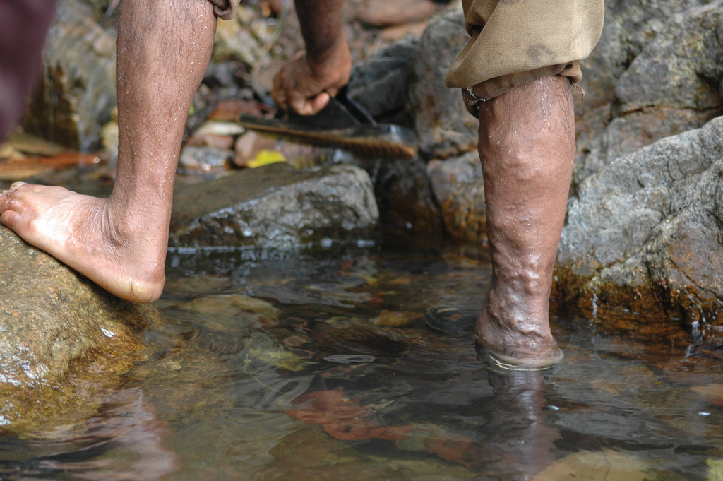
Symptom spectrum
Symptoms are related to venous congestion and skin changes, and classically worsen through the day, with prolonged standing, and in heat, improving with rest and leg elevation: heaviness, fatigue, throbbing, cramping, burning or aching, itchiness of the skin, and a restless-legs-like discomfort.[1] Many patients with visible varicosities are entirely asymptomatic and present purely for cosmetic reasons; others with only mild-appearing veins report disproportionate symptom burden — the CEAP clinical class (below) and the symptom burden do not always move together.
The CEAP Clinical Classification — a genuine, usable staging framework
CEAP is the international consensus system for describing chronic venous disorders, and while it has not been correlated with treatment outcomes directly, its clinical (C) component is genuinely useful in practice as the framework this chapter uses throughout to match compression class and treatment intensity to disease severity.[2][1]
| Component | Code | Meaning |
|---|---|---|
| Clinical findings (C) | C0 | No visible or palpable signs of venous disease |
| C1 | Telangiectasias or reticular veins | |
| C2 | Varicose veins | |
| C3 | Edema | |
| C4a | Eczema (stasis dermatitis) or pigmentation (hemosiderin staining) | |
| C4b | Lipodermatosclerosis or atrophie blanche | |
| C5 | Healed venous leg ulcer | |
| C6 | Active venous leg ulcer | |
| Etiology (E) | Ec | Congenital |
| Ep | Primary | |
| Es | Secondary (e.g., post-thrombotic) | |
| En | No venous etiology identified | |
| Anatomy (A) | As | Superficial veins |
| Ap | Perforating veins | |
| Ad | Deep veins | |
| An | No venous location identified | |
| Pathophysiology (P) | Pr | Reflux |
| Po | Obstruction | |
| Pr,o | Reflux and obstruction | |
| Pn | No venous pathophysiology identified |
Physical findings across the spectrum
| Finding | What it looks like / means |
|---|---|
| Edema | Palpable, pitting swelling, typically starting at the ankle; worsens with dependency, improves with elevation. |
| Stasis (eczematous) changes | Redness and scaling, often itchy; managed with emollients or topical corticosteroids — must be distinguished from cellulitis (below) and from allergic contact dermatitis to a topical product. |
| Hemosiderin staining | Grey-brown pigmentation in the gaiter (lower-leg) area from breakdown of red blood cells forced out of failing capillaries. |
| Corona phlebectatica | A fan-shaped pattern of small, visible blood vessels at the ankle or foot ("ankle flare") — an early sign of more advanced venous disease. |
| Acute lipodermatosclerosis | An extremely painful, red-to-purple, indurated, warm area of the lower leg — frequently misdiagnosed as cellulitis, phlebitis, or panniculitis (see Red Flags & Refer). |
| Chronic lipodermatosclerosis | Chronic inflammation and fibrosis of the skin and subcutaneous tissue, sometimes with an "inverted champagne bottle" leg shape (proximal swelling, a narrowing fibrotic band at the ankle) — a recognized sign of C4 disease. |
| Atrophie blanche | Localized, circular, whitish, atrophic "porcelain" scars surrounded by dilated capillaries, occurring in roughly a third of patients with venous disease; painful due to underlying vascular occlusion. |
| Venous ulcer | A full-thickness skin defect, most often in the gaiter/malleolar region, that fails to heal spontaneously and is sustained by the underlying venous hypertension. |
Stasis dermatitis versus cellulitis — a genuinely important distinction
| Dermatitis | Cellulitis | |
|---|---|---|
| Symptoms | Afebrile; itching | Possible fever; painful |
| Signs | Normal temperature; erythema; may be tender; vesicles/crusting; may be bilateral; often matches the area where a topical product was applied | Elevated local temperature; erythema; tenderness; no crusting; unilateral |
| Portal of entry | Loss of the epidermal barrier allows allergens/irritants to penetrate | Usually a break in the skin (ulcer, trauma, tinea pedis, intertrigo) |
Contact dermatitis (irritant in about 80% of cases, allergic in about 20%) is common in patients using multiple topical products on already-compromised skin; common allergens include lanolin, neomycin, latex, and preservatives in creams and dressing adhesives.[1]
Venous versus arterial ulcer — feeds directly into the PAD-screening safety point below
| Venous ulcer | Arterial ulcer | |
|---|---|---|
| Location | Gaiter/malleolar region | Below the ankle, tip of toes |
| Size/depth | Shallow, irregular, often larger | Deep, often smaller, "punched out" |
| Exudate | Moderate to large amount | None to minimal |
| Wound bed | Granulation or fibrin | Black and dry tissue |
| Pain pattern | Discomfort at end of day, heaviness | Pain with leg elevation or with activity (claudication/rest pain) |
| Pedal pulses | Present | Weak or absent |
| Surrounding leg | Varicose veins, edema, hemosiderin staining | Pallor, dependent rubor, hair loss, thickened nails |
Mixed arterial-venous disease is genuinely common — approximately 15-25% of patients with venous disease also have some degree of arterial insufficiency — which is exactly why every patient needs an objective ABI rather than relying on this table (or a palpated pulse) alone.[1]
Risk Factors
Risk factors map onto the three hemodynamic mechanisms described in Clinical Snapshot.[1]
Valvular reflux
- Obesity
- Multiple pregnancies (mechanical compression of pelvic veins plus hormonally mediated venous wall relaxation)
- Prolonged standing or sitting, whether occupational or recreational
- A prior history of varicose-vein stripping — recurrence is common
- Older age (prevalence and severity both rise with age, particularly past 55) and female sex
- Family history and, rarely, genetic connective-tissue conditions (e.g., Ehlers-Danlos syndrome, Klippel-Trénaunay syndrome)
Obstruction
- A history of deep vein thrombosis (post-thrombotic syndrome)
- May-Thurner syndrome (anatomic compression of the left common iliac vein) — consider in a young woman with unexplained left-leg-predominant chronic swelling
Calf-muscle pump failure
- Reduced ankle range of motion from arthritis, prior joint surgery, or trauma — normal calf-pump function needs a mobile ankle and at least about 10 degrees of dorsiflexion
- An abnormal gait pattern, particularly a shuffling gait, as seen in Parkinson disease and other neurologic conditions affecting mobility
- General immobility or a sedentary lifestyle
Additional considerations
- Smoking — not itself a direct CVI risk factor, but a major driver of concurrent peripheral arterial disease, which is exactly what makes the ABI-screening step in Choose Initial Treatment non-negotiable rather than a formality.
- Diabetes — both a PAD risk factor and, independently, a cause of falsely elevated (non-compressible) ABI readings that can mask true arterial disease (see Assessment & Diagnosis).
Red Flags & Refer
- Suspected acute deep vein thrombosis — new unilateral leg swelling, warmth, or pain, especially with recent immobility, surgery, active cancer, or hormonal therapy: obtain urgent duplex evaluation and anticoagulation assessment rather than attributing new swelling to "the usual" CVI.
- ABI <0.5, or any sign or symptom of critical limb ischemia (rest pain, non-healing digital ulceration, a cool pale foot with absent pulses) — this is an absolute contraindication to standard compression and needs urgent vascular referral, not a modified compression trial in primary care.[3][1]
- Suspected cellulitis (fever, spreading warm tender erythema, systemic symptoms) — use the dermatitis-vs-cellulitis table in Recognize the Condition; do not dismiss as "just eczema," and do not reflexively prescribe antibiotics for uncomplicated stasis dermatitis either.
- Acute lipodermatosclerosis — an extremely painful, red-to-purple, warm, indurated area that is genuinely and commonly misdiagnosed as cellulitis, phlebitis, or panniculitis; recognizing it prevents an unnecessary antibiotic course.
- A non-healing ulcer with atypical features — a friable surface, raised or rolled border, abnormal granulation tissue, or failure to improve after 4-6 weeks of genuinely appropriate management (including compression) — biopsy to exclude malignancy. A squamous cell carcinoma arising within a chronic non-healing wound is a Marjolin's ulcer; basal cell carcinoma is the most common malignancy presenting as a leg ulcer overall. Risk rises with immunosuppression, a prior scar or radiation site, and advanced age.[1]
- Suspected ascending superficial thrombophlebitis, particularly one approaching the saphenofemoral or saphenopopliteal junction — carries a real risk of extension into the deep system; refer for duplex evaluation.
- New arterial symptoms developing in a patient already using compression (increasing pain with the garment on, new claudication, a cooler or more painful foot) — reassess ABI promptly and hold compression pending that reassessment rather than assuming it is "just adjustment discomfort."
Assessment & Diagnosis
History
Cover the risk factors above, any personal history of DVT or venous surgery/procedures, current medications, and comorbidities most relevant to safe compression use — diabetes, peripheral arterial disease, heart failure, and any prior arterial intervention.
Physical examination
Examine both limbs for the physical findings described in Recognize the Condition, palpate femoral, popliteal, dorsalis pedis, and posterior tibial pulses, and assess gait and ankle range of motion (calf-muscle-pump screening). A skilled clinician should not rely on pulse palpation alone: the dorsalis pedis pulse is absent as a normal anatomic variant in roughly 8% of healthy adults, so an impalpable pulse alone neither confirms nor excludes arterial disease — an objective ABI is what actually answers the question.[1]
Ankle-brachial pressure index (ABI) — the gate before any compression recommendation
ABI measurement is recommended in every patient being considered for compression, and is the single most important safety test in this entire chapter.[1][3]
| ABI result | Interpretation and action |
|---|---|
| <0.5 | Absolute contraindication to compression; significant arterial disease — refer urgently to vascular surgery/medicine rather than trialling any compression, even a reduced class.[3] |
| 0.5-0.8 | Mixed arterial-venous disease — compression should only be used following a comprehensive vascular assessment (e.g., by a vascular surgeon), typically with a reduced-pressure, inelastic system, and with close monitoring.[1] |
| 0.8-1.3, or >0.9 by the stricter threshold some short-stretch/high-compression protocols use | Adequate arterial inflow — standard compression is generally safe; high-compression systems should still be applied by, or under the direction of, a trained practitioner.[1] |
| >1.4 | Falsely elevated — indicates calcified, non-compressible vessels rather than true reassurance about arterial flow. This is a common finding in patients with diabetes; further arterial testing (e.g., toe-brachial index) is needed rather than treating this result as normal.[1] |
Duplex ultrasound
Duplex is the reference-standard investigation for confirming venous reflux and/or obstruction, and is used both to complete the anatomic (A) and pathophysiologic (P) components of CEAP staging and to plan any procedural intervention. A reflux duration of 1 second or more in the deep veins, or 0.5 seconds or more in the superficial or perforator veins, is diagnostic of venous insufficiency at that site.[1][4] Duplex also reliably distinguishes acute venous thrombosis from chronic venous changes and evaluates arterial flow-limiting lesions where relevant.
Calf-muscle-pump screening
Because calf-pump failure is one of the three core hemodynamic mechanisms, screening for it is a genuine part of the assessment, not an afterthought: assess ankle dorsiflexion range of motion (at least about 10 degrees is needed to fully activate the pump), calf-muscle strength, and gait pattern, watching specifically for a shuffling gait. A patient with deficits in any of these should be referred to physiotherapy, since a treatment program addressing range of motion, strength, and gait can meaningfully improve calf-muscle-pump function and, with it, response to compression.[1]
Wound assessment (if an ulcer is present)
The MEASURE mnemonic structures a systematic venous ulcer assessment: Measure (length, width, depth), Exudate (amount and character), Appearance of the wound bed, Suffering (pain), Undermining, Re-evaluate at each dressing change, and Edge condition.[1]
Treatment Targets
- Reduce ambulatory venous hypertension — the mechanism underlying every manifestation of this disease — primarily through correctly selected and correctly worn compression.
- Screen for peripheral arterial disease (ABI) before recommending or dispensing standard compression, every time — this is a genuine, recurring safety gate, not a one-time check.
- Prevent progression from mild, cosmetic disease to skin changes, edema, or ulceration.
- Heal an existing venous ulcer, with compression as the treatment cornerstone.
- Prevent ulcer recurrence once healed, primarily through sustained garment adherence.
- Manage symptom burden — aching, heaviness, cramping, itching — even when disease is not severe by CEAP stage.
- Recognize when conservative management has reached its limit and escalate to sclerotherapy, endovenous ablation, or surgical ligation/stripping for significant or persistently symptomatic disease, or for recurrent ulceration.
- Set realistic expectations: even with genuinely optimized care, a meaningful proportion of venous ulcers remain unhealed at 24 weeks — this reflects disease severity, not necessarily a treatment failure at any single step.
Choose Initial Treatment
Before recommending, fitting, or dispensing any standard compression product, confirm arterial status by ABI. Guidelines agreeing across 20 reviewed sources converge on the same absolute contraindications: an ABI below 0.5, significant arterial occlusive disease, and uncontrolled or decompensated heart failure.[3] An ABI in the 0.5-0.8 range needs a comprehensive vascular assessment before any (typically reduced-pressure, inelastic) compression is used.[1] Never recommend high-compression hosiery to a patient with unassessed arterial status on the assumption that "a pulse is present" — a palpable pulse does not exclude arterial disease (see Assessment & Diagnosis).
Compression class selection by CEAP stage / indication
The table below follows the compression-class framework used in the Wounds Canada best-practice document itself.[1] Prescribing garments above 20 mmHg should be done with, or under the direction of, a trained fitter or clinician.
| Class | Pressure | Typical indication |
|---|---|---|
| — | 15-20 mmHg | Tired, achy legs; slight edema; air travel; spider veins/early varicose veins (CEAP C1) 1st-line, mild/cosmetic |
| I | 20-30 mmHg | Varicose veins, mild edema, DVT prevention (CEAP C2) 1st-line, C2 |
| II | 30-40 mmHg | Moderate varicose veins, mild-to-moderate edema, prevention of venous ulcer recurrence (CEAP C3-C4) 1st-line, C3-C4 |
| III | 40-50 mmHg | Severe varicose veins; prevention and treatment of venous leg ulcers; postphlebitic limb; more advanced CVI (CEAP C5-C6) 1st-line, C5-C6/active ulcer |
| IV | 50-60 mmHg | Lymphedema (a different primary indication — see Special Populations for phlebolymphedema) Situational |
Bandages/devices versus garments — a genuinely important distinction, not interchangeable terms
Bandaging systems and compression devices are generally used for active venous ulcers and to reduce established lower-leg edema; compression garments (stockings, or wrap-style hook-and-loop systems) are generally reserved for maintenance once edema has already been reduced.[1] A genuine, common error is measuring and fitting a stocking on a still-edematous limb — as edema resolves the limb shrinks and the garment becomes loose and ineffective. Compression garments should be fitted only after edema has been reduced through bandaging, wraps, or pumps.[1]
Multi-component bandage systems are more effective than single-component systems, and multi-component systems that include an elastic component outperform those composed mainly of inelastic materials; two-component systems perform comparably to four-layer bandaging.[5][1] Compression, in whatever form, genuinely increases ulcer-healing rates compared with no compression — this is well-established, high-confidence evidence, not a marginal effect.[5]
Escalation to procedural treatment
Situational, specialist-directed when varicose veins are significant, persistently symptomatic despite adequate compression, or when ulceration recurs despite genuinely adherent conservative management, escalation to a procedure is appropriate: sclerotherapy (chemical ablation, typically for telangiectasia, reticular veins, and smaller varicosities), endovenous thermal (laser or radiofrequency) ablation, and surgical ligation and stripping of the incompetent superficial vein all aim to remove the source of reflux and reduce the impact of venous hypertension on already-damaged skin.[4][6][1] The largest and most influential trial on this question, the ESCHAR study (500 patients with isolated superficial or mixed superficial/deep reflux), found similar healing rates whether patients received compression alone or compression plus superficial venous surgery, but significantly lower ulcer recurrence in the surgery group — meaning surgical correction of superficial reflux is genuinely worthwhile primarily for reducing recurrence, not for accelerating the initial healing itself.[1] A genuinely Canadian practical detail worth knowing when counselling patients: provincial public insurance plans typically cover saphenous vein ligation and stripping, but do not typically cover endovenous laser, radiofrequency, or chemical ablation technologies, which are usually only available through private payment.[1]
Venoactive drugs — an honest evidence-quality framing, not a substitute for compression
Evidence quality genuinely differs by agent. Pentoxifylline and micronized purified flavonoid fraction (MPFF; diosmin/hesperidin) both have randomized-trial evidence supporting improved venous ulcer healing specifically when added to compression, and are the two agents current multi-society guidance recommends considering for longstanding venous ulcers.[1][6] Phlebotonics as a broader class — which includes horse chestnut seed extract, rutosides, and other botanical agents — have a materially weaker evidence base: Cochrane-level evidence finds they probably reduce oedema slightly, make little or no difference to quality of life, do not demonstrably improve ulcer healing, and carry a higher risk of adverse events than placebo.[7] Set patient expectations accordingly — these are, at best, a modest adjunct to compression, never a substitute for it.
Non-Pharmacologic Treatment
Compression stocking counselling — the centerpiece of this chapter
Measurement and fit
Accurate measurement is what separates a therapeutic garment from a decorative one: measure the ankle circumference at its narrowest point just above the malleoli, the calf at its widest point, and the appropriate length. Garments must be re-measured and re-fitted after edema has meaningfully changed — a stocking fitted to a still-swollen limb will be loose and ineffective once the swelling resolves (see Choose Initial Treatment).[1]
"Put it on before you get out of bed"
This is a genuinely practical, high-value counselling point that is easy to skip in a rushed dispensing conversation: compression stockings should be applied first thing in the morning, while still lying down, before rising — before gravity and a day of standing or sitting allow edema to accumulate. Applying a stocking over a leg that has already swollen during the day is both harder and less effective.
Application technique and donning aids
Many patients — particularly older adults, those with reduced hand strength or dexterity, or those with a large or irregularly shaped limb — genuinely struggle to don a compression garment unassisted. Commercially available donning aids ("donners") make a real, practical difference and should be actively offered, not left for the patient to discover on their own; patients should also be shown how to remove a garment ("doffing") without damaging the garment or the skin. Newer self-adjustable hook-and-loop wrap systems are a genuine alternative for a patient who cannot manage a pull-on stocking, and are increasingly recognized in funding discussions as a legitimate option specifically for this reason.[1]
Choosing garment length
Knee-high garments are adequate for most CVI, since the relevant venous pathology is predominantly below the knee, and they are generally easier to don and better tolerated for daily long-term wear than a thigh-high or waist-high garment — real-world adherence is itself a genuine clinical variable, and a garment that goes unworn provides no benefit regardless of its labelled pressure.
Skin care around compression
Wash the leg with a pH-appropriate, non-alkaline, allergen-free cleanser, and apply a non-sensitizing moisturizer in a downward motion to maintain skin integrity; confirm with the product information that any cream or emollient used is compatible with the compression garment's material (some products degrade elastic fibres).[1] Watch for both irritant and allergic contact dermatitis to the garment fabric itself, not only to topical products.
Replacement schedule
Compression garments lose elasticity and therefore therapeutic pressure with regular wear and laundering — a garment worn daily typically needs replacement roughly every 3-6 months to maintain its labelled compression class; reassess this at every follow-up rather than assuming an old, stretched-out garment is still providing therapeutic pressure.
Leg elevation
Elevate the affected limb above heart level for about 30 minutes, three times per day, to assist venous return passively.[1]
Exercise and calf-muscle-pump activation
Because calf-pump failure is itself a core mechanism (see Risk Factors), exercise is treatment, not just general advice: ankle dorsiflexion/plantarflexion ("ankle pumps"), calf raises, and walking with a proper heel-to-toe gait all directly activate the pump. A structured, supervised exercise program in addition to compression has been associated with meaningfully faster ulcer healing than compression alone in the literature reviewed by Wounds Canada's evidence synthesis — exercise is a genuine adjunct, not an optional extra.[1]
Other general measures
- Weight management where relevant, as part of the overall care plan rather than a prerequisite for other treatment.
- Avoid prolonged static standing or sitting; take movement breaks.
- Avoid restrictive clothing and habitual leg-crossing, which can further impede venous return.
- Smoking cessation — chiefly relevant to reducing concurrent peripheral arterial disease risk, given how directly that risk shapes what compression can safely be recommended.
- Referral to physiotherapy for a shuffling gait, reduced ankle range of motion, or other calf-muscle-pump deficits identified on assessment.
Special Populations
Pregnancy
Progesterone-mediated venodilation combined with mechanical compression of pelvic veins from the gravid uterus commonly causes new or worsened varicosities during pregnancy. Compression stockings are safe and are first-line management. Most pregnancy-related varicosities regress substantially after delivery, so elective procedural treatment (sclerotherapy, ablation, surgery) is generally deferred until the postpartum period rather than pursued during pregnancy. The hypercoagulable state of pregnancy also raises the stakes of any new superficial thrombophlebitis — assess promptly rather than assuming it is routine venous congestion.
Diabetes
Two separate reasons make ABI interpretation more difficult, and more important, in diabetes: peripheral neuropathy can blunt the pain that would otherwise warn of developing ischemia, and medial arterial calcification commonly produces a falsely elevated (non-compressible) ABI reading above 1.4 — a result that must prompt further arterial testing (e.g., toe-brachial index), not reassurance (see Assessment & Diagnosis).[1] Infection risk is also higher; monitor any wound more vigilantly.
Older adults and frailty
Thinner, more fragile skin raises the risk of compression-related skin tears or pressure injury from a poorly fitted or incorrectly applied garment. Reduced hand strength and dexterity genuinely complicate donning — proactively offer donning aids, caregiver training, or a lower-force hook-and-loop wrap alternative rather than assuming non-adherence reflects a lack of motivation.
Heart failure
Uncontrolled or decompensated heart failure is an absolute contraindication to standard high compression, since mobilizing peripheral edema fluid into the central circulation can precipitate volume overload.[3] Any compression use in a patient with a heart failure history needs cardiology coordination and typically a reduced-pressure, closely monitored approach rather than the standard compression ladder applied unmodified.
Obesity
Standard-sized garments may not fit reliably; custom-fit or extra-wide compression products are often needed, and accurate measurement of an irregularly shaped limb is genuinely more difficult and may require padding to protect bony prominences and narrow areas from excessive localized pressure.[1] Obesity is independently a risk factor for both CVI and DVT, reinforcing the value of the broader weight-management conversation as part of the overall plan.
Phlebolymphedema (venous disease with coexisting lymphedema)
Combined venous and lymphatic disease typically needs higher compression classes (III-IV) and different bandaging/garment considerations than CVI alone. This chapter does not duplicate dedicated lymphedema management — refer to the Canadian Lymphedema Framework (canadalymph.ca) for lymphedema-specific guidance.[1]
Reduced mobility and neurologic gait disorders
Parkinson disease, arthritis, and other conditions producing a shuffling gait or reduced ankle range of motion directly impair calf-muscle-pump function (see Risk Factors and Assessment & Diagnosis) — refer to physiotherapy for targeted range-of-motion work, strengthening, and gait retraining rather than treating this purely as a compression-garment problem.
Monitoring & Follow-Up
- Reassess CEAP clinical stage and symptom burden periodically, and confirm the prescribed compression class still matches current status as edema and skin findings change.
- For an active ulcer, reassess using the MEASURE parameters at each dressing change, and set realistic expectations: even with genuinely optimized care, an estimated 30% of venous leg ulcers remain unhealed at 24 weeks — this is a known feature of the disease's healing trajectory, not automatically a sign that something has gone wrong.[1]
- Reassess ABI periodically, and specifically whenever pain develops or worsens with compression, when new arterial symptoms appear, or when diabetes is progressing — do not treat a single normal ABI as permanently reassuring.
- Check garment condition at follow-up and reassess the replacement schedule — a visibly stretched-out or long-worn garment may no longer be delivering its labelled pressure even if it still looks intact.
- Reinforce adherence at every visit. Recurrence after a healed ulcer or resolved edema is disproportionately linked to patients discontinuing maintenance compression once the visible problem has resolved — this is one of the most common, and most preventable, causes of relapse.[1]
- Follow post-procedural patients (sclerotherapy, ablation, surgery) per the proceduralist's specific protocol, typically including a defined period of continued compression afterward.
Common Errors to Avoid
- Recommending or dispensing standard high-compression hosiery without first confirming arterial status by ABI — the single most important safety step in this entire chapter.
- Assuming a palpable pedal pulse rules out arterial disease — it does not, and the dorsalis pedis pulse is absent as a normal variant in about 8% of healthy adults.
- Treating an ABI above 1.4 as reassuring — it commonly reflects calcified, non-compressible vessels (especially in diabetes) rather than genuinely adequate arterial flow, and needs further testing, not reassurance.
- Fitting a compression garment before edema has been adequately reduced with bandaging, wraps, or a pump — the garment will be loose and ineffective once the limb shrinks further.
- Confusing stasis dermatitis for cellulitis and prescribing an unnecessary antibiotic course — or, in the other direction, dismissing true cellulitis as "just eczema."
- Missing acute lipodermatosclerosis and treating it as cellulitis, phlebitis, or panniculitis with antibiotics it does not need.
- Recommending diuretics for pure venous edema — diuretics are indicated for volume-overload states (heart failure, hepatic or renal disease), not for edema caused by venous reflux and valvular incompetence, and do not address the underlying mechanism.
- Advising rest or inactivity for painful legs, when calf-muscle activation through walking and ankle exercises is actually beneficial and directly addresses one of the three core disease mechanisms.
- Presenting venoactive drugs (phlebotonics) generally as a proven substitute for compression, when Cochrane-level evidence shows this class does not improve ulcer healing and carries more adverse events than placebo.
- Skipping the "put it on before you get out of bed" counselling point — a garment applied over a leg that has already swollen through the day is both harder to don and less effective.
- Assuming a non-healing ulcer at some fixed follow-up point automatically represents a treatment failure, rather than recognizing that a substantial minority of venous ulcers remain unhealed at 24 weeks even with genuinely optimized care.
Clinical Decision Algorithm
Drug Tables
How to use this table: this catalogues the drug options relevant across the full CVI spectrum, organized by role — venoactive adjuncts, symptomatic/analgesic support, topical anti-inflammatory therapy for stasis dermatitis, antibiotics for genuine secondary infection, anticoagulants for concurrent thrombosis, and the sclerosant used in procedural sclerotherapy. None of these substitute for correctly selected, correctly worn compression, which remains this chapter's central intervention. Doses shown are typical adult doses only and require patient-specific confirmation. Select any drug name to open its complete Pharmasist Drug Database page.
Venoactive Agents (Phlebotonics)
Evidence quality genuinely differs within this class — see Choose Initial Treatment. Diosmin/hesperidin (MPFF) and horse chestnut seed extract have no Drug Database entry under any plausible slug (independently verified); pentoxifylline does.[1][7]
| Drug | Role | Typical dose | Adverse effects | Comments |
|---|---|---|---|---|
| Pentoxifylline | Adjunct to compression for venous ulcer healing | 400 mg PO three times daily with food | GI upset (most common, often dose-limiting), dizziness, rare hepatotoxicity | The best-evidenced pharmacologic adjunct for venous ulcer healing; continue alongside, not instead of, compression. |
| Diosmin / Hesperidin (MPFF) | Adjunct to compression for longstanding venous ulcer healing | Product-specific, per label | GI upset, headache | No Drug Database entry — verify the specific product before recommending. Recently available OTC in Canada. |
| Horse Chestnut Seed Extract (aescin) | Symptomatic relief only (heaviness, edema) — not ulcer healing | Product-specific, per label | GI upset, headache, dizziness — more adverse events than placebo as a class | No Drug Database entry. Cochrane-level evidence does not support an ulcer-healing benefit for phlebotonics as a class. |
Anti-Inflammatory and Analgesic Adjuncts
For pain related to superficial thrombophlebitis, acute lipodermatosclerosis, or general symptom control; none of these treat the underlying venous pathology.
| Drug | Role | Typical dose | Adverse effects | Comments |
|---|---|---|---|---|
| Acetylsalicylic Acid | Anti-inflammatory adjunct, investigated for ulcer-healing benefit with compression | Low dose, per prescriber direction | GI bleeding/ulceration risk, bruising | Preliminary evidence only — confirm no bleeding-risk contraindication; not established standard therapy. |
| Ibuprofen | Pain relief for superficial thrombophlebitis or general discomfort | Standard adult OTC/prescription dosing, per product | GI upset/bleeding risk, renal effects, cardiovascular risk with prolonged use | Reasonable first-line analgesic option; assess standard NSAID cautions. |
| Naproxen | Alternative NSAID for pain relief | Standard adult dosing, per product | GI upset/bleeding risk, renal effects, cardiovascular risk with prolonged use | Longer duration of action than ibuprofen; same general NSAID cautions apply. |
| Acetaminophen | General analgesia when an NSAID is not appropriate | Standard adult dosing, per product, respecting maximum daily dose | Hepatotoxicity in overdose or with chronic excess/alcohol use | A reasonable default when NSAID cautions (GI, renal, cardiovascular, anticoagulant use) apply. |
| Lidocaine / Prilocaine (eutectic mixture) | Topical local anesthesia before sharp debridement of a painful ulcer | Applied topically prior to the debridement procedure, per product labelling | Local skin blanching/redness, rare methemoglobinemia with excessive application | A genuinely useful pre-procedure option when debridement pain is a barrier to care. |
Topical Corticosteroids for Stasis Dermatitis and Lipodermatosclerosis
Treat the eczematous/inflammatory skin changes, not the underlying venous hypertension — compression remains the definitive treatment.
| Drug | Role | Typical dose | Adverse effects | Comments |
|---|---|---|---|---|
| Betamethasone Dipropionate | Higher-potency option for more significant stasis dermatitis or painful acute lipodermatosclerosis | Thin layer once-twice daily, short defined course | Skin atrophy/thinning with prolonged use, contact sensitization to the vehicle | Step down to a lower-potency agent for maintenance once the acute flare is controlled. |
| Clobetasol Propionate | Super-potent option reserved for severe, refractory flares | Thin layer, short course only (typically under 2 weeks continuous) | Marked skin atrophy risk with prolonged use, HPA-axis suppression with extensive/prolonged use | Reserve for short, defined courses under close follow-up — not a maintenance agent. |
| Triamcinolone | Mid-potency option for moderate stasis dermatitis | Thin layer once-twice daily | Skin atrophy with prolonged use, contact sensitization | A reasonable mid-potency step between hydrocortisone and betamethasone/clobetasol. |
| Hydrocortisone | Lower-potency option for mild disease or longer maintenance use | Thin layer once-twice daily | Generally well tolerated; contact sensitization still possible | Appropriate for milder flares or as a maintenance step-down from a higher-potency course. |
| Desonide | Low-potency alternative, particularly where thinner or more fragile skin is a concern | Thin layer once-twice daily | Generally well tolerated; contact sensitization still possible | A reasonable low-potency alternative to hydrocortisone in a patient with fragile periwound skin. |
Antibiotics for Secondarily Infected Ulcers and Cellulitis
Reserved for clinical infection (spreading erythema, warmth, fever, purulence) — not for bacterial colonization of a chronic wound, which does not itself need systemic antibiotics.[1]
| Drug | Role | Typical dose | Adverse effects | Comments |
|---|---|---|---|---|
| Cephalexin | First-line oral option for uncomplicated cellulitis/infected ulcer | Standard adult dosing, per product and severity | GI upset, hypersensitivity (cross-reactivity risk in severe penicillin allergy) | A standard first-choice covering typical skin/soft-tissue pathogens. |
| Cefadroxil | Alternative first-generation cephalosporin, once- or twice-daily dosing | Standard adult dosing, per product | GI upset, hypersensitivity | A dosing-convenience alternative to cephalexin. |
| Doxycycline | Alternative when MRSA coverage is needed or penicillin/cephalosporin allergy precludes the above | Standard adult dosing, per product | Photosensitivity, GI upset, esophageal irritation | A reasonable option when broader gram-positive coverage including MRSA risk is a concern. |
| Clindamycin | Alternative in significant penicillin/cephalosporin allergy | Standard adult dosing, per product | GI upset, risk of C. difficile infection | Reserve for genuine allergy rather than using routinely given the C. difficile risk. |
| Sulfamethoxazole-Trimethoprim | Alternative with MRSA coverage | Standard adult dosing, per product and renal function | GI upset, hyperkalemia, rash, hematologic effects; potentiates warfarin | Check for a concurrent warfarin prescription given the significant interaction (see Anticoagulants below). |
| Mupirocin | Topical option for a small, localized, clinically infected area | Applied topically per product labelling | Local irritation, rare contact sensitization | Appropriate for a small localized infection; a spreading or systemic infection needs oral or IV therapy instead. |
Anticoagulants for Concurrent Venous Thromboembolism
Relevant when CVI coexists with, or is complicated by, deep or extending superficial vein thrombosis — not a treatment for CVI itself.
| Drug | Role | Typical dose | Adverse effects | Comments |
|---|---|---|---|---|
| Enoxaparin | Low-molecular-weight heparin for acute DVT treatment or bridging | Weight-based dosing, per protocol | Bleeding, injection-site bruising, rare heparin-induced thrombocytopenia | Typically an inpatient/specialist-initiated therapy for confirmed DVT. |
| Dalteparin Sodium | Alternative low-molecular-weight heparin | Weight-based dosing, per protocol | Bleeding, injection-site bruising | Similar role and cautions to enoxaparin. |
| Tinzaparin Sodium | Alternative low-molecular-weight heparin | Weight-based dosing, per protocol | Bleeding, injection-site bruising | Similar role and cautions to enoxaparin. |
| Rivaroxaban | Direct oral anticoagulant for confirmed DVT treatment | Per product labelling and indication-specific dosing | Bleeding risk | A common oral maintenance option once acute anticoagulation is established. |
| Apixaban | Alternative direct oral anticoagulant for confirmed DVT treatment | Per product labelling and indication-specific dosing | Bleeding risk | Similar role to rivaroxaban; agent choice is largely prescriber/patient-factor driven. |
| Warfarin | Alternative oral anticoagulant, particularly where a DOAC is not suitable | Individualized, INR-titrated | Bleeding risk; numerous drug and dietary interactions | Requires INR monitoring; flag interacting antibiotics (e.g., sulfamethoxazole-trimethoprim above) explicitly. |
Sclerosing Agent for Procedural Management
Administered by a trained proceduralist as part of sclerotherapy — not a community-pharmacy-dispensed product, but worth knowing to counsel patients accurately about a referral.[6]
| Drug | Role | Typical dose | Adverse effects | Comments |
|---|---|---|---|---|
| Sodium Tetradecyl Sulfate | Sclerosing agent for telangiectasia, reticular veins, and smaller varicosities | Injection, concentration/volume per procedure | Local skin staining/hyperpigmentation, thrombophlebitis, rare anaphylaxis | Polidocanol, a sclerosant used elsewhere, has no Drug Database entry — do not assume it does. |
References
- Best practice recommendations for the prevention and management of venous leg ulcers — Wounds Canada — Canadian Association of Wound Care (Evans R, Kuhnke JL, Burrows C, Kayssi A, Labreque C, O'Sullivan-Drombolis D, Houghton P). In: Foundations of Best Practice for Skin and Wound Management, 2019 (updated 2021-02-11)
- Revision of the CEAP classification for chronic venous disorders: consensus statement — Journal of Vascular Surgery, American Venous Forum International Ad Hoc Committee (Eklöf B, Rutherford RB, Bergan JJ, Carpentier PH, Gloviczki P, Kistner RL, et al.). J Vasc Surg 2004;40(6):1248-1252
- Compression therapy for venous leg ulcers: risk factors for adverse events and complications, contraindications – a review of present guidelines — Journal of the European Academy of Dermatology and Venereology (Andriessen A, Apelqvist J, Mosti G, Partsch H, Gonska C, Abel M). J Eur Acad Dermatol Venereol 2017;31(9):1562-1568
- The 2022 Society for Vascular Surgery, American Venous Forum, and American Vein and Lymphatic Society clinical practice guidelines for the management of varicose veins of the lower extremities. Part I. Duplex Scanning and Treatment of Superficial Truncal Reflux — Journal of Vascular Surgery: Venous and Lymphatic Disorders (Gloviczki P, Lawrence PF, Wasan SM, Meissner MH, Almeida J, et al.). J Vasc Surg Venous Lymphat Disord 2023;11(2):231-261
- Compression for venous leg ulcers — Cochrane Database of Systematic Reviews (O'Meara S, Cullum N, Nelson EA, Dumville JC). Cochrane Database Syst Rev 2012;11:CD000265
- The 2023 Society for Vascular Surgery, American Venous Forum, and American Vein and Lymphatic Society clinical practice guidelines for the management of varicose veins of the lower extremities. Part II — Journal of Vascular Surgery: Venous and Lymphatic Disorders (Gloviczki P, Lawrence PF, Wasan SM, Meissner MH, Almeida J, et al.). J Vasc Surg Venous Lymphat Disord 2024;12(1):101670
- Phlebotonics for venous insufficiency — Cochrane Database of Systematic Reviews (Martinez-Zapata MJ, Vernooij RWM, Simancas-Racines D, Uriona Tuma SM, Stein AT, Moreno Carriles RM, et al.). Cochrane Database Syst Rev 2020;11:CD003229
Professional reference only. Apply clinical judgment and verify patient-specific decisions against current authoritative guidance and product information.